Mg-Zn-Ca Alloy (ZX00) Screws Are Resorbed at a Mean of 2.5 Years After Medial Malleolar Fracture Fixation: Follow-up of a First-in-humans Application and Insights From a Sheep Model
- PMID: 37603369
- PMCID: PMC10723859
- DOI: 10.1097/CORR.0000000000002799
Mg-Zn-Ca Alloy (ZX00) Screws Are Resorbed at a Mean of 2.5 Years After Medial Malleolar Fracture Fixation: Follow-up of a First-in-humans Application and Insights From a Sheep Model
Abstract
Background: In the ongoing development of bioresorbable implants, there has been a particular focus on magnesium (Mg)-based alloys. Several Mg alloys have shown promising properties, including a lean, bioresorbable magnesium-zinc-calcium (Mg-Zn-Ca) alloy designated as ZX00. To our knowledge, this is the first clinically tested Mg-based alloy free from rare-earth elements or other elements. Its use in medial malleolar fractures has allowed for bone healing without requiring surgical removal. It is thus of interest to assess the resorption behavior of this novel bioresorbable implant.
Questions/purposes: (1) What is the behavior of implanted Mg-alloy (ZX00) screws in terms of resorption (implant volume, implant surface, and gas volume) and bone response (histologic evaluation) in a sheep model after 13 months and 25 months? (2) What are the radiographic changes and clinical outcomes, including patient-reported outcome measures, at a mean of 2.5 years after Mg-alloy (ZX00) screw fixation in patients with medial malleolar fractures?
Methods: A sheep model was used to assess 18 Mg-alloy (ZX00) different-length screws (29 mm, 24 mm, and 16 mm) implanted in the tibiae and compared with six titanium-alloy screws. Micro-CT was performed at 13 and 25 months to quantify the implant volume, implant surface, and gas volume at the implant sites, as well as histology at both timepoints. Between July 2018 and October 2019, we treated 20 patients with ZX00 screws for medial malleolar fractures in a first-in-humans study. We considered isolated, bimalleolar, or trimalleolar fractures potentially eligible. Thus, 20 patients were eligible for follow-up. However, 5% (one patient) of patients were excluded from the analysis because of an unplanned surgery for a pre-existing osteochondral lesion of the talus performed 17 months after ZX00 implantation. Additionally, another 5% (one patient) of patients were lost before reaching the minimum study follow-up period. Our required minimum follow-up period was 18 months to ensure sufficient time to observe the outcomes of interest. At this timepoint, 10% (two patients) of patients were either missing or lost to follow-up. The follow-up time was a mean of 2.5 ± 0.6 years and a median of 2.4 years (range 18 to 43 months).
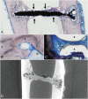
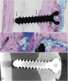
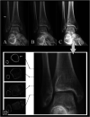
Results: In this sheep model, after 13 months, the 29-mm screws (initial volume: 198 ± 1 mm 3 ) degraded by 41% (116 ± 6 mm 3 , mean difference 82 [95% CI 71 to 92]; p < 0.001), and after 25 months by 65% (69 ± 7 mm 3 , mean difference 130 [95% CI 117 to 142]; p < 0.001). After 13 months, the 24-mm screws (initial volume: 174 ± 0.2 mm 3 ) degraded by 51% (86 ± 21 mm 3 , mean difference 88 [95% CI 52 to 123]; p = 0.004), and after 25 months by 72% (49 ± 25 mm 3 , mean difference 125 [95% CI 83 to 167]; p = 0.003). After 13 months, the 16-mm screws (initial volume: 112 ± 5 mm 3 ) degraded by 57% (49 ± 8 mm 3 , mean difference 63 [95% CI 50 to 76]; p < 0.001), and after 25 months by 61% (45 ± 10 mm 3 , mean difference 67 [95% CI 52 to 82]; p < 0.001). Histologic evaluation qualitatively showed ongoing resorption with new bone formation closely connected to the resorbing screw without an inflammatory reaction. In patients treated with Mg-alloy screws after a mean of 2.5 years, the implants were radiographically not visible in 17 of 18 patients and the bone had homogenous texture in 15 of 18 patients. No clinical or patient-reported complications were observed.
Conclusion: In this sheep model, Mg-alloy (ZX00) screws showed a resorption to one-third of the original volume after 25 months, without eliciting adverse immunologic reactions, supporting biocompatibility during this period. Mg-alloy (ZX00) implants were not detectable on radiographs after a mean of 2.5 years, suggesting full resorption, but further studies are needed to assess environmental changes regarding bone quality at the implantation site after implant resorption.
Clinical relevance: The study demonstrated successful healing of medial malleolar fractures using bioresorbable Mg-alloy screws without clinical complications or revision surgery, resulting in pain-free ankle function after 2.5 years. Future prospective studies with larger samples and extended follow-up periods are necessary to comprehensively assess the long-term effectiveness and safety of ZX00 screws, including an exploration of limitations when there is altered bone integrity, such as in those with osteoporosis. Additional use of advanced imaging techniques, such as high-resolution CT, can enhance evaluation accuracy.
Copyright © 2023 The Author(s). Published by Wolters Kluwer Health, Inc. on behalf of the Association of Bone and Joint Surgeons.
Conflict of interest statement
All ICMJE Conflict of Interest Forms for authors and Clinical Orthopaedics and Related Research ® editors and board members are on file with the publication and can be viewed on request.
Figures


Comment in
-
CORR Insights®: Mg-Zn-Ca Alloy (ZX00) Screws Are Resorbed at a Mean of 2.5 Years After Medial Malleolar Fracture Fixation: Follow-up of a First-in-humans Application and Insights From a Sheep Model.Clin Orthop Relat Res. 2024 Jan 1;482(1):198-200. doi: 10.1097/CORR.0000000000002866. Epub 2023 Sep 28. Clin Orthop Relat Res. 2024. PMID: 37768868 Free PMC article. No abstract available.
References
-
- Amerstorfer F, Fischerauer SF, Fischer L, et al. Long-term in vivo degradation behavior and near-implant distribution of resorbed elements for magnesium alloys WZ21 and ZX50. Acta Biomater. 2016;42:440-450. - PubMed
-
- Brunner P, Brumbauer F, Steyskal EM, et al. Influence of high-pressure torsion deformation on the corrosion behaviour of a bioresorbable Mg-based alloy studied by positron annihilation. Biomater Sci. 2021;9:4099-4109. - PubMed
-
- Castellani C, Lindtner RA, Hausbrandt P, et al. Bone-implant interface strength and osseointegration: biodegradable magnesium alloy versus standard titanium control. Acta Biomater. 2011;7:432-440. - PubMed
-
- Cihova M, Martinelli E, Schmutz P, et al. The role of zinc in the biocorrosion behavior of resorbable Mg‒Zn‒Ca alloys. Acta Biomater. 2019;100:398-414. - PubMed
Publication types
MeSH terms
Substances
LinkOut - more resources
Full Text Sources
Medical

