. 2023 Oct;27(19):2983-2994.
doi: 10.1111/jcmm.17924. Epub 2023 Aug 21.

Epigenetic histone modification by butyrate downregulates KIT and attenuates mast cell function

Affiliations

Epigenetic histone modification by butyrate downregulates KIT and attenuates mast cell function

Ravindra Gudneppanavar et al. J Cell Mol Med. 2023 Oct.

Abstract

Short-chain fatty acid butyrate is produced from the bacterial fermentation of indigestible fiber in the intestinal lumen, and it has been shown to attenuate lung inflammation in murine asthma models. Mast cells (MCs) are initiators of inflammatory response to allergens, and they play an important role in asthma. MC survival and proliferation is regulated by its growth factor stem cell factor (SCF), which acts through the receptor, KIT. It has previously been shown that butyrate attenuates the activation of MCs by allergen stimulation. However, how butyrate mechanistically influences SCF signalling to impact MC function remains unknown. Here, we report that butyrate treatment triggered the modification of MC histones via butyrylation and acetylation, and inhibition of histone deacetylase (HDAC) activity. Further, butyrate treatment caused downregulation of SCF receptor KIT and associated phosphorylation, leading to significant attenuation of SCF-mediated MC proliferation, and pro-inflammatory cytokine secretion. Mechanistically, butyrate inhibited MC function by suppressing KIT and downstream p38 and Erk phosphorylation, and it mediated these effects via modification of histones, acting as an HDAC inhibitor and not via its traditional GPR41 (FFAR3) or GPR43 (FFAR2) butyrate receptors. In agreement, the pharmacological inhibition of Class I HDAC (HDAC1/3) mirrored butyrate's effects, suggesting that butyrate impacts MC function by HDAC1/3 inhibition. Taken together, butyrate epigenetically modifies histones and downregulates the SCF/KIT/p38/Erk signalling axis, leading to the attenuation of MC function, validating its ability to suppress MC-mediated inflammation. Therefore, butyrate supplementations could offer a potential treatment strategy for allergy and asthma via epigenetic alterations in MCs.

Keywords: HDAC; KIT; MAPK; MC; SCF; asthma; butyrate; proliferation; viability.


Conflict of interest statement

Authors declare no conflict of interest.

Figures
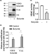
FIGURE 1
FIGURE 1
Epigenetic modification of histones by butyrate. BMCs were isolated and differentiated into BMMCs using murine interleukin‐3 (IL‐3; 30 ng/mL) for 5 weeks. BMMCs (1 × 106) were treated with sodium butyrate (5 mM) for 24 h, and histones were extracted. Histone proteins were subjected to western blotting using H3K9 butyrylation, pan lysine acetylation and total histone H3 antibodies. (A) Western blot of histones using butyrylation, acetylation, and total Histone antibodies. (B) Densitometric analysis of data shown in (A). (C) Nuclear extracts were prepared from untreated and butyrate (5 mM) treated cells and 2 μg of nuclear protein was used to analyse HDAC activity. HDAC activity was measured colorimetrically by measuring the absorbance at 450 nm using Microplate reader. Data are represented as mean ± SEM of three separate experiments. The significance was tested using one‐way anova and post hoc analysis. *p ≤ 0.05, **p ≤ 0.01, ****p ≤ 0.0001, ns = not significant.
FIGURE 2
FIGURE 2
Butyrate downregulates KIT and attenuate KIT phosphorylation. (A) Effect of butyrate on internalization of KIT receptors in BMMCs. BMMCs were treated with butyrate (5 mM, 24 h) and surface expression of KIT receptor was analysed by flow cytometry. (B) Quantification of data shown in (A) and expressed as net mean fluorescence intensity (MFI). (C) BMMCs were treated with butyrate (5 mM, 6, 12 and 24 h) followed by treatment with SCF (100 ng/mL) for 30 min. KIT phosphorylation was assessed by western blotting using phospho‐specific KIT (Y719) antibody. Blots were re‐probed for total KIT and GAPDH. (D) BMMCs were treated with butyrate (5 mM, 24 h) followed by treatment with SCF (100 ng/mL) for 30 min. KIT phosphorylation was assessed by western blotting using phospho‐specific KIT (Y719) antibody. Blots were re‐probed for total KIT and GAPDH. (E) and (F) represent densitometric analysis of data shown in C. Data are represented as mean ± SEM of three separate experiments. The significance was tested using one‐way anova and post hoc analysis. *p ≤ 0.05, ***p ≤ 0.001, ****p ≤ 0.0001, ns = not significant.
FIGURE 3
FIGURE 3
Butyrate attenuates SCF‐mediated BMMC viability and proliferation. (A). Cartoon depicting that SCF/KIT axis promotes MC activation. BMMCs were plated in triplicate at a density of (1 × 104) in each well of a 96 well plate suspended in fresh medium without IL‐3 and in the presence or absence of SCF (100 ng/mL), pretreated or not with butyrate (5 mM, 24 h). (B) Cell viability was analysed by XTT assay after 72 h. (C) Cell proliferation was measured after 72 h by BrdU ELISA. BrdU label was added 24 h before the assay. (D) Cell proliferation was measured by BrdU ELISA as described above in BMMCs pretreated with 5 mM of other SCFA; acetate, propionate and butyrate. Viability and proliferation data are represented as percentage over control and mean ± SEM of three separate experiments. The significance was tested using one‐way anova and post hoc analysis. **p ≤ 0.01, ***p ≤ 0.001, ****p ≤ 0.0001, ns = not significant.
FIGURE 4
FIGURE 4
Butyrate attenuates SCF‐mediated inflammatory mediator release. BMMCs were pretreated or not with butyrate (5 mM) for 24 h, and stimulated in the presence or absence of SCF (100 ng/mL) for 4 h. RNA was extracted followed by cDNA synthesis and transcript levels of MIP1β (A), MCP‐1 (B), TNFα (C), COX‐2 (D), and COX‐1 (E) were measured by qPCR and analysed compared to GAPDH. Culture supernatants were collected and analysed for MIP1β (F), MCP‐1 (G), TNFα (H) proteins by ELISA. Data are represented as mean ± SEM of three separate experiments. The significance was tested using one‐way anova and post hoc analysis. **p ≤ 0.01, ***p ≤ 0.001, ****p ≤ 0.0001, NS = not significant.
FIGURE 5
FIGURE 5
Butyrate attenuates SCF‐mediated phosphorylation of p38 and Erk‐ BMMCs were pretreated or not with butyrate (5 mM) for 24 h, and stimulated in the presence or absence of SCF (100 ng/mL) for 30 min. (A) p38 and Erk phosphorylation and expression was determined in the cell lysates by western blotting. Blots were re‐probed for total KIT and GAPDH. (B, C) represent densitometric analysis of data shown in (A). BMMCs were stimulated in the presence or absence of SCF (100 ng/mL; 4 h) with or without BIRB0796 (0.1 μM) and PD98059 (50 μM) and MIP1β (D) and MCP‐1(E) expression in the culture medium was determined by ELISA. Data are represented as mean ± SEM of three separate experiments. The significance was tested using one‐way anova and post hoc analysis. *p ≤ 0.05, **p ≤ 0.01, ***p ≤ 0.001, ****p ≤ 0.0001, NS = not significant.
FIGURE 6
FIGURE 6
Butyrate induces MC function independent of GPR41 or GPR43. (A) BMMCs were analysed for the expression of GPR41, GPR43, and GPR109A by qPCR. BMMCs were plated in triplicate at a density of (1 × 104) in each well of a 96 well plate suspended in fresh medium without IL‐3 and in the presence or absence of SCF (100 ng/mL), pretreated or not with butyrate ± BHB (both 5 mM, 24 h) (B) and butyrate ± GLPG (5 mM and 0.1 μM respectively, 24 h) (C). Cell proliferation was measured after 72 h by BrdU ELISA. BrdU label was added 24 h before the assay. Viability and proliferation data are represented as percentage over control and mean ± SEM of three separate experiments. The significance was tested using one‐way anova and posthoc analysis. *p ≤ 0.05, ns = not significant.
FIGURE 7
FIGURE 7
HDAC1/3 inhibitors mirror butyrate effect on SCF‐mediated MC activation. BMMCs were treated with SCF with or without and SBHA (20 μM), and SAHA (1 μM) and Cell viability was analysed by XTT assay after 72 h (A). Cell proliferation was measured after 72 h by BrdU ELISA. BrdU label was added 24 h before the assay. Viability and proliferation data are represented as percentage over control (B). BMMCs were pretreated or not with SBHA (20 μM), and SAHA (1 μM) for 24 h, and stimulated in the presence or absence of SCF (100 ng/mL) for 30 min. (C) KIT, Erk and p38 phosphorylation and expression was determined in the cell lysates by western blotting. Blots were re‐probed for total KIT and GAPDH. BMMCs were pretreated or not with butyrate (5 mM) for 24 h, and stimulated in the presence or absence of SCF (100 ng/mL) for 4 h. Culture supernatants were collected and analysed for MIP1β (D), MCP‐1 (E), TNFα (F) proteins by ELISA. Data are represented as mean ± SEM of three separate experiments. The significance was tested using one‐way anova and post hoc analysis. *p ≤ 0.05, **p ≤ 0.01, ***p ≤ 0.001, ****p ≤ 0.0001, NS = not significant.
FIGURE 8
FIGURE 8
Schematic depicting putative mechanisms of epigenetic regulation of KIT and SCF‐mediated mast cell (MC) function. In untreated MC, binding of SCF to KIT induces its tyrosine phosphorylation at Y719 which in turn activates p38, Erk‐dependent expression of KIT and inflammatory genes leading to MC proliferation and secretion of inflammatory mediators. However, butyrate treatment reduces KIT expression, tyrosine phosphorylation and downstream signalling (p38 MAPK, Erk, proliferation and inflammatory mediator secretion) via epigenetic modification of histones leading to suppression of KIT‐mediated MC activation and proliferation.

Similar articles

Cited by

References

    1. Gurish MF, Boyce JA. Mast cells: ontogeny, homing, and recruitment of a unique innate effector cell. J Allergy Clin Immunol. 2006;117(6):1285‐1291. - PubMed
    1. Galli SJ, Kalesnikoff J, Grimbaldeston MA, Piliponsky AM, Williams CM, Tsai M. Mast cells as "tunable" effector and immunoregulatory cells: recent advances. Annu Rev Immunol. 2005;23:749‐786. - PubMed
    1. Galli SJ, Tsai M, Wershil BK, Tam SY, Costa JJ. Regulation of mouse and human mast cell development, survival and function by stem cell factor, the ligand for the c‐kit receptor. Int Arch Allergy Immunol. 1995;107(1–3):51‐53. - PubMed
    1. Metcalfe DD, Baram D, Mekori YA. Mast cells. Physiol Rev. 1997;77(4):1033‐1079. - PubMed
    1. Teegala LR, Elshoweikh Y, Gudneppanavar R, et al. Protein Kinase C alpha and beta compensate for each other to promote stem cell factor‐mediated KIT phosphorylation, mast cell viability and proliferation. FASEB J. 2022;36(5):e22273. doi: 10.1096/fj.202101838RRR - DOI - PMC - PubMed

Publication types