. 2023 Aug 29;120(35):e2302070120.
doi: 10.1073/pnas.2302070120. Epub 2023 Aug 21.

Glucocorticoids regulate lipid mediator networks by reciprocal modulation of 15-lipoxygenase isoforms affecting inflammation resolution

Affiliations

Glucocorticoids regulate lipid mediator networks by reciprocal modulation of 15-lipoxygenase isoforms affecting inflammation resolution

Zhigang Rao et al. Proc Natl Acad Sci U S A. .

Abstract

Glucocorticoids (GC) are potent anti-inflammatory agents, broadly used to treat acute and chronic inflammatory diseases, e.g., critically ill COVID-19 patients or patients with chronic inflammatory bowel diseases. GC not only limit inflammation but also promote its resolution although the underlying mechanisms are obscure. Here, we reveal reciprocal regulation of 15-lipoxygenase (LOX) isoform expression in human monocyte/macrophage lineages by GC with respective consequences for the biosynthesis of specialized proresolving mediators (SPM) and their 15-LOX-derived monohydroxylated precursors (mono-15-OH). Dexamethasone robustly up-regulated pre-mRNA, mRNA, and protein levels of ALOX15B/15-LOX-2 in blood monocyte-derived macrophage (MDM) phenotypes, causing elevated SPM and mono-15-OH production in inflammatory cell types. In sharp contrast, dexamethasone blocked ALOX15/15-LOX-1 expression and impaired SPM formation in proresolving M2-MDM. These dexamethasone actions were mimicked by prednisolone and hydrocortisone but not by progesterone, and they were counteracted by the GC receptor (GR) antagonist RU486. Chromatin immunoprecipitation (ChIP) assays revealed robust GR recruitment to a putative enhancer region within intron 3 of the ALOX15B gene but not to the transcription start site. Knockdown of 15-LOX-2 in M1-MDM abolished GC-induced SPM formation and mono-15-OH production. Finally, ALOX15B/15-LOX-2 upregulation was evident in human monocytes from patients with GC-treated COVID-19 or patients with IBD. Our findings may explain the proresolving GC actions and offer opportunities for optimizing GC pharmacotherapy and proresolving mediator production.

Keywords: glucocorticoid; inflammation; lipid mediators; lipoxygenases.


Conflict of interest statement

The authors declare no competing interest.

Figures

Fig. 1.
Fig. 1.
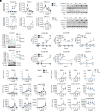
Dexamethasone (Dex) causes reciprocal modulation of 15-LOX isoform expression and dictates LM signature profiles. (A) Schematic illustration of the LM-biosynthetic pathways with relevance for this study. (B) Human M0GM-CSF and M0M-CSF (2 × 10⁶ cells/mL, each) were preincubated with 100 nM Dex or vehicle (0.1% DMSO) for 15 min and then polarized for 48 h to M1/M1Dex or M2/M2Dex, respectively. Protein expression of 15-LOX-1 and 15-LOX-2 normalized to β-actin was determined by Western blot; n = 3 separate donors, ratio paired t-test vs. vehicle. (C) MDM (2 × 10⁶ cells/mL; differentiated with 10 ng/mL GM-SCF and 10 ng/mL M-CSF for 6 d) or (D) monocytes (2 × 10⁶ cells/mL) were incubated with Dex (100 nM) or vehicle (0.1% DMSO) for 48 h. Protein expression of 15-LOX-2 normalized to β-actin was determined by Western blot; n = 3 separate donors, ratio paired t-test vs. vehicle. (E) M0GM-CSF and M0M-CSF (2 × 10⁶ cells/ml, each) were preincubated with 100 nM Dex or vehicle (0.1% DMSO) for 15 min and then polarized for 48 h to M1/M1Dex or M2/M2Dex, respectively. Protein expression of COX-2, COX-1, 5-LOX, and FLAP normalized to β-actin was determined by Western blot; n = 3 separate donors, ratio paired t-test vs. vehicle. (F) Secreted cytokines were quantified by ELISA; n = 3-5 separate donors, ratio paired t-test vs. vehicle. (G) Expression of CD54 and CD80 (M1) as well as CD163 and CD206 (M2) among viable CD14+ cells was analyzed, the mean fluorescence intensity (MFI) is shown as mean + S.E.M, n = 3-4, ratio paired t-test vs. vehicle control. (H) M0GM-CSF and M0M-CSF (2 × 10⁶ cells/ml, each) were preincubated with 100 nM Dex or vehicle (0.1% DMSO) for 15 min and then polarized for 48 h to M1/M1Dex or M2/M2Dex, respectively. MDM (2 × 10⁶ cells/ml; differentiated with 10 ng/ml GM-CSF and 10 ng/ml M-CSF for six days) or monocytes (2 × 10⁶ cells/ml) were incubated with Dex (100 nM) or vehicle (0.1% DMSO) for 48 h. Cells were incubated with E. coli (O6:K2:H1; ratio 1:50) for another 180 min. Formed LM were analyzed and normalized to total protein amounts given as pg/0.15 mg protein. LOD for LM analysis was 3 pg/0.15 mg protein. Data in the radar blots are means of n = 3-4 separate donors for different LM. Statistics: ratio paired t-test Dex-group vs. vehicle group. (B-H) Level of significances: *P < 0.05, **P < 0.01, and ***P < 0.001.
Fig. 2.
Fig. 2.
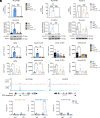
Temporal modulation of LM pathways by dexamethasone (Dex) during M1- and M2-polarization. (A) Human M0GM-CSF and M0M-CSF (2 × 10⁶ cells/mL, each) were preincubated with Dex (100 nM) or vehicle (0.1% DMSO) for 15 min and then polarized for the indicated times to M1/M1Dex or M2/M2Dex, respectively. Protein expression of 15-LOX-1, 15-LOX-2 and cPLA2α, normalized to β-actin, was determined by western blot; n = 3-4 separate donors, two-way ANOVA–Bonferroni multiple comparison test. (B) Human M0M-CSF (2 × 10⁶ cells/mL) were polarized to M2 for 48 h and then treated with Dex (100 nM) or vehicle (0.1% DMSO) for another 48 h. 15-LOX-1 and 15-LOX-2 protein expression, normalized to β-actin, was determined by western blot; n = 3 separate donors, ratio paired t-test vs. vehicle. (CE) M0GM-CSF and M0M-CSF (2 × 10⁶ cells/ml, each) were preincubated with Dex (100 nM) or vehicle (0.1% DMSO) for 15 min and then polarized for the indicated times to M1/M1Dex or M2/M2Dex, respectively. (C and D) Total RNA, including mRNA (C) and pre-mRNA (D), was isolated, transcribed into cDNA and quantified by qPCR; fold increase vs. control (vehicle, t=0 h) is given; n = 3 separate donors, two-way ANOVA-Bonferroni multiple comparison test. (E) Cells were incubated with E. coli (O6:K2:H1; ratio 1:50) for 180 min and formed LM were analyzed. LM were normalized to total protein amounts; n = 4 separate donors, two-way ANOVA-Bonferroni multiple comparison test. (AE) Level of significances: *P < 0.05, **P < 0.01, and ***P < 0.001.
Fig. 3.
Fig. 3.
Modulation of LM pathways by dexamethasone (Dex) is mediated by the GC receptor. Human M0GM-CSF and M0M-CSF (2 × 10⁶ cells/mL, each) were preincubated with (AC) RU486 (1 µM) for 15 min before treatment with 100 nM Dex or vehicle (0.1% DMSO) for another 15 min, and then cells were polarized for 48 h to M1 or M2, respectively. (A) Total RNA was isolated, transcribed into cDNA, and quantified via qPCR; data are given as the -fold increase vs. vehicle; n = 3 separate donors, RM one-way ANOVA-Bonferroni multiple comparison test (vehicle vs. Dex, vehicle vs. RU486, Dex vs. RU486 + Dex). (B) Protein expression of 15-LOX isoforms, normalized to β-actin, was determined by western blot; n = 3 to 4 separate donors, RM one-way ANOVA–Bonferroni multiple comparison test (vehicle vs. Dex, vehicle vs. RU486, Dex vs. RU486 + Dex). (C) MDM were incubated with E. coli (O6:K2:H1; ratio 1:50) for another 180 min. Formed LM were extracted and analyzed. LM were normalized to total protein amounts. Data are mean ± SEM from n = 4 separate donors, RM one-way ANOVA-Bonferroni multiple comparison test (vehicle vs. Dex, vehicle vs. RU486, Dex vs. RU486 + Dex). SPM include PDX, PD1, MaR1 and RvD5. (D, Upper) GR occupancy on ALOX15B and ALOX15 genes from representative ChIP-seq data from Wang et al. (26) in vehicle (veh) and GC (triamcinolone; TA)-treated human macrophages. Lower: ChIP-qPCR data showing occupancy of GR at ALOX15B TSS and intron 3, and at ALOX15 TSS (amplicons indicated in orange, blue and purple, respectively) in MDM treated with IL-4 (M2) or Dex (100 nM)/IL-4 (M2Dex) for 6 h. Data are presented as percentage of input DNA. IgG IP served as negative control. Bars represent mean ± SEM (n = 3 donors), independent ratio paired t test Dex vs. vehicle for IgG and GR IP. Level of significances: *P < 0.05, **P < 0.01 and ***P < 0.001.
Fig. 4.
Fig. 4.
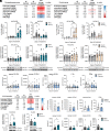
Impact of genetic interference with ALOX15B mRNA on Dex-modulated LM formation. (A and B) Human M0GM-CSF and M0M-CSF were subjected to electroporation with sequence-specific siRNA against ALOX15B or nontarget siRNA (200 nM, each) and then preincubated with Dex (100 nM) for 15 min before polarization for 48 h to M1/M1Dex or M2/M2Dex, respectively. (A) Protein expression of 15-LOX-1, 15-LOX-2, and COX-2, normalized to β-actin, was determined by western blot; n = 3 separate donors, RM one-way ANOVA-Bonferroni multiple comparison test (nontarget vs. ALOX15B, nontarget vs. nontarget + Dex, ALOX15B vs. ALOX15B + Dex, nontarget + Dex vs. ALOX15B + Dex). (B) Cells were incubated with E. coli (O6:K2:H1; ratio 1:50) for another 180 min. Formed LM were analyzed and normalized to total protein amount. In the heatmap, the amounts of each LM in nontarget control group are shown in pg/0.15 mg protein given as means, and additionally the -fold change of each LM after different treatments vs. treatment with nontarget siRNA is given. For values with LOD ≤ 3 pg/0.15 mg protein, the -fold change was calculated on the basis of 3 pg/0.15 mg protein.
Fig. 5.
Fig. 5.
Upregulation of 15-LOX-2 in monocytes during clinical GC therapy in acute (COVID-19) and chronic (IBD) inflammation. (AD) COVID-19 study. (A) Blood samples were tested for clinical parameters before and after a median of 4 d (IQR 3/6). Median absolute values and median -fold changes are shown, Wilcoxon matched-pairs signed rank test vs. control (day 0). (B and C) Monocytes (CD14+) and neutrophils (CD16+) were isolated by magnetic beads cell sorting. (B) mRNA expression of ALOX15 and ALOX15B, given as -fold change vs. control (day 0, n = 5 to 7). (C) Protein expression of 15-LOX-2 and 5-LOX, normalized to β-actin (n = 6 to 7 separate donors). Statistics: Friedman test with Dunn multiple comparison test vs. control (day 0). (D) Plasma (1 mL) was prepared followed by LM analysis (n = 6 to 7); Friedman test with Dunn multiple comparison test vs. control (day 0). (EH) IBD study. (E) Blood was tested for clinical parameters. Median absolute values and median -fold changes are shown from n = 6 to 11 patients, Wilcoxon matched-pairs signed rank test day 4 vs. control (day 0). (F) Plasma was prepared from blood at day 0 and day 4 and analyzed for LM. LM are given in pg/2 mL of plasma; n = 11 separate donors, Wilcoxon matched-pairs signed rank test day 4 vs. control (day 0). (G and H) Monocytes were isolated from blood (18 mL) at day 0 and day 4, cultivated for 24 h at 37 °C, and (G) used for determination of protein expression of 15-LOX-2 (n = 11) and 5-LOX (n = 7), normalized to β-actin, using western blot, or (H) stimulated with E. coli-conditioned medium (0.5%) for 90 min, followed by LM analysis. LM are given as mean ± SEM in pg/mg total protein (n = 8); Wilcoxon matched-pairs signed rank test day 4 vs. control (day 0). Level of significances: *P < 0.05, **P < 0.01 and ***P < 0.001.

References

    1. Cain D. W., Cidlowski J. A., Immune regulation by glucocorticoids. Nat. Rev. Immunol. 17, 233–247 (2017). - PMC - PubMed
    1. Perretti M., D’Acquisto F., Annexin A1 and glucocorticoids as effectors of the resolution of inflammation. Nat. Rev. Immunol. 9, 62–70 (2009). - PubMed
    1. Horby P., et al. , Dexamethasone in hospitalized patients with Covid-19. N Engl. J. Med. 384, 693–704 (2021). - PMC - PubMed
    1. Ehrchen J. M., Roth J., Barczyk-Kahlert K., More than suppression: Glucocorticoid action on monocytes and macrophages. Front. Immunol. 10, 2028 (2019). - PMC - PubMed
    1. Barnes P. J., Glucocorticosteroids: Current and future directions. Br. J. Pharmacol. 163, 29–43 (2011). - PMC - PubMed

Publication types