. 2023 Aug 29;120(35):e2303814120.
doi: 10.1073/pnas.2303814120. Epub 2023 Aug 21.

NLRP3 is essential for neutrophil polarization and chemotaxis in response to leukotriene B4 gradient

Affiliations

NLRP3 is essential for neutrophil polarization and chemotaxis in response to leukotriene B4 gradient

Stijn Van Bruggen et al. Proc Natl Acad Sci U S A. .

Abstract

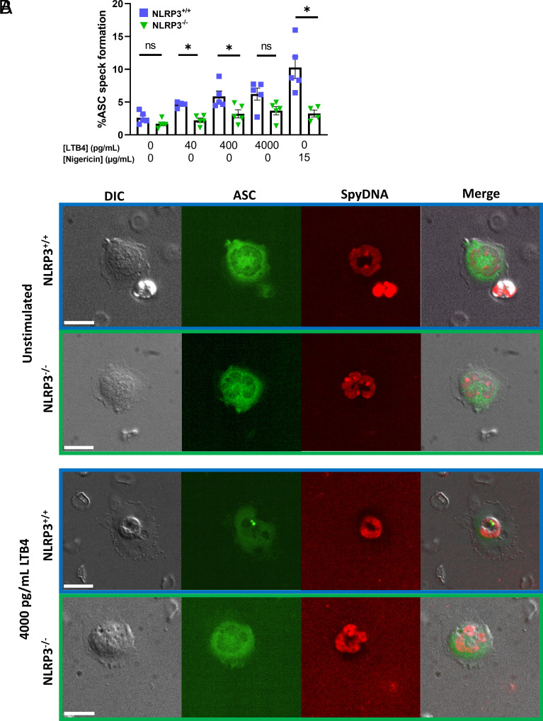
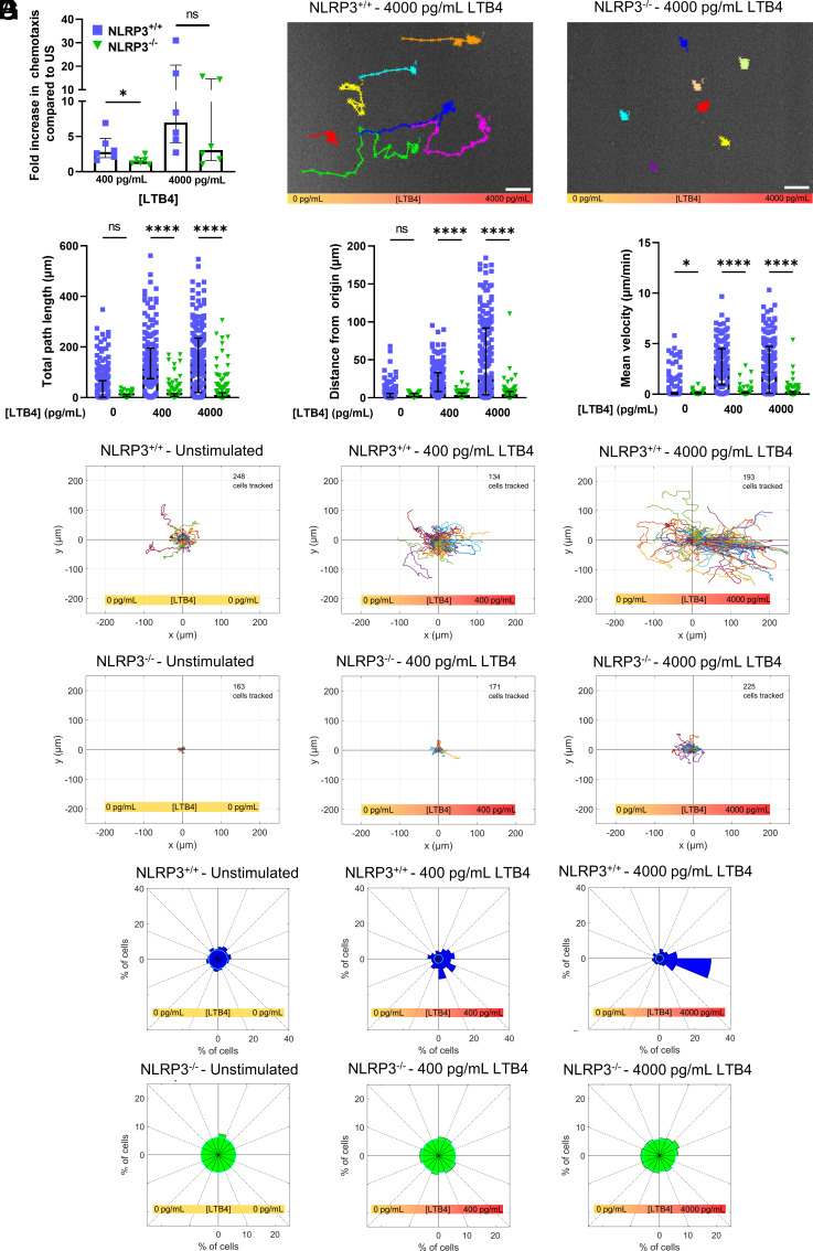
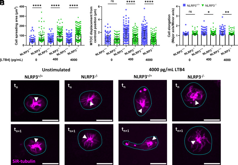
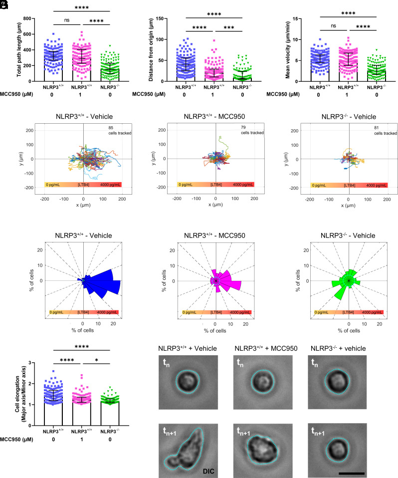
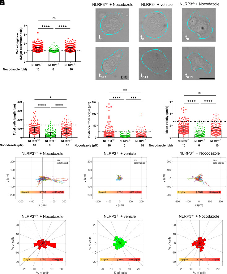
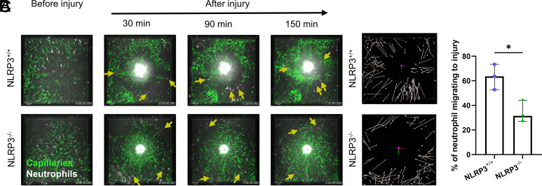
Neutrophil recruitment to sites of infection and inflammation is an essential process in the early innate immune response. Upon activation, a subset of neutrophils rapidly assembles the multiprotein complex known as the NLRP3 inflammasome. The NLRP3 inflammasome forms at the microtubule organizing center, which promotes the formation of interleukin (IL)-1β and IL-18, essential cytokines in the immune response. We recently showed that mice deficient in NLRP3 (NLRP3-/-) have reduced neutrophil recruitment to the peritoneum in a model of thioglycolate-induced peritonitis. Here, we tested the hypothesis that this diminished recruitment could be, in part, the result of defects in neutrophil chemotaxis. We find that NLRP3-/- neutrophils show loss of cell polarization, as well as reduced directionality and velocity of migration toward increasing concentrations of leukotriene B4 (LTB4) in a chemotaxis assay in vitro, which was confirmed through intravital microscopy of neutrophil migration toward a laser-induced burn injury of the liver. Furthermore, pharmacologically blocking NLRP3 inflammasome assembly with MCC950 in vitro reduced directionality but preserved nondirectional movement, indicating that inflammasome assembly is specifically required for polarization and directional chemotaxis, but not cell motility per se. In support of this, pharmacological breakdown of the microtubule cytoskeleton via nocodazole treatment induced cell polarization and restored nondirectional cell migration in NLRP3-deficient neutrophils in the LTB4 gradient. Therefore, NLRP3 inflammasome assembly is required for establishment of cell polarity to guide the directional chemotactic migration of neutrophils.

Keywords: NLRP3 inflammasome; cell polarization; chemotaxis; neutrophils.


Conflict of interest statement

The authors declare no competing interest.

Figures

Fig. 1.
Fig. 1.
Stimulation of primary murine neutrophils through leukotriene B4 results in the formation of NLRP3 inflammasome. (A) Stimulation of primary isolated murine neutrophils with increasing concentrations of LTB4 resulted in increased occurrence of NLRP3 inflammasome formation, as visualized as ASC-speck in NLRP3+/+ neutrophils. In neutrophils deficient in NLRP3 (NLRP3−/−), ASC-speck formation was visibly reduced upon LTB4 stimulation. Stimulation of neutrophils through potassium ionophore nigericin was taken as a positive control for inflammasome formation. (B) Representative images of isolated neutrophils in absence and presence of LTB4 stimulation (4,000 pg/mL), scale bar represents 10 µm. Mann–Whitney U test used for statistical analysis of two groups (P < 0.05 *, n = 4 to 5).
Fig. 2.
Fig. 2.
NLRP3 deficiency impairs proper neutrophil chemotaxis in response to LTB4 gradient. (A) Transwell migration of isolated neutrophils through Boyden chamber set-up resulted in reduced migration of NLRP3−/− neutrophils compared to NLRP3+/+ cells in the presence of 400 pg/mL LTB4, but not 4,000 pg/mL. (B) representative images of NLRP3+/+ (Left) and NLRP3−/− neutrophils (Right) in the presence of a stable 4,000 pg/mL LTB4 gradient. Cells were tracked for a period of 1 h, scale bar represents 50 µm. (CE) Quantification of cellular movement as a result of stimulation using different concentration gradients of LTB4. (C) Quantification of total distance traveled of cells in a period of 1 h. (D) Distance from the starting point was quantified as the length at which the neutrophil was located at the end of the 1 h follow through live-cell imaging. (E) Quantification of the mean velocity at which cells displaced was taken as the average of cellular velocity every minute, taken for a period of 1 h in presence of LTB4 stimulation. (F) Rose plots of cellular displacement after 1 h of stimulation using the designated concentration gradients of LTB4. Cellular movement is visualized by bringing the starting point of each cell to the center of the Cartesian coordinate system. Cells were followed for a period of 1 h, the number of cells followed in each condition is depicted in the top right corner of the corresponding rose plot. (G) Directionality plots give an unbiased depiction of the direction in which cells traveled upon stimulation through exogenous LTB4. Cells which stayed in the confinement of 10 µm in any direction of the starting point were taken in the central circle of the plot, of which the radius represents the percentage of cells. Percentage of cells which ended up in any of the 16 segments of the directionality plots are represented by the length of the plot in the respective segment. Kruskal–Wallis test was used for statistical analysis of two groups (P < 0.05 *, < 0.0001 ****, n = 3 to 6).
Fig. 3.
Fig. 3.
Deficiency in NLRP3 results in an inability of neutrophils to polarize in response to the presence of an LTB4 gradient. (A) Cellular spreading, as quantified by the surface area of the cell in the field of view, is significantly increased in the absence of NLRP3, both in the presence and absence of an LTB4 gradient. (B and C) quantification of neutrophil polarization through microtubule organizing center displacement (MTOC) and cellular elongation. (B) Displacement of the MTOC compared to the cell’s centroid position was taken as a measure for cellular polarization. NLRP3−/− neutrophils showed decreased capability for MTOC displacement away from the centroid position upon contact with a extracellular LTB4 gradient. (C) Cellular elongation, determined as the ratio of major and minor axis of the best fit ellipse on the cell. Again, a clear decrease in the propensity to elongate was observed in the NLRP3−/− neutrophils. (D) representative images of neutrophils, stained through SiR-tubulin, allowing for real-time visualization of the microtubule cytoskeleton. The shape of the cell is indicated through a blue dotted line, and the position of the MTOC is given by white arrowheads. A comparison between neutrophils at timepoint tn and 1 min later (tn+1) shows a clear polarization of NLRP3+/+ but not NLRP3−/− neutrophils, scale bar represents 10 µm. Kruskal–Wallis test was used for statistical analysis of two groups (P < 0.05 *, < 0.01 **, < 0.0001 ****, n = 3 to 4).
Fig. 4.
Fig. 4.
Inhibition of inflammasome assembly through treatment with MCC950 decreases directional migration of NLRP3+/+ neutrophils. (AC) Quantification of neutrophil movement in response to a 4,000 pg/mL concentration gradient of LTB4, in the presence or absence of MCC950 pretreatment. (A) Quantification of total path length of neutrophils pretreated with either vehicle or MCC950, a small molecule inflammasome assembly inhibitor. (B) Distance from origin quantified as the shortest distance to the starting position at the 1-h mark of neutrophil follow-up. Cells were tracked for 1 h following preincubation with MCC950 or vehicle. (C) Quantification of mean cellular velocity over a 1-h follow-up period of neutrophils through live-cell imaging. (D) Rose plots of neutrophil migration in response to a 4,000 pg/mL LTB4 gradient, with the total number of cells depicted in the top right corner of the corresponding plot. (E) Directionality plots of 1-h neutrophil tracking in a stable LTB4 concentration gradient. (F) Cellular polarization, quantified through the effect ratio of the best fit ellipse. (G) Representative images of cellular elongation in response to LTB4 gradient. Images were taken randomly during the 1-h follow-up (tn) and 1 min late (tn+1) to show the dynamic motion of cells in response to external LTB4 gradient of 4,000 pg/mL. MCC950 drastically reduced distance traveled from the origin and the extent of NLRP3+/+ neutrophil elongation. Kruskal–Wallis test was used for statistical analysis of two groups (P < 0.001 ***, < 0.0001 ****, n = 4).
Fig. 5.
Fig. 5.
Pharmacological break-down of the microtubule cytoskeleton by nocodazole rescues cellular movement but not directionality of NLRP3-deficient neutrophils. (A) Vehicle-treated NLRP3−/− neutrophils showed a clear impairment of cellular elongation, quantified through the effect ratio of the best fit ellipse. (B) Representative images of cellular elongation in response to LTB4 gradient. Images were taken randomly during the 1-h follow-up (tn) and 1 min late (tn+1) to show the dynamic motion of cells in response to external LTB4 gradient of 4,000 pg/mL. While nocodazole promoted NLRP3−/− cell elongation and migration, it did not improve chemotaxis toward the LTB4 gradient. (CE) Quantification of cellular movement of neutrophils following real-time tracking of cells in the presence of a 4,000 pg/mL gradient of LTB4. Prior to exposure to LTB4 gradients, neutrophils were pretreated for 1 h with nocodazole or vehicle in order to break down the microtubule cytoskeleton. The mean of NLRP+/+ neutrophils, taken from (Fig. 2 C and D) are depicted as a dotted line. Total distance traveled of neutrophils over a 1-h time period in an LTB4 gradient. Treatment of NLRP3−/− neutrophils with nocodazole increased their migration significantly as compared to vehicle-treated NLRP3−/− neutrophils. (F) Rose plots of cellular movement confirm that NLRP3−/− neutrophils show increased movement as compared to vehicle-treated NLRP3−/− cells. However, cellular migration of NLRP3+/+ cells, treated with nocodazole, was still significantly higher. (G) Directionality plots show that the treatment of NLRP3−/− neutrophils with nocodazole resulted in a migration without a bias as regard to direction, directionality was also reduced in NLRP3+/+ neutrophils treated with nocodazole. Kruskal–Wallis test was used for statistical analysis of two groups (P < 0.05 *, < 0.01 **, < 0.001 ***, < 0.0001 ****, n = 6).
Fig. 6.
Fig. 6.
NLRP3 deficiency reduces neutrophil chemotaxis toward a laser-induced liver burn injury. (A) Representative intravital microscopy images acquired every 30 s for a period of 150 min following laser-induced liver burn injury. Neutrophil migration toward the burn was tracked during this period. Neutrophils are highlighted by yellow arrows, directionality of the arrow depicts the direction of neutrophil migration in the next frame. We observed an increase in blood vessel dextran leakage in the livers of NLRP3+/+ mice following injury, seen as an increasing area of green fluorescence outside the liver microvasculature. (B) Visualization of neutrophil tracking during the 150-min follow-up period. Arrows depict the start and end point of the neutrophil during the 150 min of follow-up. (C) Quantification of the fraction of neutrophils that migrated toward the site of injury over the 150-min follow-up period by a blinded investigator, presented as the percentage of neutrophils from the total number of neutrophils inside the field of view during the follow-up period. Unpaired t test was used for statistical analysis of two groups (P < 0.05 *, n = 3).

References

    1. Takeuchi O., Akira S., Pattern recognition receptors and inflammation. Cell 140, 805–820 (2010). - PubMed
    1. Franchi L., Eigenbrod T., Munoz-Planillo R., Nunez G., The inflammasome: A caspase-1-activation platform that regulates immune responses and disease pathogenesis. Nat. Immunol. 10, 241–247 (2009). - PMC - PubMed
    1. Sharma D., Kanneganti T. D., The cell biology of inflammasomes: Mechanisms of inflammasome activation and regulation. J. Cell Biol. 213, 617–629 (2016). - PMC - PubMed
    1. Lamkanfi M., Dixit V. M., Mechanisms and functions of inflammasomes. Cell 157, 1013–1022 (2014). - PubMed
    1. Fernandes-Alnemri T., et al. , The pyroptosome: A supramolecular assembly of ASC dimers mediating inflammatory cell death via caspase-1 activation. Cell Death Differ 14, 1590–1604 (2007). - PMC - PubMed

Publication types

LinkOut - more resources