. 2023 Aug 1:16:1176823.
doi: 10.3389/fnmol.2023.1176823. eCollection 2023.

A new Hoxb8FlpO mouse line for intersectional approaches to dissect developmentally defined adult sensorimotor circuits

Affiliations

A new Hoxb8FlpO mouse line for intersectional approaches to dissect developmentally defined adult sensorimotor circuits

Manon Bohic et al. Front Mol Neurosci. .

Abstract

Improvements in the speed and cost of expression profiling of neuronal tissues offer an unprecedented opportunity to define ever finer subgroups of neurons for functional studies. In the spinal cord, single cell RNA sequencing studies support decades of work on spinal cord lineage studies, offering a unique opportunity to probe adult function based on developmental lineage. While Cre/Flp recombinase intersectional strategies remain a powerful tool to manipulate spinal neurons, the field lacks genetic tools and strategies to restrict manipulations to the adult mouse spinal cord at the speed at which new tools develop. This study establishes a new workflow for intersectional mouse-viral strategies to dissect adult spinal function based on developmental lineages in a modular fashion. To restrict manipulations to the spinal cord, we generate a brain-sparing Hoxb8FlpO mouse line restricting Flp recombinase expression to caudal tissue. Recapitulating endogenous Hoxb8 gene expression, Flp-dependent reporter expression is present in the caudal embryo starting day 9.5. This expression restricts Flp activity in the adult to the caudal brainstem and below. Hoxb8FlpO heterozygous and homozygous mice do not develop any of the sensory or locomotor phenotypes evident in Hoxb8 heterozygous or mutant animals, suggesting normal developmental function of the Hoxb8 gene and protein in Hoxb8FlpO mice. Compared to the variability of brain recombination in available caudal Cre and Flp lines, Hoxb8FlpO activity is not present in the brain above the caudal brainstem, independent of mouse genetic background. Lastly, we combine the Hoxb8FlpO mouse line with dorsal horn developmental lineage Cre mouse lines to express GFP in developmentally determined dorsal horn populations. Using GFP-dependent Cre recombinase viruses and Cre recombinase-dependent inhibitory chemogenetics, we target developmentally defined lineages in the adult. We show how developmental knock-out versus transient adult silencing of the same ROR𝛃 lineage neurons affects adult sensorimotor behavior. In summary, this new mouse line and viral approach provides a blueprint to dissect adult somatosensory circuit function using Cre/Flp genetic tools to target spinal cord interneurons based on genetic lineage.

Keywords: dorsal root ganglion (DRG); genetic tools; neuroscience; sensorimotor system; spinal cord.


Conflict of interest statement

The authors declare that the research was conducted in the absence of any commercial or financial relationships that could be construed as a potential conflict of interest.

Figures

Figure 1
Figure 1
Generation of Hoxb8FlpO mouse. (A) Donor plasmid construct for Hoxb8FlpO containing a 5′ homology arm (blue), a T2a-FlpO sequence (yellow), and a 3′ homology arm (blue). The construct was targeted for insertion right before the stop codon of Hoxb8 exon 2 (green). (B) Cotransfection of mouse NIH/3 T3 cells by two different Hoxb8FlpO plasmids along with a Flp-dependent GFP reporter (pCMVDsred-FRT-GFP-FRT). A FlpE plasmid (pCAGGS-FLPe) was used as a positive control. Cells cotransfected with the Hoxb8FlpO plasmids or FlpE plasmid expressed Flp-mediated GFP recombination, while cells lacking the Hoxb8FlpO plasmid did not express GFP. Scale bar = 400 μm. (C) PCR genotyping results from heterozygous F1 progeny of the founder Hoxb8FlpO male. One common forward primer, one reverse wild type primer, and one reverse mutant primer were used for the reaction. Results displayed a 494 bp wild type band (top) and a 298 bp mutant band (bottom).
Figure 2
Figure 2
Hoxb8FlpO mediated transgene expression is restricted to caudal embryonic structures starting at E10.5 of gestation. (A) Schematic of breeding scheme for generation of Hoxb8FlpO;FSF-TdTomato embryos. Hoxb8FlpO/O males were crossed to FSF-TdTomato/Tomato females, and embryos were dissected from pregnant mothers at E9.5 showing the start of caudal expression pattern of Hoxb8FlpO during embryonic development. (B,C) Hoxb8FlpO-induced expression of TdTomato in E10.5 (B) and E11.5 (C) embryos showing caudal expression pattern of Hoxb8FlpO during embryonic development. At E10.5, the rostral expression boundary of Hoxb8FlpO ends around somite 10 (arrow), while at E11.5 the rostral boundary extends to around somite 1/2 (arrow). (D) Transverse cryosection of E11.5 Hoxb8FlpO;FSF-TdTomato embryo showing TdTomato fluorescence (red) in viscera, DRG (dashed line), and spinal cord (dashed line). DAPI (blue) was used as a counterstain.
Figure 3
Figure 3
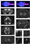
Hoxb8FlpO expression is restricted to the caudal brainstem and below. Bulbo- and spinofugal axons in the brain of Hoxb8 reporter mice. (A,A′) TdTomato reporter in an adult brain and cervical spinal cord visualized under UV illumination. The reporter expression boundary is marked by the arrows. (B,B′) Transverse section of the medulla at the level of the obex illustrating positively-labeled neurons in the spinal nucleus of the trigeminal tract (SpV), the nucleus of the solitary tract (NTS), and gracile nucleus (GN, arrows in B′). (C,D) Transverse sections at the level of the cerebellum (Cb) and superior colliculus (SC, D), in which axons are apparent in the periaqueductal gray matter (PAG). (E,F) Transverse sections of the caudal (E) and rostral (F) thalamus showing a dense network of axons in the ventral posterolateral nucleus (VPLN). (G) Transverse section at the level of the anterior commissure (AC). Labeled structures include mesenchymal cells in the choroid plexus (Chp, G′), scattered neurons in the lateral septal nuclei (Sp, G′′) and axons in the fusiform nucleus of the bed nucleus of the stria terminalis (BNST; G′′′).
Figure 4
Figure 4
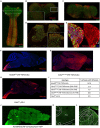
Hoxb8FlpO mediated transgene expression in the adult is restricted to the spinal cord. (A) Sagittal section of cervical spinal cord and brainstem of adult (6 weeks) Hoxb8FlpO;FSF-TdTomato mouse showing Tdtomato fluorescence (red) largely restricted to the spinal cord. NeuN (green) was used as a counterstain. (B) Transverse section of caudal brainstem from an adult Hoxb8FlpO;FSF-TdTomato mouse showing sparse TdTomato fluorescence (red) in neuronal cell bodies (green) of the spinal trigeminal nucleus. (C) Transverse section of rostral brainstem from an adult Hoxb8FlpO;FSF-TdTomato mouse showing TdTomato fluorescence (red) in neuronal tracts, but not in cell bodies (green). (D) Transverse section of lumbar spinal cord from an adult Hoxb8FlpO;FSF-TdTomato mouse showing TdTomato fluorescence (red) in a large majority of spinal neurons (green). Zoomed in image of spinal neurons is shown in the bottom right. (E) Cryosection of an L2 dorsal root ganglia from a Hoxb8FlpO;FSF-TdTomato mouse showing TdTomato fluorescence (red) in a large majority of DRG neurons, including CGRP+ (green) and IB4+ (blue) neurons. (F) Sagittal section of brain and brainstem from an adult Hoxb8FlpO;FSF-TdTomato mouse showing TdTomato fluorescence (red) in tracts in the brainstem, but absence of TdTomato in neurons of the brain. DAPI (blue) was used as a counterstain. (G) Sagittal section of brain and brainstem from an adult Cdx2NSE-FlpO;FSF-TdTomato mouse on a 25% FVB background showing widespread neuronal TdTomato fluorescence (red) in the brain. DAPI (blue) was used as a counterstain. (H) Sagittal section of brain and brainstem from an adult CdxCre;LSL-TdTomato mouse showing TdTomato fluorescence (red) in the brain. DAPI (blue) was used as a counterstain. (I) Quantification of the number of brains containing neuronal TdTomato fluorescence in different caudal-targeting mouse lines. Notably, Hoxb8 tissue had no observable neuronal TdTomato fluorescence, while neuronal TdTomato fluorescence was observed in all Cdx2NSE-FlpO and Cdx2Cre samples. (J) Image of sagittal brain from adult Hoxb8FlpO;FSF-Synaptophysin-GFP mice, showing projections from Hoxb8+ neurons (green) into the brain. Notably, no GFP+ neurons are observed in the brain. (K,L) Hoxb8FlpO+ projections (green) were observed in the thalamus (K) and parabrachial nucleus (L), likely reflecting spinothalamic and spinoparabrachial projections, respectively.
Figure 5
Figure 5
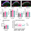
Hoxb8FlpO mice exhibit normal spinal cord development and somatosensory function. (A–C) Immunostaining for different laminae markers in transverse lumbar sections from Hoxb8FlpO/O (A), Hoxb8FlpO/+ (B), and wild type (WT; C) mice reveals no obvious differences in laminae formation defined by immunostaining for lamina markers. Staining was conducted for CGRP (lamina I-red), IB4 (lamina IIo-blue), and VGLUT3 (lamina IIi-green). n = 3 mice per group. (D–G) Sensory assays used to probe mechanical and heat sensitivity in Hoxb8FlpO/+ mice. Hoxb8FlpO/O and Hoxb8FlpO/+ mice have no differences in withdrawal response to dynamic brush (D), noxious pinprick (E), von Frey (F), or radiant heat (G) compared to wild type littermate controls, suggesting normal mechanical and heat sensory processing. Data are represented as mean ± SEM (n = 11–15 mice per group), and significance was assessed using a one-way ANOVA with Tukey’s multiple comparisons. (H) Hoxb8FlpO/+ and Hoxb8FlpO/+ mice have no differences in time spent self-grooming compared to wild type littermates, suggesting normal grooming behavior. Data are represented as mean ± SEM (n = 10–11 mice per group), and significance was assessed using a one-way ANOVA with Tukey’s multiple comparisons. (I) Hoxb8FlpO/O and Hoxb8FlpO/+ mice have no difference in stride frequency compared to wild type littermates. Data are represented as mean + individual data points (n = 9–15 mice per group), and significance was assessed using a one-way ANOVA with Tukey’s multiple comparisons.
Figure 6
Figure 6
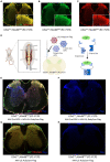
Cre-DOG virus to label spinal cord lineages defined by Cdx2 and Hoxb8. (A,B,C) Expression of GFP and Tomato in a transverse lumbar section of a Cdx2Cre;Hoxb8FlpO;RC::FLTG mouse. (D) Schematic of Cre-DOG virus strategy in the Cdx2Cre;Hoxb8FlpO;RC::FLTG mouse. The Cre-DOG virus consists of N- and C-terminal Cre fragments, which combine and become active in the presence of GFP. In Cdx2Cre;Hoxb8FlpO; RC::FLTG mice, endogenous Cdx2Cre expression is absent in the adult. Therefore, injection of the Cre-DOG virus drives expression of Cre in GFP+ cells, in the absence of endogenous Cdx2Cre expression. (E,F,G,H) Transverse lumbar image from Cdx2Cre;Hoxb8FlpO;RC::FLTG mice co-injected with AAV-Cre-DOG and AAV-LSL-Ruby2sm-Flag (E,F) or AAV-LSL-Ruby2sm-Flag only (G,H). Cre-DOG-mediated Cre expression becomes active in GFP+ cells, which drives expression of the viral LSL-Ruby2sm-Flag, labelled in blue in E,F but not in G,H where there is no Cre expression. (G,H) Transverse image from a Cdx2Cre;Hoxb8FlpO;RC::FLTG mouse injected with AAV-LSL-Ruby2sm-Flag. Cre-dependent Flag expression is not observed in this tissue due to the lack of cdx2-driven Cre expression in the adult mouse.
Figure 7
Figure 7
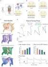
CRE-DOG and inhibitory chemogenetics viruses to assess developmentally determined ROR𝛽 lineage neurons to sensorimotor integration in adult. (A). Schematic of Cre-DOG virus strategy in the Ror𝛽Cre;Hoxb8FlpO;RC::FLTG mouse. The Cre-DOG virus consists of N- and C-terminal Cre fragments, which combine and become active in the presence of GFP, and a Cre-dependent LSL-Di-mCherry virus. (B–E) Schematic of the Ror𝛽 populations silenced in each experimental group. (B) Control animals with dorsal horn injection of AAV-Cre-DOG and control reporter virus LSL-eGFP (C). The Ror𝛽Cre gene is expressed during development more broadly than in adult mice in the superficial and deeper dorsal horn. As a consequence, Ror𝛽Cre;RC::FLTG + AAV Cre-DOG + AAV LSL-Di-mCherry silences the adult Ror𝛽 populations (ad-Ror𝛽-Di). (D) Ror𝛽Cre;Hoxb8FlpO;RC::FLTG + AAV Cre-DOG + AAV LSL-Di-mCherry silences the adult and developmental lineage Ror𝛽 populations (ad+dev-Ror𝛽-Di). (E) Ror𝛽GFP/GFP is a developmental total Knock-Out of the Ror𝛽Cre gene. (F) Stick model representations (two step cycles) of control (tan), ad-Ror𝛽-Di (purple), ad+dev-Ror𝛽-Di (green), and Ror𝛽GFP/GFP (blue) mutant groups. Locomotor parameters were quantified in the peak of the swing phase (red). (G; Top) Example stick model representation of the peak of the swing phase. (Bottom) The following locomotor parameters were quantified: (1) Ankle Joint Height during the swing phase; (2) Knee Joint Angle (red arrow points to the peak of swing phase); and (3) Iliac Crest to MTP Distance. Control mice exhibit the smallest Ankle Height, largest Knee Angle, and largest Iliac Crest to MTP Distance. Ror𝛽GFP/GFP mice exhibit the highest Ankle Height, smallest Knee Angle, and smallest Iliac Crest to MTP Distance due to their characteristic “duck gait” phenotype. Following administration of DREADDs-agonist CNO, ad-Ror𝛽-Di and ad+dev-Ror𝛽-Di mice display intermediate phenotypes, with ad+dev-Ror𝛽-Di mice exhibiting the more severe phenotype of the two.

Similar articles

Cited by

References

    1. Abraira V. E., Ginty D. D. (2013). The sensory neurons of touch. Neuron 79, 618–639. doi: 10.1016/j.neuron.2013.07.051, PMID: - DOI - PMC - PubMed
    1. Abraira V. E., Kuehn E. D., Chirila A. M., Springel M. W., Toliver A. A., Zimmerman A. L., et al. . (2017). The cellular and synaptic architecture of the Mechanosensory dorsal horn. Cells 168, 295–310.e19. doi: 10.1016/j.cell.2016.12.010, PMID: - DOI - PMC - PubMed
    1. Adrienne E. D., Ardem P. (2010). Nociceptors: the sensors of the pain pathway. J. Clin. Invest. 120, 3760–3772. doi: 10.1172/JCI42843, PMID: - DOI - PMC - PubMed
    1. Awatramani R., Soriano P., Rodriguez C., Mai J. J., Dymecki S. M. (2003). Cryptic boundaries in roof plate and choroid plexus identified by intersectional gene activation. Nat. Genet. 35, 70–75. doi: 10.1038/ng1228, PMID: - DOI - PubMed
    1. Bang S. J., Jensen P., Dymecki S. M., Commons K. G. (2012). Projections and interconnections of genetically defined serotonin neurons in mice. Eur. J. Neurosci. 35, 85–96. doi: 10.1111/j.1460-9568.2011.07936.x, PMID: - DOI - PMC - PubMed