. 2023 Aug 21;6(11):e202301965.
doi: 10.26508/lsa.202301965. Print 2023 Nov.

Allotopic expression of COX6 elucidates Atco-driven co-assembly of cytochrome oxidase and ATP synthase

Affiliations

Allotopic expression of COX6 elucidates Atco-driven co-assembly of cytochrome oxidase and ATP synthase

Leticia Veloso R Franco et al. Life Sci Alliance. .

Abstract

The Cox6 subunit of Saccharomyces cerevisiae cytochrome oxidase (COX) and the Atp9 subunit of the ATP synthase are encoded in nuclear and mitochondrial DNA, respectively. The two proteins interact to form Atco complexes that serve as the source of Atp9 for ATP synthase assembly. To determine if Atco is also a precursor of COX, we pulse-labeled Cox6 in isolated mitochondria of a cox6 nuclear mutant with COX6 in mitochondrial DNA. Only a small fraction of the newly translated Cox6 was found to be present in Atco, which can explain the low concentration of COX and poor complementation of the cox6 mutation by the allotopic gene. This and other pieces of evidence presented in this study indicate that Atco is an obligatory source of Cox6 for COX biogenesis. Together with our finding that atp9 mutants fail to assemble COX, we propose a regulatory model in which Atco unidirectionally couples the biogenesis of COX to that of the ATP synthase to maintain a proper ratio of these two complexes of oxidative phosphorylation.


Conflict of interest statement

The authors declare that they have no conflict of interest.

Figures
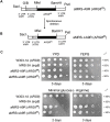
Figure 1.
Figure 1.. Maps and expression of ARG8m under the control of the ATP9 promoter.
(A) Map of ARG8m fused to the 5′ UTR of ATP9 and inserted at the BamH1 site downstream of VAR1. G/B indicates a ligation made from BamHI and BglII fragments with compatible ends. A complete description of the allele can be found at the Materials and Methods section. (B) Map of the MRS/sA9R in which part of the ATP9 promoter region of MRS/A9R was spontaneously deleted. (C) Spot growth tests of the WT strain W303-1A, the arg8 mutant MRS-3A, aMRS-A9R, an arg8 mutant with a mitochondrial copy of ARG8m under the control of the full ATP9 promoter, and aMRS/sA9R with a partially deleted ATP9 promoter. In the upper panels, cells grown on solid ethanol–glycerol (YPEG) were serially diluted and spotted on rich glucose YPD and on rich non-fermentable ethanol/glycerol media YPEG. On the lower panels, cells grown on solid YPEG were serially diluted and spotted on minimal glucose medium-lacking arginine. The plates were incubated for the indicated number of days at 30°C.
Figure 2.
Figure 2.. Expression of ARG8m.
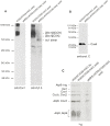
(A) aMRS-A9R and aMRS-sA9R that contained mitochondrial DNA with ARG8m fused to the complete and partially deleted ATP9 promoter region, respectively, were grown on rich liquid galactose. Mitochondria (250 μg protein) isolated from each strain were labeled with 35S-methionine/cysteine for 20 min and extracted with 2% digitonin. Proteins were separated by SDS–PAGE on a 12% polyacrylamide gel, transferred to nitrocellulose, and exposed to an X-ray film. The radiolabeled mitochondrial gene products are identified in the margin. (B) Mitochondria (50 μg of protein) from the WT strain W303-1B, MRS/COX3-HAC, and MRS/COX2-pH both harboring the arg8 null mutation, and aMRS-A9R and aMRS-sA9R, were separated by SDS–PAGE in a 12% polyacrylamide gel and transferred to a nitrocellulose membrane for Western blot analysis. The blot was reacted with a primary rabbit polyclonal antibody against yeast acetylornithine aminotransferase followed by a secondary antibody against rabbit IgG conjugated to peroxidase. (C) Mitochondria (50 μg protein) from the arg8 mutant strain MRS-3A and from aMRS-A9R and aMRS-sA9R were extracted with 2% digitonin and separated on a non-denaturing 4–13% polyacrylamide gel by BN–PAGE. Proteins were transferred to a PVDF membrane and reacted with a primary polyclonal antibody against the β-subunit of F1 ATPase and separately against the Cox1 subunit of COX. Proteins were detected with SuperSignal chemiluminescent substrate kit (Pierce). In all experiments, aMRS-A9R was estimated to consist of 40% ρ+ cells.
Figure 3.
Figure 3.. Complementation of cox6 mutants by the COX6-Cm allele.
(A) Structure of the COX6-Cm allele. COX6-Cm is preceded by the promoter region of ATP9 and ends with three methionine codons (3 met) followed by the protein C tag. G/B indicates a ligation made from BamHI and BglII fragments with compatible ends. A complete description of the allele can be found in the Materials and Methods section. (B) Serial dilutions of the respiratory competent diploid a/αW303 and haploid W303-1A strains and of the diploid and haploid cox6 mutants expressing COX6-Cm. Cells grown on solid ethanol–glycerol were spotted on rich glucose (YPD) and rich ethanol–glycerol (YPEG) and incubated at 30°C for 3 d. The percentages of ρ+ cells in each culture are indicated in the right margin.
Figure S1.
Figure S1.. Properties of the strain expressing a nuclear-tagged COX6-C allele.
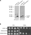
(A) Mitochondria isolated from cells grown on rich galactose liquid media of WT strain W303-1B and of the strain W303/COX6-C expressing COX6 with three methionine codons and a protein C tag were extracted with digitonin and separated on a 12% polyacrylamide gel by SDS–PAGE. Proteins were transferred to a nitrocellulose membrane and reacted with a primary rabbit antibody against protein C and against Cox6 followed by a secondary antibody against rabbit IgG conjugated to peroxidase. (B) The WT strain W303-1B, the cox6 null mutant (W303ΔCOX6), and W303/COX6-C were grown in liquid YPD and serial dilutions were spotted on rich glucose (YPD) and on rich ethanol glycerol media (YPEG) and grown at 30°C for 2 d.
Figure 4.
Figure 4.. Newly translated and steady-state levels of mitochondrially encoded Cox6.
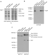
(A) Mitochondria (250 μg protein) of the WT diploid (a/αW303) and a/αW303ΔCOX6/COX6-Cm, a homozygous cox6 mutant with a mitochondrial copy of COX6-Cm were labeled with 35S-methionine/cysteine for 20 min, extracted with 2% digitonin, and purified on protein C antibody beads. The digitonin extracts and purified fractions (PC elution) were separated by SDS–PAGE on a 12% polyacrylamide gel. Proteins were transferred to a PVDF membrane and exposed to an X-ray film. The radiolabeled mitochondrial gene products are identified in the margins. (B) Total mitochondrial proteins (50 μg) of WT W303 and of αW303ΔCOX6/COX6-Cm were separated by SDS–PAGE on a 12% polyacrylamide gel. Proteins were transferred to a nitrocellulose membrane and blotted with a rabbit polyclonal antibody against Cox6 followed by a secondary antibody against rabbit IgG conjugated to peroxidase and visualized as in Fig 2C. Mitochondria in (A, B) were isolated from cells grown on solid rich ethanol/glycerol media.
Figure 5.
Figure 5.. Steady-state levels of the oxphos complexes.
Mitochondria (50 μg protein) of the WT W303-1B and of a/αW303ΔCOX6/COX6-Cm were isolated from cells grown on solid ethanol/glycerol media. The mitochondria were extracted with 2% digitonin and separated on a non-denaturing 4–13% polyacrylamide gel by BN–PAGE. (A, B, C) Proteins were transferred to a PVDF membrane and reacted with primary antibodies against Cox1 (A), cytochrome b (B), and the F1 β subunit of the ATP synthase (C). Proteins were visualized with a secondary antibody conjugated to peroxidase as in Fig 2C.
Figure 6.
Figure 6.. Steady-state levels of the mitochondrially encoded subunits of the respiratory chain.
Mitochondria (50 μg protein) of the WT W303-1B and of αW303ΔCOX6/COX6-Cm were isolated from cells grown on solid rich ethanol/glycerol media. The mitochondria were separated on a denaturing 12% polyacrylamide gel by SDS–PAGE. (A) Proteins were transferred to a nitrocellulose membrane and reacted with primary monoclonal mouse antibodies against Cox1, Cox2, and Cox3 followed by a secondary antibody against mouse IgG conjugated to peroxidase (A). (B) Cytochrome b was detected with a primary polyclonal rabbit antibody against cytochrome b followed by a secondary antibody against rabbit IgG conjugated to peroxidase. Proteins were visualized as in 2C.
Figure 7.
Figure 7.. Analysis of Atco and Cox6 by two-dimensional electrophoresis.
Digitonin extracts of aW303ΔCOX6/COX6-Cm mitochondria (250 μg protein) isolated from cells grown on solid ethanol/glycerol media were labeled with 35S-methionine/cysteine, purified on protein C beads, and separated on the first dimension in a non-denaturing 4–13% polyacrylamide gel by BN–PAGE followed by a second dimension on a 12% polyacrylamide gel by SDS–PAGE. Proteins were transferred to a PVDF membrane and exposed to the X-ray film. A sample of the fraction eluted from the protein C beads (EL 1D) was separated directly on the SDS gel.
Figure 8.
Figure 8.. Intra-mitochondrial localization of mitochondrially encoded Cox6.
Mitochondria of aW303ΔCOX6/COX6-Cm were isolated from cells grown on solid rich ethanol/glycerol media. Mitochondria (M) were sonicated and centrifuged at 105,000g to separate the soluble matrix proteins (S) and inverted submitochondrial particles (P). The distribution of mitochondrially encoded Cox6 in the different fractions was assessed using an anti-Cox6 antibody. Mitochondrial breakage was checked using an antibody against the soluble matrix Kgd1.
Figure 9.
Figure 9.. Analysis of the mitochondrial gene products and supercomplexes in the atp9 null mutant.
(A) Mitochondria were isolated from cultures of W303/COX6-HAC, a strain expressing Cox6 tagged with hemagglutinin followed by a protein C epitope from the nuclear COX6-HAC gene, and of the atp9 null mutant aMRSΔATP9/COX6-HAC. Both strains were grown on solid rich galactose media. Mitochondria were extracted with 2% digitonin and separated under non-denaturing conditions by BN–PAGE on a 4–13% polyacrylamide gel. Proteins were transferred to PVDF membranes and reacted with the indicated primary mouse antibody against Cox1 and separately with an antibody against cytochrome b. Proteins were visualized after a reaction with a secondary antibody conjugated to peroxidase and visualized as in Fig 2C. (B) Total mitochondria (50 μg proteins) of W303/COX6-HAC and aMRSΔATP9/COX6-HAC, both grown on solid rich galactose medium, were separated by SDS–PAGE. Proteins were transferred to a nitrocellulose membrane and Cox6 was visualized by reacting the membrane with a primary rabbit antibody against protein C, followed by a secondary antibody against rabbit IgG conjugated to peroxidase and visualized as in 2C. (C) Mitochondria (250 μg protein) from W303/COX6-HAC grown either on liquid of solid rich galactose media, and from aMRSΔATP9/COX6-HAC, grown on solid rich galactose medium, were labeled with 35S- methionine/cysteine for 20 min, extracted with 2% digitonin, and separated by SDS–PAGE on a 12% polyacrylamide gel. Proteins were transferred to nitrocellulose and the blot exposed to an X-ray film. The radiolabeled mitochondrial gene products are identified in the margins. In all experiments, aMRSΔATP9/COX6-HAC was estimated to consist of 50% ρ+ cells.
Figure 10.
Figure 10.. Model of Atco in coupling the biogenesis of cytochrome oxidase (COX) to that of the ATP synthase.
(A) In WT, Cox6 precursor synthesized on cytoplasmic ribosomes is transported (open arrow) into the mitochondrial inner membrane where it interacts with oligomeric Atp9 to form Atco. The interactions of the Atp9 in the oligomer are similar to those in the Atp9 ring (Franco et al, 2020a), but at present, it is not known if the Atp9 of Atco is present as a 10-membered ring as shown in the diagram or some other oligomeric form. Atco provides Cox6 for the Cox1 assembly module of COX, and the Atp9 oligomer to the ATP synthase assembly pathway (shown above). Not all the subunits of the membrane unit (F0) are shown in the diagram of the ATP synthase. (B) Co-translational membrane insertion of Cox6 from mitochondrial ribosomes bound to the matrix side of the inner membrane allows some Cox6 to enter the normal COX assembly pathway. Because of the less efficient COX assembly, some of the Cox6 that failed to be incorporated into Atco is proteolytically cleaved into a smaller fragment. The retarded rate of Cox6 entry into the normal COX assembly pathway elicits substantial turnover of Cox1, but not Cox2 or Cox3. (C) In the cox6 mutant, COX is not assembled. However, the Atp9 monomer synthesized on mitochondrial ribosomes can assemble into a ring and be recruited for ATP synthase assembly. (D) In the atp9 mutant, cytoplasmically translated Cox6 is transport in the matrix as shown in the figure or into the inner membrane. The free non-Atco-associated subunit does not interact with the Cox1 intermediate resulting in proteolysis of the Cox1 causing the complete loss of COX.

References

    1. Barrientos A, Zambrano A, Tzagoloff A (2004) Mss51p and Cox14p jointly regulate mitochondrial Cox1p expression in Saccharomyces cerevisiae. EMBO J 23: 3472–3482. 10.1038/sj.emboj.7600358 - DOI - PMC - PubMed
    1. Barros MH, McStay GP (2020) Modular biogenesis of mitochondrial respiratory complexes. Mitochondrion 50: 94–114. 10.1016/j.mito.2019.10.008 - DOI - PubMed
    1. Bonitz SG, Berlani R, Coruzzi G, Li MacinoG, Macino G, Nobrega FG, Nobrega MP, Thalenfeld BE, Tzagoloff A (1980) Codon recognition rules in yeast mitochondria. Proc Natl Acad Sci U S A 77: 3167–3170. 10.1073/pnas.77.6.3167 - DOI - PMC - PubMed
    1. Bonnefoy N, Fox TD (2007) Directed alteration of Saccharomyces cerevisiae mitochondrial DNA by biolistic transformation and homologous recombination. Methods Mol Biol 372: 153–166. 10.1007/978-1-59745-365-3_11 - DOI - PMC - PubMed
    1. Böttinger L, Guiard B, Oeljeklaus S, Kulawiak B, Zufall N, Wiedemann N, Warscheid B, van der Laan M, Becker T (2013) A complex of Cox4 and mitochondrial Hsp70 plays an important role in the assembly of the cytochrome c oxidase. Mol Biol Cell 24: 2609–2619. 10.1091/mbc.E13-02-0106 - DOI - PMC - PubMed

Publication types

MeSH terms

Substances

LinkOut - more resources