

Retinal Optical Coherence Tomography Features Associated With Incident and Prevalent Parkinson Disease
- PMID: 37604659
- PMCID: PMC10585674
- DOI: 10.1212/WNL.0000000000207727
Retinal Optical Coherence Tomography Features Associated With Incident and Prevalent Parkinson Disease
Abstract
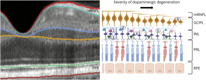
Background and objectives: Cadaveric studies have shown disease-related neurodegeneration and other morphological abnormalities in the retina of individuals with Parkinson disease (PD); however, it remains unclear whether this can be reliably detected with in vivo imaging. We investigated inner retinal anatomy, measured using optical coherence tomography (OCT), in prevalent PD and subsequently assessed the association of these markers with the development of PD using a prospective research cohort.
Methods: This cross-sectional analysis used data from 2 studies. For the detection of retinal markers in prevalent PD, we used data from AlzEye, a retrospective cohort of 154,830 patients aged 40 years and older attending secondary care ophthalmic hospitals in London, United Kingdom, between 2008 and 2018. For the evaluation of retinal markers in incident PD, we used data from UK Biobank, a prospective population-based cohort where 67,311 volunteers aged 40-69 years were recruited between 2006 and 2010 and underwent retinal imaging. Macular retinal nerve fiber layer (mRNFL), ganglion cell-inner plexiform layer (GCIPL), and inner nuclear layer (INL) thicknesses were extracted from fovea-centered OCT. Linear mixed-effects models were fitted to examine the association between prevalent PD and retinal thicknesses. Hazard ratios for the association between time to PD diagnosis and retinal thicknesses were estimated using frailty models.
Results: Within the AlzEye cohort, there were 700 individuals with prevalent PD and 105,770 controls (mean age 65.5 ± 13.5 years, 51.7% female). Individuals with prevalent PD had thinner GCIPL (-2.12 μm, 95% CI -3.17 to -1.07, p = 8.2 × 10⁻⁵) and INL (-0.99 μm, 95% CI -1.52 to -0.47, p = 2.1 × 10⁻⁴). The UK Biobank included 50,405 participants (mean age 56.1 ± 8.2 years, 54.7% female), of whom 53 developed PD at a mean of 2,653 ± 851 days. Thinner GCIPL (hazard ratio [HR] 0.62 per SD increase, 95% CI 0.46-0.84, p = 0.002) and thinner INL (HR 0.70, 95% CI 0.51-0.96, p = 0.026) were also associated with incident PD.
Discussion: Individuals with PD have reduced thickness of the INL and GCIPL of the retina. Involvement of these layers several years before clinical presentation highlight a potential role for retinal imaging for at-risk stratification of PD.
Copyright © 2023 The Author(s). Published by Wolters Kluwer Health, Inc. on behalf of the American Academy of Neurology.
Conflict of interest statement
S.K. Wagner is funded by the Medical Research Council (MR/TR000953/1) and the Rank Prize. D. Romero-Bascones received funding from the Basque Health Department (2022333011). M. Cortina-Borja, D.J. Williamson, R.R. Struyven, Y. Zhou, and S. Patel report no disclosures. R.S. Weil is supported by a Wellcome Clinical Research Career Development Fellowship (205167/Z/16/Z). C.A. Antoniades receives funding from the NIHR, Biomedical Research Centre (BRC), UCB-OXford collaborative grant, and Merck. E.J. Topol and E. Korot report no disclosures. P.J. Foster receives salary support from NIHR through a grant to the Biomedical Research Centre at Moorfields Eye Hospital and UCL Institute of Ophthalmology. K. Balaskas reports no disclosures. U. Ayala receives funding from the Basque Health Department (2022333011). M. Barrenechea receives funding from the Basque Health Department (2022333011). I. Gabilondo receives funding from the Basque Health Department (2022333011). A.H.V. Schapira reports no disclosures. A.P. Khawaja is supported by a UK Research & Innovation Future Leaders Fellowship (MR/T040912/1), an Alcon Research Institute Young Investigator Award and a Lister Institute Fellowship. A.P. Khawaja receives salary support from NIHR through a grant to the Biomedical Research Centre at Moorfields Eye Hospital and UCL Institute of Ophthalmology. P.J. Patel, J.S. Rahi, and A.K. Denniston report no disclosures. A. Petzold receives salary support from NIHR through a grant to the Biomedical Research Centre at Moorfields Eye Hospital and UCL Institute of Ophthalmology. P.A. Keane is supported by a UK Research & Innovation Future Leaders Fellowship (MR/T019050/1). P.A. Keane receives salary support from NIHR through a grant to the Biomedical Research Centre at Moorfields Eye Hospital and UCL Institute of Ophthalmology. Additional support was from the NIHR Birmingham, Great Ormond Street, Moorfields, Oxford, and University College London Hospitals Biomedical Research Centres. Go to
Figures


References
Publication types
MeSH terms
Grants and funding
LinkOut - more resources
Full Text Sources
Medical