. 2023 Aug 21;13(1):13589.
doi: 10.1038/s41598-023-40049-0.

Insights into cellular behavior and micromolecular communication in urothelial micrografts

Affiliations

Insights into cellular behavior and micromolecular communication in urothelial micrografts

Nikolai Juul et al. Sci Rep. .

Abstract

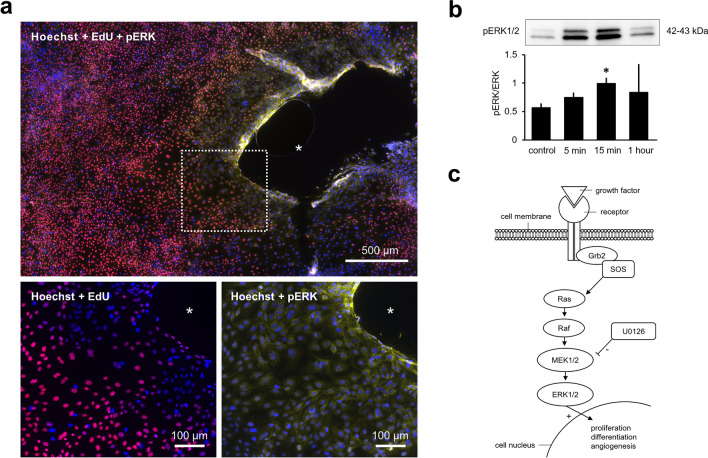
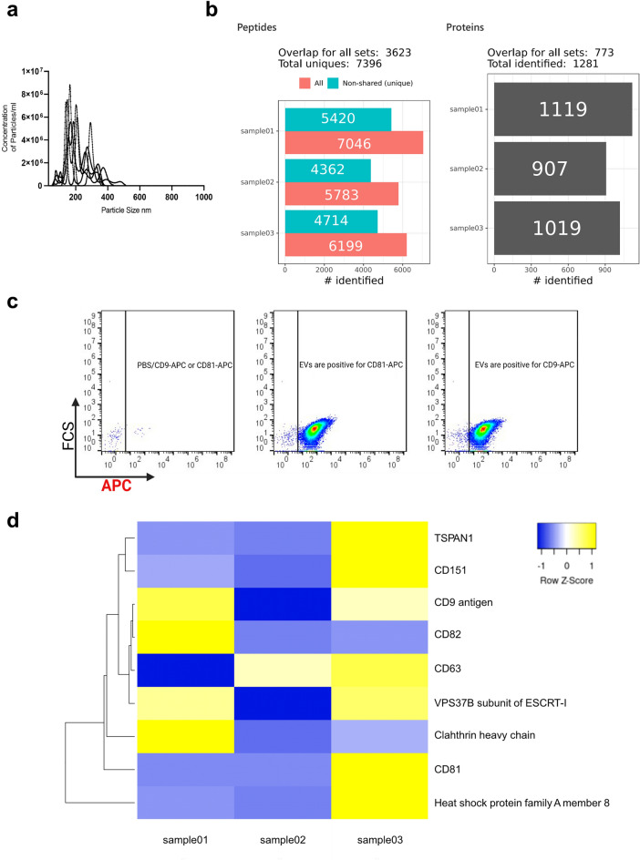
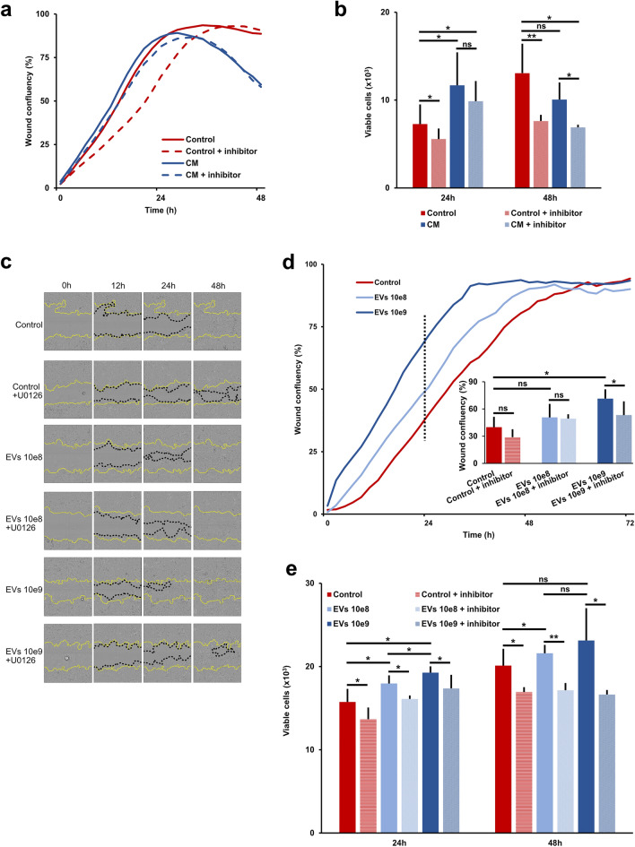
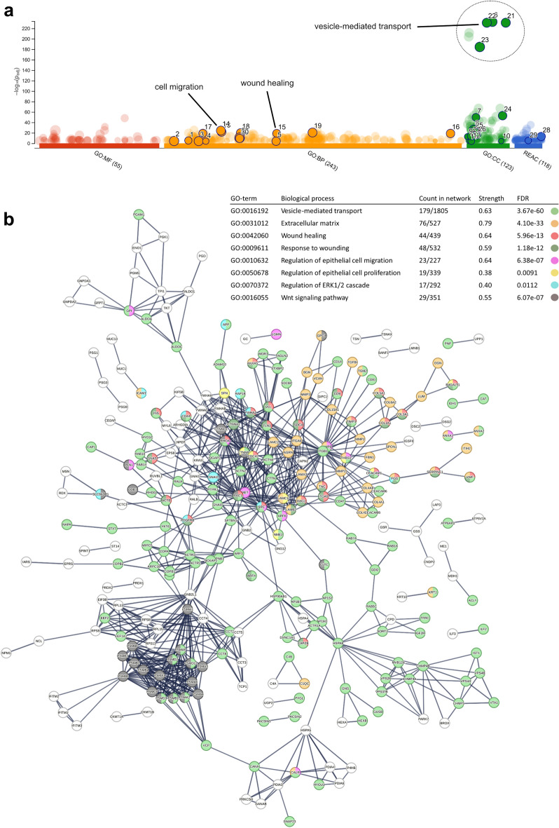
Autologous micrografting is a technique currently applied within skin wound healing, however, the potential use for surgical correction of other organs with epithelial lining, including the urinary bladder, remains largely unexplored. Currently, little is known about the micrograft expansion potential and the micromolecular events that occur in micrografted urothelial cells. In this study, we aimed to evaluate the proliferative potential of different porcine urothelial micrograft sizes in vitro, and, furthermore, to explore how urothelial micrografts communicate and which microcellular events are triggered. We demonstrated that increased tissue fragmentation subsequently potentiated the yield of proliferative cells and the cellular expansion potential, which confirms, that the micrografting principles of skin epithelium also apply to uroepithelium. Furthermore, we targeted the expression of the extracellular signal-regulated kinase (ERK) pathway and demonstrated that ERK activation occurred predominately at the micrograft borders and that ERK inhibition led to decreased urothelial migration and proliferation. Finally, we successfully isolated extracellular vesicles from the micrograft culture medium and evaluated their contents and relevance within various enriched biological processes. Our findings substantiate the potential of applying urothelial micrografting in future tissue-engineering models for reconstructive urological surgery, and, furthermore, highlights certain mechanisms as potential targets for future wound healing treatments.


Conflict of interest statement

The authors declare no competing interests.

Figures

Figure 1
Figure 1
Micrograft expansion potential. Schematic illustration of the fundamental principles of micrografting according to the theory of Meek, and the numerical relationship between tissue fragmentation and expansion potential.
Figure 2
Figure 2
Experimental design and the relation of tissue fragmentation and cell expansion. (a) Schematic study representation and total cell colony quantification. Porcine bladder dissection and subdivision at different fragmentation ratios. Flowchart depicting different subsequent experiments performed from the tissue. EV: extracellular vesicle, MS: mass spectrometry. (b) Macroscopic image of different micrograft study conditions (left), microscopic image of urothelial cells expanding from a micrograft after 5 days (middle), and urothelial cells stained with crystal violet from a micrograft colony (right). (c) Examples of individual cell colony sizes from different micrograft study conditions stained with crystal violet after two weeks in culture. (d) Mean summative cell counts and standard deviations for each study condition after two weeks, statistically referenced to the 1:1 condition (* for p < 0.05). (e) Mean summative cell colony areas and standard deviations in cm2 for each condition after two weeks, statistically referenced to the 1:1 condition.
Figure 3
Figure 3
ERK expression in urothelial micrograft cultures. (a) Immunofluorescent stains of a urothelial micrograft colony stained with: Hoechst® 33342 cell nucleic marker (blue), EdU (5-ethynyl-2’-deoyuridine) coupled with Alexa Flour® (picolyl azide) as a proliferative marker (red), and pERK (phosphorylated extracellular signal-regulated kinase) antibody (yellow). (b) Normalized expression of pERK (phosphorylated extracellular signal-regulated kinase) proportional to the expression of total ERK at three timepoints after stimulation with conditioned culture medium, statistically referenced to the stimulation with fresh keratinocyte culture medium used as control. Examples of cropped bands from the Western blots analyzed at different timepoints after stimulation with conditioned medium and with fresh medium as the control represented above (original blots are presented in Supplementary Fig. S1). (c) Schematic representation of the ERK signaling pathway; SOS son of sevenless guanine nucleotide exchange factor, Grb2 growth factor receptor-bound protein 2, MEK1/2 mitogen-activated protein kinase kinase 1 and 2, ERK1/2 extracellular signal-regulated kinase 1 and 2, Ras Ras GTPase, Raf Raf kinase kinase, U0126 1,4-diamino-2,3-dicyano-1.4-bis[2-aminophenylthio]butadiene.
Figure 4
Figure 4
Isolation and characterization of micrograft extracellular vesicles. (a) Nanoparticle tracking analysis was used to measure the size and concentration of the particles. (b) Mass spectrometry summary of the EV cargo and summary of total number of identified peptides and proteins. (c) EVs markers were detected according to forward scatter signal (FCS) using high-resolution imaging flow cytometry (Amnis CellStream) and tetraspanin-labeled-allophycocyanine (APC) anti-Big. (d) Heat map representation of EVS markers identified by MS; the intensity represents Signal intensity of the precursor peptides identified. Clustering based on average linkage and Euclidean distance measurement.
Figure 5
Figure 5
Stimulatory effects of micrograft extracellular vesicle contents. (a) Wound confluency rates of monolayered urothelial cell cultures after standardized scratching and stimulation with either fresh culture medium (control) with or without ERK inhibitor (+ inhibitor) or conditioned culture medium (CM) with or without inhibitor. (b) Mean number and standard deviation of monolayered urothelial cells per 20 mm2 after 24 and 48 h of stimulation with either CM or control medium, with or without additional inhibitor (* for p < 0.05, ** for p < 0.0001 and ns: not significant). (c) Examples of wound confluency at different timepoints after wounding and stimulation with control medium with either additional EVs at 10e8 or 10e9 µM concentration and with or without additional ERK inhibitor, respectively. (d) Wound confluency rates of monolayered urothelial cell cultures after standardized scratching and stimulation with either control or different concentrations of EVs and with or without additional inhibitor. Compared mean wound confluency (%) with standard deviations 24 h after wounding (lower right). (e) Mean number and standard deviation of monolayered urothelial cells per 20 mm2 after 24 and 48 h of stimulation with either control or different concentrations of EVs and with or without additional inhibitor (* for p < 0.05, ** for p < 0.0001, and ns: not significant).
Figure 6
Figure 6
Biological characterization of the extracellular vesicle proteome. (a) Manhattan plot showing results from functional profiling in g:Profiler. Gene Ontology (GO) terms under the categories molecular function (MF), biological process (BP), cellular component (CC), and Reactome pathways (REAC), displayed in color coding. (b) A detailed result view on enriched GO terms from protein–protein interaction network generated using the STRING resource including proteins that fall into several of the GO enriched terms, with respective false detection rates (FDR).

References

    1. Cranidis A, Nestoridis G. Bladder augmentation. Int. Urogynecol. J. Pelvic Floor Dysfunct. 2000;11:33–40. - PubMed
    1. Husmann DA. Mortality following augmentation cystoplasty: A transitional urologist’s viewpoint. J. Pediatr. Urol. 2017;13:358–364. doi: 10.1016/j.jpurol.2017.05.008. - DOI - PubMed
    1. McDougal WS. Metabolic complications of urinary intestinal diversion. J. Urol. 1992;147:1199–1208. doi: 10.1016/S0022-5347(17)37517-1. - DOI - PubMed
    1. Atala A, Bauer SB, Hendren WH, Retik AB. The effect of gastric augmentation on bladder function. J. Urol. 1993;149:1099–1102. doi: 10.1016/S0022-5347(17)36307-3. - DOI - PubMed
    1. Szymanski KM, Misseri R, Whittam B, Lingeman JE, Amstutz S, Ring JD, et al. Bladder stones after bladder augmentation are not what they seem. J. Pediatr. Urol. 2016;12:98.e1–98.e6. doi: 10.1016/j.jpurol.2015.06.021. - DOI - PubMed

Publication types

Substances