. 2023 Aug 21;14(8):537.
doi: 10.1038/s41419-023-06023-4.

Targeting SphK1/2 by SKI-178 inhibits prostate cancer cell growth

Affiliations

Targeting SphK1/2 by SKI-178 inhibits prostate cancer cell growth

Lu Jin et al. Cell Death Dis. .

Abstract

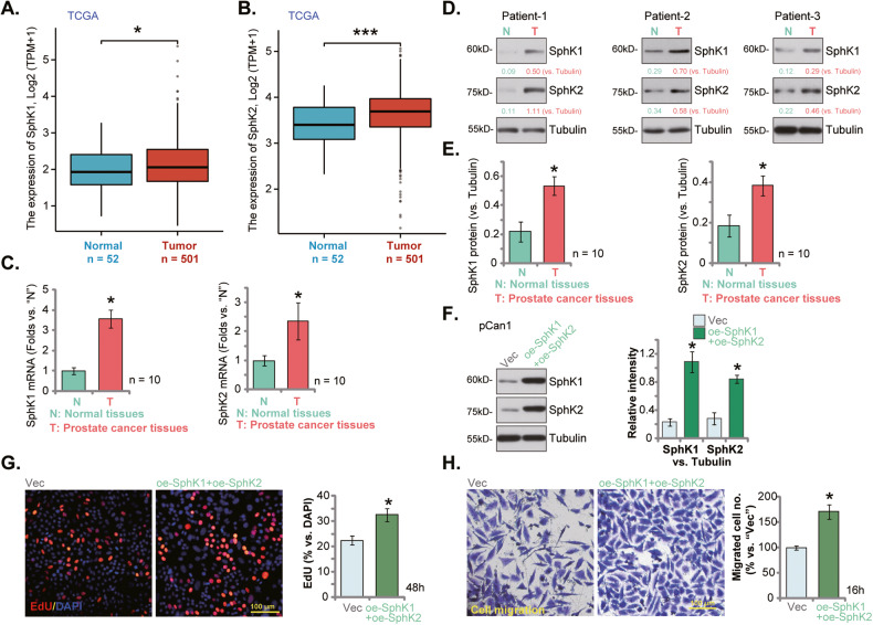
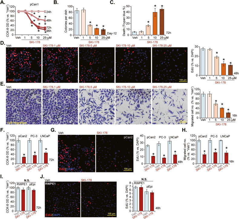
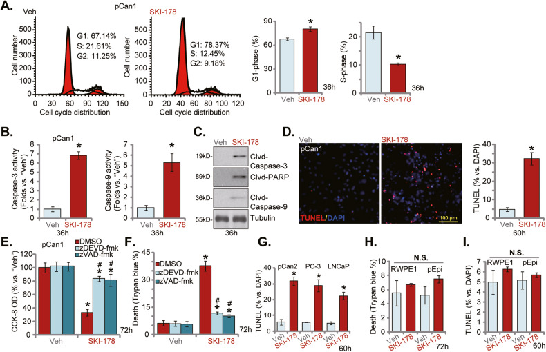
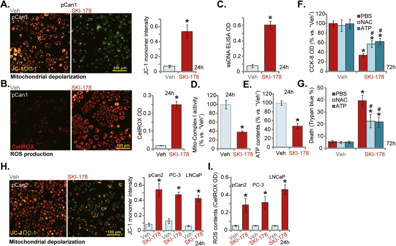
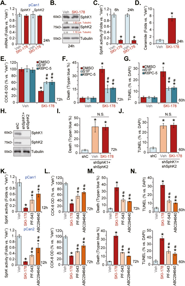
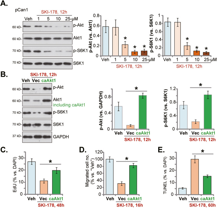
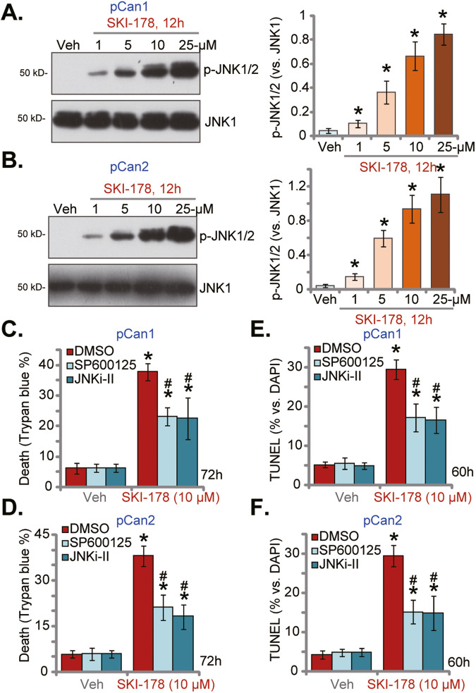
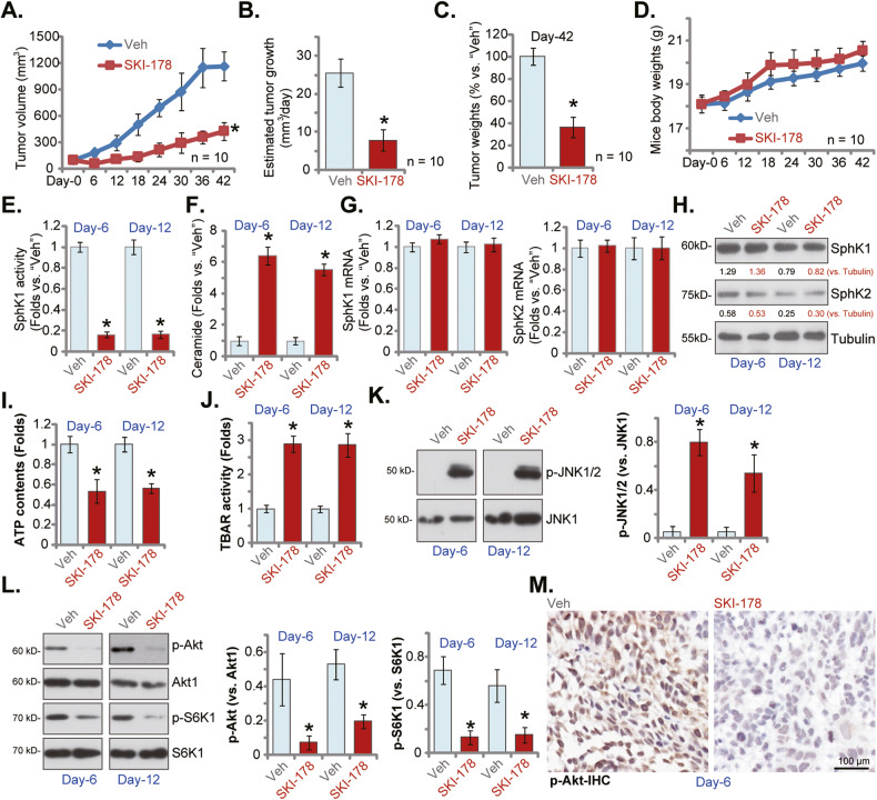
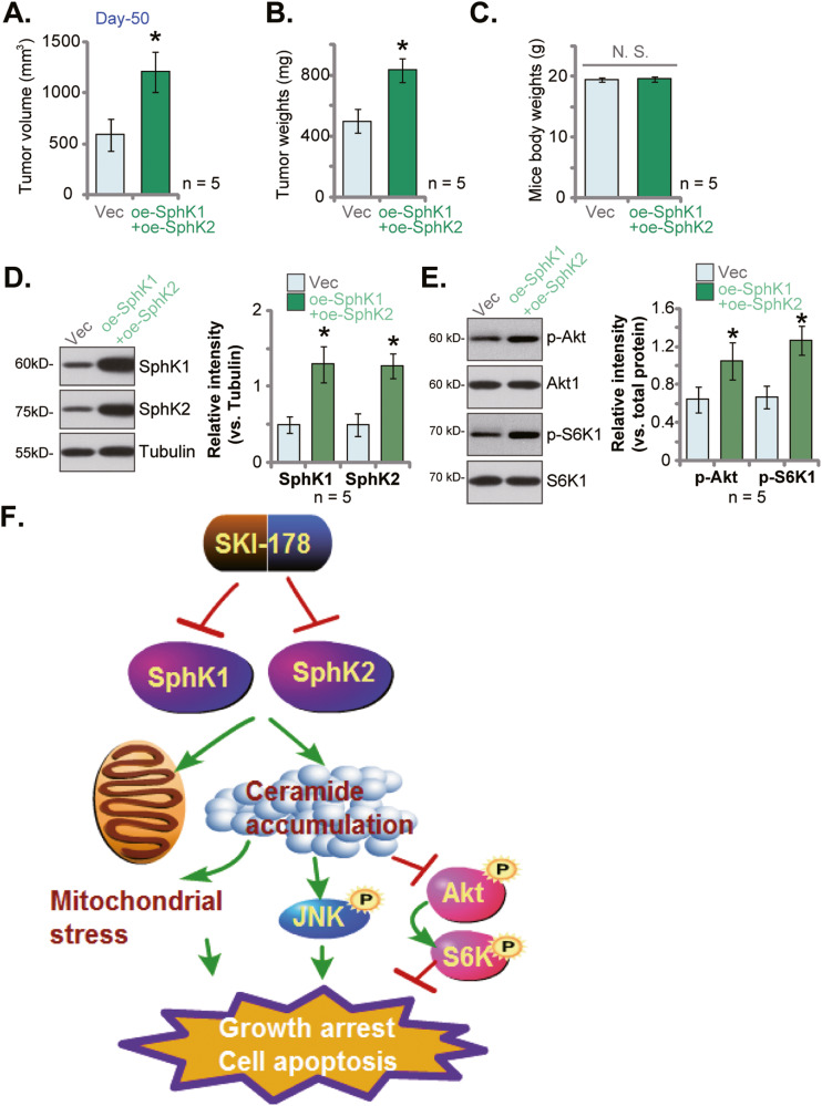
Sphingosine kinases (SphK), including SphK1 and SphK2, are important enzymes promoting progression of prostate cancer. SKI-178 is a novel and highly potent SphK1/2 dual inhibitor. We here tested the potential anti-prostate cancer cell activity of SKI-178. Bioinformatics analyses and results from local tissues demonstrated that that both SphK1 and SphK2 are upregulated in human prostate cancer tissues. Ectopic overexpression of SphK1 and SphK2, by lentiviral constructs, promoted primary prostate cancer cell proliferation and migration. In primary human prostate cancer cells and immortalized cell lines, SKI-178 potently inhibited cell viability, proliferation, cell cycle progression and cell migration, causing robust cell death and apoptosis. SKI-178 impaired mitochondrial functions, causing mitochondrial depolarization, reactive oxygen species production and ATP depletion.SKI-178 potently inhibited SphK activity and induced ceramide production, without affecting SphK1/2 expression in prostate cancer cells. Further, SKI-178 inhibited Akt-mTOR activation and induced JNK activation in prostate cancer cells. Contrarily, a constitutively-active Akt1 construct or the pharmacological JNK inhibitors attenuated SKI-178-induced cytotoxicity in prostate cancer cells. In vivo, daily intraperitoneal injection of a single dose of SKI-178 potently inhibited PC-3 xenograft growth in nude mice. SphK inhibition, ceramide production, ATP depletion and lipid peroxidation as well as Akt-mTOR inactivation and JNK activation were detected in PC-3 xenograft tissues with SKI-178 administration. Together, targeting SphK1/2 by SKI-178 potently inhibited prostate cancer cell growth in vitro and in vivo.


Conflict of interest statement

The authors declare no competing interests.

Figures

Fig. 1
Fig. 1. SphK1 and SphK2 are upregulated in human prostate cancer tissues.
TCGA database shows the number of SphK1 transcripts (A) and the number of SphK2 transcript (B) in 501 prostate cancer tissue samples (“Tumor”) and 52 para-cancerous normal prostate tissue samples (“Normal”). Expression of SphK1 and SphK2 mRNA (C) and listed proteins (D, E) in human prostate cancer tissues (“T”, derived from 10 different CRPC patients, n = 10) or cancer-surrounding normal prostate tissues (“N”) were shown. The primary human prostate cancer cells pCan1, with the lentiviral SphK1-overexpressing construct plus the lentiviral SphK2-overexpressing construct (“oe-SphK1+oe-SphK2”) or the empty vector (“Vec”), were established and expression of listed proteins was tested (F); Cells were further cultivated for indicated time periods, cell proliferation (by measuring nuclear EdU staining, G) and cell migration (“Transwell” assays, H) were measured. Data were expressed as the mean ± standard deviation (SD). “TPM” stands for transcripts per million. ***P < 0.001 versus “Normal” tissues. *P < 0.05 versus “Normal” or “N” tissues (CE). *P < 0.05 versus “Vec” cells (FH). Scale Bar = 100 μm.
Fig. 2
Fig. 2. SKI-178 exerts significant anti-cancer activity in cultured prostate cancer cells.
The primary human prostate cancer cells (“pCan1” and “pCan2”) (AH), the established cell lines (PC-3 and LNCaP) (FH), the primary human prostate epithelial cells (“pEpi”) (I and J) or the established RWPE-1 epithelial cells (I, J) were cultivated in complete medium and treated with SKI-178 (10 μM or at designated concentrations for A–E) for indicated time periods; Cell viability (CCK-8 assay, A, F and I), colony formation (B), cell death (Trypan blue staining assays, C) proliferation (nuclear EdU staining assays, D, G, and J), and cell migration (“Transwell” assays, E and H) were tested by the assays mentioned in the text. “Veh” stands for vehicle control. Values represented the mean ± SD (n = 5). *P < 0.05 versus “Veh” group. Experiments in this figure were repeated five times, with similar results achieved. Scale Bar = 100 μm.
Fig. 3
Fig. 3. SKI-178 induces cell cycle arrest and apoptosis in prostate cancer cells.
The primary human prostate cancer cells (“pCan1” and “pCan2”) (AD, G), the established cell lines (PC-3 and LNCaP, G), the primary human prostate epithelial cells (“pEpi”) (H, I) or the established RWPE-1 epithelial cells (H and I) were cultivated in complete medium and treated with SKI-178 (10 μM) for indicated time periods; cell cycle progression (PI-FACS assays, A) and caspase-PARP activation (B, C) were tested; Cell apoptosis was examined by the nuclear TUNEL staining (D, G and I) assays, with cell death measured by Trypan blue staining (H). The pCan1 primary cells were pretreated for 1 h with z-DEVD-fmk (40 μM) or z-VAD-fmk (40 μM), followed by SKI-178 (10 μM) stimulation for 72 h; Cell viability and death were tested by CCK-8 (E) and Trypan blue staining (F) assays, respectively. “Veh” stands for vehicle control. Values represented the mean ± SD (n = 5). *P < 0.05 versus “Veh” group. #P < 0.05 versus SKI-178 only treatment (E, F). Experiments in this figure were repeated five times, with similar results achieved. Scale Bar = 100 μm.
Fig. 4
Fig. 4. The mitochondrial functions are impaired following SKI-178 treatment in prostate cancer cells.
The primary human prostate cancer cells (“pCan1”) cells were cultivated in complete medium and treated with SKI-178 (10 μM) for 24 h, mitochondrial depolarization (by analyzing JC-1 green monomers, A), ROS contents (by examining CellROX fluorescence intensity, B), and single strand DNA (ssDNA) contents (ELISA OD, C) as well as the mitochondrial complex I activity (D) and the cellular ATP contents (E) were tested. The pCan1 primary cells were pretreated for 1 h with NAC (500 μM) or ATP (2 mM), followed by SKI-178 (10 μM) stimulation for72h; Cell viability and death were tested by CCK-8 (F) and Trypan blue staining (G) assays, respectively. The primary human prostate cancer cells (“pCan2”) or established cell lines (PC-3 and LNCaP) were cultivated in complete medium and treated with SKI-178 (10 μM) for 48 h; Mitochondrial depolarization (by analyzing JC-1 green monomers, H) and ROS contents (by examining CellROX fluorescence intensity, I) were tested. “Veh” stands for vehicle control. Values represented the mean ± SD (n = 5). *P < 0.05 versus “Veh” group. #P < 0.05 versus SKI-178 only treatment (F, G). Experiments in this figure were repeated five times, with similar results achieved. Scale Bar = 100 μm.
Fig. 5
Fig. 5. SKI-178 inhibits SphK1 and SphK2 activity in prostate cancer cells.
The primary human prostate cancer cells (“pCan1”) were treated with SKI-178 (10 μM) for applied time periods, mRNA and protein expression of SphK1 and SphK2 were tested by qRT-PCR (A) and Western blotting (B) assays, and SphK activity tested using the described methods (C). Cellular ceramide contents were examined as well (D). The pCan1 primary cells were pretreated for 1 h with S1P (25 μM), K6PC-5 (10 μM) or the vehicle control (“DMSO”), followed by SKI-178 (10 μM) stimulation for applied time periods, cell viability, death and apoptosis were tested by CCK-8 (E), Trypan blue staining (F) and nuclear TUNEL staining (G) assays, respectively. The pCan1 primary cells bearing the lentiviral SphK1 shRNA and the lentiviral SphK2 shRNA (“shSphK1+shSphK2”) were treated with SKI-178 (10 μM) or vehicle, control cells were transduced with the scramble control shRNA (“shC”), expression of listed proteins were shown (H). Cells were further cultured for applied time periods, cell death and apoptosis were tested by Trypan blue staining (I) and nuclear TUNEL staining (J) assays, respectively. The primary human prostate cancer cells (“pCan1” and “pCan2”) were treated with 10 μM of SKI-178, PF-543 or ABC294640 and cultured for applied time periods, the SphK1 activity was shown (K); Cell viability, cell death and apoptosis were tested by CCK-8 (L), Trypan blue staining (M) and TUNEL staining (N) assays, respectively. “Veh” stands for vehicle control. Values represented the mean ± SD (n = 5). *P < 0.05 versus “Veh”/“shC” group. #P < 0.05 versus SKI-178 group. “N.S.” stands for non-statistical difference. Experiments in this figure were repeated five times, with similar results achieved.
Fig. 6
Fig. 6. SKI-178 inhibits Akt-mTOR activation in prostate cancer cells.
The primary human prostate cancer cells (“pCan1”) cells were cultivated in complete medium and treated with SKI-178 (at designated concentrations) for 12 h, expression of listed proteins was shown (A). pCan1 cells, expressing the lentivirus-packed constitutively-active mutant Akt (S473D, “caAkt1”) or the empty vector (“Vec”), were treated with SKI-178 (10 μM) for 12 h, expression of listed proteins was shown (B). Alternatively, cells were cultivated for indicated time periods, cell proliferation (by measuring nuclear EdU ratio, C) and cell migration (“Transwell” assays, D) were tested. Cell apoptosis was examined via measuring nuclear TUNEL ratio (E) assays. Values represented the mean ± SD (n = 5). *P < 0.05 versus “Veh” group (A). *P < 0.05 (BE). Experiments in this figure were repeated five times, with similar results achieved.
Fig. 7
Fig. 7. SKI-178 activates JNK cascade in prostate cancer cells.
The primary human prostate cancer cells (“pCan1/pCan2”) cells were cultivated in complete medium and treated with SKI-178 (at designated concentrations) for 12 h, expression of listed proteins was shown (A, B). The pCan1 and pCan2 primary cells were pretreated for 1 h with SP600125 (10 μM) or JNKi-II (10 μM), followed by SKI-178 (10 μM) stimulation for 60 h/72 h; Cell death and apoptosis were Trypan blue staining (C, D) and TUNEL nuclei staining (E, F) assays, respectively. Values represented the mean ± SD (n = 5). *P < 0.05 versus “Veh” group. #P < 0.05 versus SKI-178 only treatment (CF). Experiments in this figure were repeated five times, with similar results achieved.
Fig. 8
Fig. 8. SKI-178 administration inhibits PC-3 xenograft growth in nude mice.
PC-3 xenograft-bearing nude mice were administrated with SKI-178 (intraperitoneal injection, 25 mg/kg body weight, daily for 18 days) or vehicle control (“Veh”); Tumor volumes (A) and mice body weights (D) were recorded every 6 days for a total of 42 days (“Day-0” to “Day-42”). Estimated daily tumor growth, in mm3 per day, was calculated as described (B). At “Day-42” all tumors were isolated and weighted (C). At Day-6 and Day-12, one tumor of each group was isolated carefully, and tumor tissues from the total four tumors were obtained; Each tumor was cut into five pieces, SphK activity (E), ceramide contents (F) and expression of listed mRNAs and proteins (G, H, K and L) as well as ATP contents (I) and TBAR activity (J) were tested. Alternatively, p-Akt (Ser-473) in the PC3 xenograft slides was tested by immunohistochemistry (IHC) staining (M). “Veh” stands for vehicle control. Values represented the mean ± SD. *P < 0.05 versus “Veh” group.
Fig. 9
Fig. 9. SphK1 and SphK2 overexpression promotes primary human prostate cancer cell growth in vivo.
The primary human prostate cancer cells pCan1, with the lentiviral SphK1-overexpressing construct plus the lentiviral SphK2-overexpressing construct (“oe-SphK1+oe-SphK2”) or the empty vector (“Vec”), were s.c. injected to the flanks of the nude mice (at six million cells per mouse). After 50 days, the tumor volumes (A), the tumor weights (B) and the mice body weights (C) were recorded. Expression of listed proteins in tumor tissues were tested (D, E). The proposed signaling cartoon of this study (F). Values represented the mean ± SD (five xenografts per group, n = 5). *P < 0.05 versus “Vec” group. “N.S.” stands for non-statistical difference.

Similar articles

Cited by

References

    1. Siegel RL, Miller KD, Fuchs HE, Jemal A. Cancer statistics, 2021. CA Cancer J Clin. 2021;71:7–33. doi: 10.3322/caac.21654. - DOI - PubMed
    1. Siegel RL, Miller KD, Jemal A. Cancer statistics, 2020. CA Cancer J Clin. 2020;70:7–30. doi: 10.3322/caac.21590. - DOI - PubMed
    1. Lei JH, Liu LR, Wei Q, Yan SB, Song TR, Lin FS, et al. Systematic review and meta-analysis of the survival outcomes of first-line treatment options in high-risk prostate cancer. Sci Rep. 2015;5:7713. doi: 10.1038/srep07713. - DOI - PMC - PubMed
    1. Eeles R, Goh C, Castro E, Bancroft E, Guy M, Al Olama AA, et al. The genetic epidemiology of prostate cancer and its clinical implications. Nat Rev Urol. 2014;11:18–31. doi: 10.1038/nrurol.2013.266. - DOI - PubMed
    1. Sturge J, Caley MP, Waxman J. Bone metastasis in prostate cancer: emerging therapeutic strategies. Nat Rev Clin Oncol. 2011;8:357–68. doi: 10.1038/nrclinonc.2011.67. - DOI - PubMed

Publication types

Substances