. 2023 Aug 21;13(1):13586.
doi: 10.1038/s41598-023-40742-0.

Amyloid-β slows cilia movement along the ventricle, impairs fluid flow, and exacerbates its neurotoxicity in explant culture

Affiliations

Amyloid-β slows cilia movement along the ventricle, impairs fluid flow, and exacerbates its neurotoxicity in explant culture

Ryota Makibatake et al. Sci Rep. .

Abstract

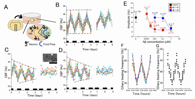
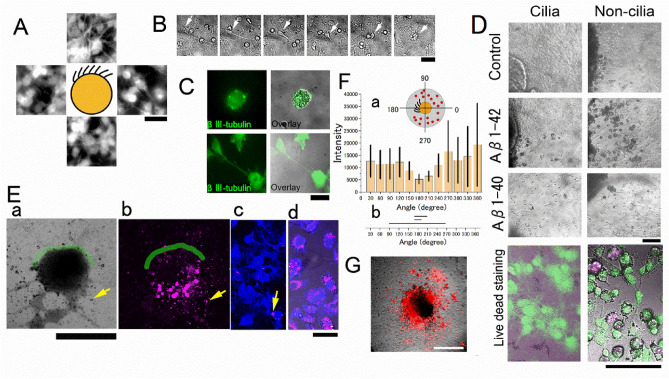
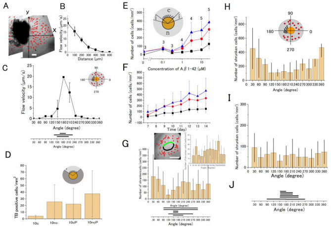
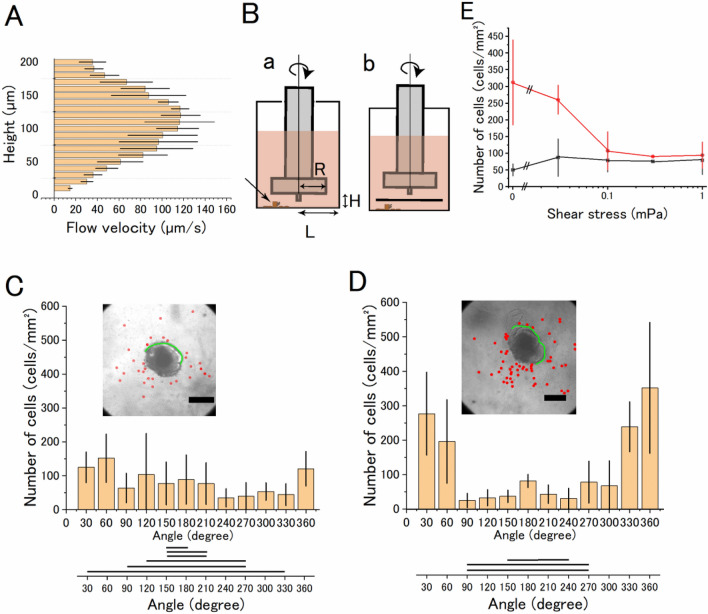
Alzheimer's disease (AD) is characterized by extensive and selective death of neurons and deterioration of synapses and circuits in the brain. The Aβ1-42 concentration is higher in an AD brain than in cognitively normal elderly individuals, and Aβ1-42 exhibits neurotoxicity. Brain-derived Aβ is transported into the cerebrospinal fluid (CSF), and CSF flow is driven in part by the beating of cilia and CSF secretion into ventricles. Ventricles are lined with ependyma whose apical surface is covered with motile cilia. Herein, we constructed an experimental system to measure the movement of ependymal cilia and examined the effects of Aβ1-42 to the beating of cilia and neurons. The circadian rhythm of the beating frequency of ependymal cilia was detected using brain wall explant-cultures containing ependymal cilia and neurons; the beating frequency was high at midday and low at midnight. Aβ1-42 decreased the peak frequency of ciliary beating at midday and slightly increased it at midnight. Aβ1-42 exhibited neurotoxicity to neurons on the non-ciliated side of the explant culture, while the neurotoxicity was less evident in neurons on the ciliated side. The neurotoxic effect of Aβ1-42 was diminished when 1 mPa of shear stress was generated using a flow chamber system that mimicked the flow by cilia. These results indicate that Aβ1-42 affects the circadian rhythm of ciliary beating, decreases the medium flow by the cilia-beating, and enhances the neurotoxic action of Aβ1-42 in the brain explant culture.


Conflict of interest statement

The authors declare no competing interests.

Figures

Figure 1
Figure 1
Inhibitory action of Aβs on the circadian rhythm of the ciliary beating frequency. (A) A schematic drawing of the brain explant culture from the brain wall. Neurons migrated from the brain explant are illustrated schematically. CS, ciliated side; NS, non-ciliated side. (B) The circadian oscillation of the frequency of the ciliary beating in the control medium. (C) The circadian oscillation in 10 nM Aβ1–42. (D) The circadian oscillation and in 1 µM Aβ1–40. The vertical axis is the ciliary beating frequency (CBF) and the horizontal axis is the time (days) from the start of imaging (2–3 days after making the explant culture). Black bars denote the night-time (6:00 p.m.–6:00 a.m.). The ependymal cilia were kept under a constant condition (35 °C, 5% CO2 concentration, in total darkness) and the frequency of beating was measured every 4 h with the optical microscope. Each color denotes an individual explant culture. The CBF of 16 explant cultures was recorded on day 0 and 14 cultures on day 7 in panel (B). The inset in panel C shows typical ciliary cells on the wall of a chemically fixed explant culture. Bar, 10 µm. (E) The concentration dependent inhibitory action of Aβs on the amplitude of the circadian rhythm. The amplitude of the circadian rhythm (i.e., the difference between the maximum and the minimum frequency of the ciliary beating) was measured on day 7 in the control medium and under different concentrations of Aβ1–42 (red) and Aβ1–40 (blue). Error bars denote the standard deviation of the mean (the number of data points shown near the bar). (F) The CBF recorded from these pieces of acutely dissected brain tissue showed a circadian rhythm with a 24-h period. Each color corresponds to the CBF of a piece of dissected brain tissue with ependymal cilia prepared at 12:00. (G)The distribution of CBF recorded from acutely dissected brain tissue with ependymal cilia when the dissection was made every four hours. Each point corresponds to a piece of dissected brain tissue. Timepoints of the first 12:00–12:00 (24 h) have been duplicated to facilitate viewing of the time curve. N denotes the number of culture wells. Image analysis by HCimage and ImageJ.
Figure 2
Figure 2
Neurotoxic effects of Aβ1–42 observed in the explant culture. (A) Fluorescence images of tubulin βIII positive neurons migrated from the explant; images are from the area 0.5 mm up and down, left and right of the schematically illustrated explant. (B) Typical time-lapse images of neurites retracted in 10 µM Aβ1–42 (time-lapse images were taken at 1, 2, 3, 4, 5, and 13 h from left to right). The soma is shown by the arrows. (C) A typical shrunken neuron in 3 µM Aβ1–42 for three days is positive for tubulin βIII (left) and superimposed on the DIC image (right). Neurons in the control medium in the same notation (lower panels). (D) TB-positive shrunken cells in the explant culture in the Aβs. DIC images of explant culture of the control (upper), Aβ1–42 (3 µM, middle), and Aβ1–40 (3 µM, lower). Live (green) and dead (red) cell staining of neurons superimposed on the DIC images (10 µM Aβ1–42 for four days, bottom) on the cilia side (left column) and non-cilia side (right column). (E) A typical explant culture treated with Aβ1–42 (10 µM) and RAβ1–42 (0.4 µM). (a) DIC image of an explant culture. The yellow arrow shows a typical shrunken cell. (b) Fluorescence image of RAβ1–42. The shrunken cell positive for RAβ1–42 is pointed by the arrow. (c) Neurons on the ciliated side are positive for MAP-2 (blue). Small fraction of neurons positive for RAβ1–42 (magenta) shown by an arrow. (d) Neurons on the non-ciliated side are positive for MAP-2 and RAβ1–42. The green line shows the ependymal cilia cells (a and b). (F) (a)The distribution of RAβ1–42 plotted in the polar coordinate system of the explant culture; the fluorescence intensity of RAβ1–42 in the area 100 μm from the edge of the explant culture was plotted. N = 3 culture wells. The inset is an illustration of an explant culture and RAβ1–42 positive cells (red dots) and the assignment of the angle; the polar coordinate system of the explant culture is divided into 12 sections [ 0, 30), [ 30, 60) …, [ 330, 360); the center of the ciliated area is assigned 180 degrees. (b) The individual bar denotes the distribution of beating cilia. The fluorescence intensity of RAβ1–42 on the non-ciliated area is significantly higher than that on the ciliated area control (p = 0.006, one-way ANOVA test, Origin ver. 2020b). (G) fluorescence image of RAβ1–42 positive cells of an explant culture that had no ciliated cells. Bars are 50 µm (panel a) and 30 µm (panels C), 100 µm (panels B and D), 500 µm (panels E a and b), 50 µm (panels E c and d), and 300 µm (panel G). N denotes the number of culture wells.
Figure 3
Figure 3
Analysis of the toxic effect of Aβ1–42 diminished on the ciliated side of the explant culture. (A) A flow map of the medium around a brain explant with beating cilia. The flow directions are indicated by arrows. The length of the arrow denotes the speed of flow estimated by particle tracking at 110 µm above the substrate for 1 s. (B) The speed of the flow along the x-axis at 10 µm above the substrate; the speed is high near the beating cilia (x = 0) and declines with distance from the explant. N = 3. Bars denote the standard deviation of the mean. (C) The distribution of the speed of the flow at 10 µm above the substrate plotted in the polar coordinate system of the explant culture (inset). N = 3. Bars denote the standard deviation of the mean. (D) The neurotoxic effect of Aβ1–42 was augmented by phosphoramidon (20 µM). The number of TB-positive cells in mm2 in 10 µM Aβ1–42 on the ciliated side of the explant culture (10c), in 10 µM Aβ1–42 on the non-ciliated side of the explant culture (10nc), the number of TB-positive cells in 10 µM Aβ1–42 with 20 µM phosphoramidon on the ciliated side (10cP), and that on the non-ciliated side (10ncP). The number of TB-positive cells in 1 mm2 on the non-ciliated side is higher than that on the ciliated side, and that in phosphoramidon is significantly higher than that in the control (p = 0.04, two-way ANOVA test). (E) The dose-dependent increase in the neurotoxicity of Aβ1–42 on the non-ciliated (blue triangle), ciliated side (black squares), and the intermediate area (red circles) between the ciliated and non-ciliated sides of the explant culture. These regions are shown in the inset; “c” cilia, “i” intermediate (20 degrees in the polar coordinate system), and “n” non-cilia regions. The number of data points is shown in the figure. The number of shrunken cells in 1 mm2 on the ciliated, non-ciliated, and intermediate area was significantly different (p = 9.6 × 10−7, two-way ANOVA test), supporting the idea that the flow affects the Aβ1–42 neurotoxic action. (F) Time-dependent increase in the neurotoxicity of Aβ1–42 on the non-ciliated (blue triangle), ciliated side (black squares), and the intermediate (red circles) region. The number of data points is 5 (except 3 on day 12). The interaction between the time-dependent increase in the toxic effect of Aβ1–42 and the flow level was not significant, suggesting that the time delay of the Aβ1–42 neurotoxic action was not apparently affected by the medium flow. (G) When the explant brain tissue was removed from the bottom and 25 µM Aβ1–42 was applied, a nearly uniform distribution of shrunken cells was seen in the polar coordinate system of the explant culture. Inset image, the circle denotes the position of the pre-existing explant, and the green line denotes the distribution of pre-existing beating cilia. Bar denotes 500 μm. The number of shrunken cells within the area 100 μm from the edge of the explant culture was counted. N = 6. Inset graph, the number of shrunken cells increased from 7 to 14 days culture within 350 μm from the edge. The horizontal bars at the bottom denote the distribution of beating cilia in the polar coordinate system. These distributions of the number of shrunken cells are not dependent on the pre-existing cilia of the removed explant. (H) The distribution of shrunken cells in the polar coordinate system of the explant culture in 25 µM Aβ1–42. The number of shrunken cells in the area 100 μm from the edge of the explant culture was counted. The inset shows the polar coordinate system and the assignment of the angle. (I) The distribution of shrunken cells in the area 100–350 μm from the edge of the same set of explant cultures in (H). N = 6. (J) The horizontal bars denote the distribution of beating cilia in the polar coordinate system. The number of shrunken cells in the ciliated area is lower than that of the non-ciliated direction in panel (H) (p = 5.8 × 10−5, two-way ANOVA test). N denotes the number of culture wells. Statistical analyses by Origin ver. 2020b.
Figure 4
Figure 4
Effects of artificially generated medium flow to neurons migrated from the explant brain tissue culture with Aβ1–42. (A) The flow map of the medium along the z-axis of the explant brain tissue culture with beating cilia shows that the speed of flow in the vicinity (ca. 10 μm) of the beating cilia is high at around 100 μm from the bottom where the beating of cilia was detected and is low at the bottom of the culture plate. N = 3. (B)(a) A schematic drawing of the parallel plate flow chamber and an explant culture. H = 1 mm, R = 2 mm, and L = 3 mm. The arrow shows the position of the brain explant. (b) A 4-mm square coverslip was placed 1 mm above the neurons. (C) The distribution of shrunken cells in 10 µM Aβ1–42 under 1 mPa shear stress in the area 100 μm from the edge of the explant culture in the polar coordinate system. The inset in panel (C) shows a typical DIC image of the explant culture in 10 µM Aβ1–42 under shear stress for four days. The inset in (D) shows the explant culture of the sham control. Red dots denote the location of the shrunken cells. The green line denotes the area of ciliary cells found. Bar, 500 μm. The distribution of shrunken cells on the ciliated or on the non-ciliated side was significantly different. The distribution of shrunken cells in the presence or absence of the artificial flow was also significantly different (p = 0.04, two-way ANOVA test, Origin ver. 2020b). (E) Shear force dependent decrease in the neurotoxic action of Aβ1–42 (10 µM). Vertical axis, number of dead cells/mm2 on the non-ciliated side (red circles) and ciliated-side (black circles), and horizontal axis, shear stress. N = 3, except N  = 1 at 0.3 mPa (N denotes the number of culture wells).

Similar articles

Cited by

References

    1. Toledo JB, et al. Factors affecting Abeta plasma levels and their utility as biomarkers in ADNI. Acta Neuropathol. 2011;122:401–413. doi: 10.1007/s00401-011-0861-8. - DOI - PMC - PubMed
    1. Dahlgren KN, et al. Oligomeric and fibrillar species of amyloid-beta peptides differentially affect neuronal viability. J. Biol. Chem. 2002;277:32046–32053. doi: 10.1074/jbc.M201750200. - DOI - PubMed
    1. Wang J, Gu BJ, Masters CL, Wang YJ. A systemic view of Alzheimer disease—Insights from amyloid-beta metabolism beyond the brain. Nat. Rev. Neurol. 2017;13:612–623. doi: 10.1038/nrneurol.2017.111. - DOI - PubMed
    1. Shaw LM, et al. Cerebrospinal fluid biomarker signature in Alzheimer's disease neuroimaging initiative subjects. Ann. Neurol. 2009;65:403–413. doi: 10.1002/ana.21610. - DOI - PMC - PubMed
    1. Sunderland T, et al. Decreased beta-amyloid1-42 and increased tau levels in cerebrospinal fluid of patients with Alzheimer disease. JAMA. 2003;289:2094–2103. doi: 10.1001/jama.289.16.2094. - DOI - PubMed

Publication types

Substances