Review
. 2023 Dec 11;214(1):79-93.
doi: 10.1093/cei/uxad101.

Difference in respiratory syncytial virus-specific Fc-mediated antibody effector functions between children and adults

Affiliations
Review

Difference in respiratory syncytial virus-specific Fc-mediated antibody effector functions between children and adults

Anke J Lakerveld et al. Clin Exp Immunol. .

Abstract

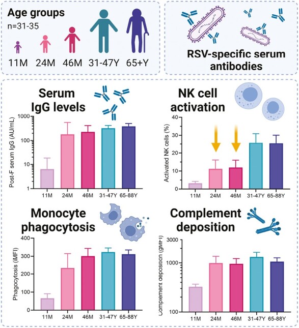
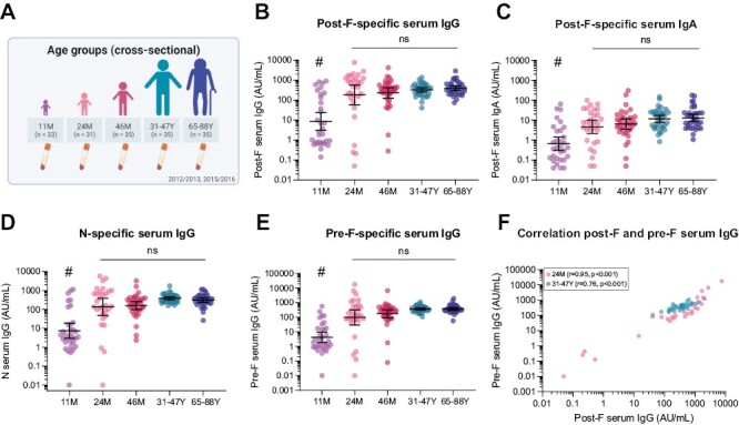
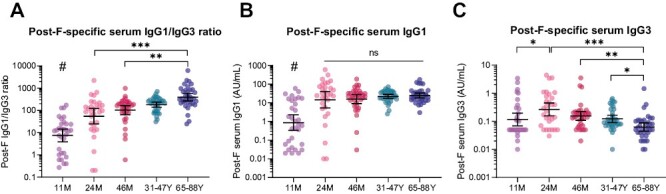
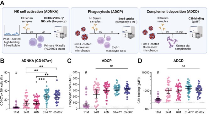
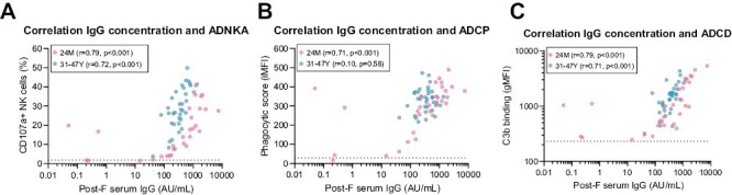
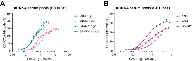
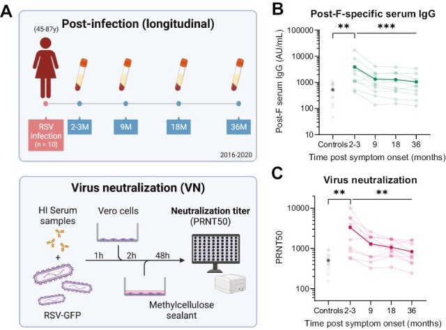
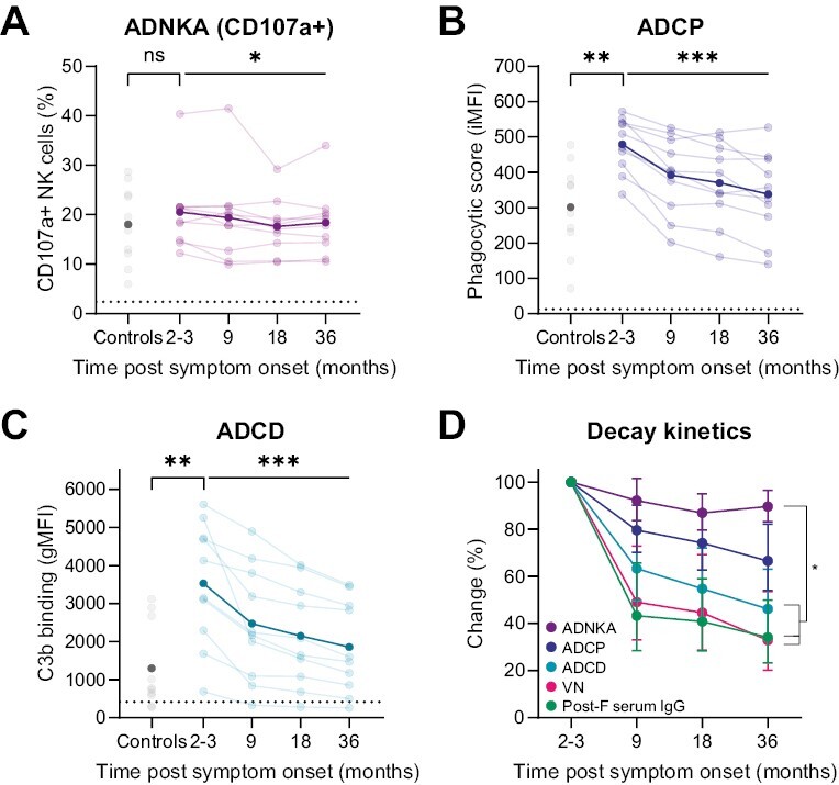
Respiratory syncytial virus (RSV) infections are a major cause of bronchiolitis and pneumonia in infants and older adults, for which there is no known correlate of protection. Increasing evidence suggests that Fc-mediated antibody effector functions have an important role, but little is known about the development, heterogeneity, and durability of these functional responses. In light of future vaccine strategies, a clear view of the immunological background and differences between various target populations is of crucial importance. In this study, we have assessed both quantitative and qualitative aspects of RSV-specific serum antibodies, including IgG/IgA levels, IgG subclasses, antibody-dependent complement deposition, cellular phagocytosis, and NK cell activation (ADNKA). Samples were collected cross-sectionally in different age groups (11-, 24-, and 46-month-old children, adults, and older adults; n = 31-35 per group) and longitudinally following natural RSV infection in (older) adults (2-36 months post-infection; n = 10). We found that serum of 24-month-old children induces significantly lower ADNKA than the serum of adults (P < 0.01), which is not explained by antibody levels. Furthermore, in (older) adults we observed boosting of antibody levels and functionality at 2-3 months after RSV infection, except for ADNKA. The strongest decrease was subsequently observed within the first 9 months, after which levels remained relatively stable up to three years post-infection. Together, these data provide a comprehensive overview of the functional landscape of RSV-specific serum antibodies in the human population, highlighting that while antibodies reach adult levels already at a young age, ADNKA requires more time to fully develop.

Keywords: anti-viral immunity; antibodies; complement; cytotoxicity; phagocytosis.


Conflict of interest statement

The authors declare no conflicts of interest.

Figures

Graphical Abstract
Graphical Abstract
Figure 1.
Figure 1.
RSV-specific serum antibody levels in different age groups. RSV-specific serum IgG and IgA levels were measured with a multiplex immunoassay in 11-month (n = 32), 24-month (n = 30), and 46-month-old children (n = 35), adults (n = 35), and older adults (n = 35). (A) Schematic overview of the cross-sectional clinical study cohort used to assess differences between age groups. (B) Post-F-specific IgG levels. (C) Post-F-specific IgA levels. (D) N-specific IgG levels. (E) Pre-F-specific IgG levels. (F) Correlation of pre-F- and post-F-specific IgG levels for 24-month-old children (light pink) and adults (teal). All data points represent individual participants, and geometric mean concentrations (GMCs) with 95% confidence intervals are depicted. Data is analyzed by Kruskal–Wallis test and correlations are assessed using the Spearman method. # indicates statistical significance of at least P < 0.01 compared to all other age groups. AU/ml, arbitrary units per milliliter; ns, not significant.
Figure 2.
Figure 2.
RSV-specific serum IgG subclasses in different age groups. Post-F-specific serum IgG subclass levels were measured with a multiplex immunoassay in 11-month (n = 32), 24-month (n = 30), and 46-month-old children (n = 35), adults (n = 35), and older adults (n = 35). (A) Post-F-specific IgG1/IgG3 ratio. (B) Post-F-specific IgG1 levels. (C) Post-F-specific IgG3 levels. All data points represent individual participants, and geometric mean concentrations (GMCs) with 95% confidence intervals are depicted. Data is analyzed by Kruskal–Wallis test. # indicates statistical significance of at least P < 0.01 compared to all other age groups. *P < 0.05; **P < 0.01; ***P < 0.001; ns, not significant. AU/ml, arbitrary units per milliliter.
Figure 3.
Figure 3.
Fc-mediated antibody effector functions in different age groups. (A) Schematic overview of the methods used to assess ADNKA, ADCP, and ADCD in serum from 11-month (n = 33), 24-month (n = 31), and 46-month-old children (n = 35), adults (n = 35), and older adults (n = 35). (B) ADNKA with percentage of CD107a + NK cells as read-out; the average of three healthy NK cell donors is depicted for each participant. (C) ADCP by THP-1 cells with phagocytic score as read-out; data for each participant consists of the average of technical duplicates. (D) ADCD with gMFI as read-out; data for each participant consists of the average of technical duplicates. The dotted lines indicate the level of the negative control. All data points represent individual participants and geometric means with 95% confidence intervals are depicted. Data is analyzed by Kruskal–Wallis test. # indicates statistical significance of at least P < 0.01 compared to all other age groups. **P < 0.01; ***P < 0.001; ns, not significant. ADCD: antibody-dependent complement deposition; ADCP: antibody-dependent cellular phagocytosis; ADNKA: antibody-dependent NK cell activation; gMFI: geometric mean fluorescence intensity; HI: heat-inactivated; iMFI, integrated mean fluorescence intensity; NK cells: natural killer cells.
Figure 4.
Figure 4.
Correlation analysis between serum IgG levels and antibody functionalities for 24-month-old children and adults. Post-F-specific serum IgG levels and ADNKA, ADCP, and ADCD were determined for the 24-month-old group (light pink, n = 30) and adults (teal, n = 35). Correlations are shown between post-F serum IgG levels and ADNKA measured as percentage CD107a + NK cells (A), ADCP by THP-1 cells (B), and ADCD (C). Dotted lines indicate negative control (ADNKA, ADCP, ADCD). All data points represent individual participants. Correlations are assessed using the Spearman method. ADCD, antibody-dependent complement deposition; ADCP, antibody-dependent cellular phagocytosis; ADNKA, antibody-dependent NK cell activation; AU/mL, arbitrary units per milliliter; gMFI, geometric mean fluorescence intensity; iMFI, integrated mean fluorescence intensity; NK cells, natural killer cells.
Figure 5.
Figure 5.
ADNKA titration curves of serum pools from different age groups. Serum pools were prepared from the 10 individuals with the highest post-F-specific IgG concentration for each age group (“high”) as well as pools of the 10 (adults) or 8 (24M) individuals with post-F-specific IgG levels around the GMC (“middle”). Post-F-specific IgG levels for each pool were measured with a multiplex immunoassay and ADNKA was assessed on a serial dilution range of each pool. (A) ADNKA titration of 24-month-old “high” (pink), 24-month-old “middle” (light pink), adults “high” (teal), and adults “middle” (light teal) serum pools, with percentage of CD107a + NK cells as read-out. (B) ADNKA titration of 11-month-old “high” (purple), 46-month-old “high” (dark pink), and older adults “high” (blue) serum pools, with percentage of CD107a + NK cells as read-out. The average of three healthy NK cell donors is depicted. The curves are fitted based on a 4-parameter nonlinear regression model. The dotted lines indicate the level of the negative control. ADNKA: antibody-dependent NK cell activation; AU/ml: arbitrary units per milliliter; GMC: geometric mean concentration; NK cells: natural killer cells.
Figure 6.
Figure 6.
Serum antibody and neutralization titer kinetics post RSV infection. (A) Upper panel: schematic overview of the longitudinal clinical study cohort to assess antibody kinetics 2–36 months post-RSV infection in (older) adults (47–87 years old, n = 10). Lower panel: schematic representation of workflow for virus neutralization assay. (B) Post-F-specific serum IgG concentrations measured with a multiplex immunoassay in controls (n = 10) and convalescent individuals following RSV infection over time. (C) Virus neutralization titers in controls and convalescent individuals following RSV infection over time. Each line represents a unique individual, dark lines indicate the geometric mean of all participants. Light grey dots represent individual age-matched controls, dark grey dots represent the geometric mean level of the control samples. Differences between controls and 2–3 months post-symptom onset was assessed with a Mann–Whitney test. Differences over the complete follow-up time post-infection were assessed with a Friedman test. **P < 0.01; ***P < 0.001. AU/ml, arbitrary units per milliliter; PRNT50, 50% plaque reduction neutralization test.
Figure 7.
Figure 7.
Fc-mediated antibody effector function kinetics post RSV infection. Serum Fc-functionality was assessed in longitudinal samples taken from (older) adults (n = 10) at 2–3, 9, 18, and 36 months post-RSV infection and from age-matched healthy controls (n = 10). Shown are antibody functions of samples for each timepoint, as indicated, assessed by (A) ADNKA with percentage of CD107a + NK cells as read-out, the average of three healthy NK cell donors is depicted for each participant; (B) ADCP by THP-1 cells with phagocytic score as read-out, data for each participant consists of the average of technical duplicates; (C) ADCD with geometric mean fluorescent intensity as read-out, data for each participant consists of the average of technical duplicates. The dotted lines indicate the level of the negative control. Each line represents a unique individual, dark lines indicate the geometric mean of all participants. Light grey dots represent individual age-matched controls, dark grey dots represent the geometric mean level of the control samples. Differences between controls and 2-3 months post-symptom onset was assessed with a Mann-Whitney test. Differences over the complete follow-up time post-infection were assessed with a Friedman test. (D) Overview of decay kinetics for antibody concentration and functionality normalized to 2-3 months post-infection with 95% confidence interval. A two-way ANOVA with Tukey’s multiple comparisons test was used to assess statistical significance of the difference in kinetics between antibody functions. Significance is indicated when there was a statistically significant difference for each timepoint between the two compared functions. *P < 0.05; **P < 0.01; ***P < 0.001; ns, not significant. ADNKA, antibody-dependent NK cell activation; ADCP, antibody-dependent cellular phagocytosis; ADCD: antibody-dependent complement deposition; gMFI: geometric mean fluorescence intensity; iMFI: integrated mean fluorescence intensity; NK cells: natural killer cells; VN: virus neutralization.

Similar articles

Cited by

References

    1. Shi T, Denouel A, Tietjen AK, Campbell I, Moran E, Li X, et al. . Global disease burden estimates of respiratory syncytial virus-associated acute respiratory infection in older adults in 2015: a systematic review and meta-analysis. J Infect Dis 2019,222 (Suppl 7), S577–S583. - PubMed
    1. Shi T, McAllister DA, O’Brien KL, Simoes EAF, Madhi SA, Gessner BD, et al. .; RSV Global Epidemiology Network. Global, regional, and national disease burden estimates of acute lower respiratory infections due to respiratory syncytial virus in young children in 2015: a systematic review and modelling study. Lancet 2017, 390, 946–58. doi:10.1016/S0140-6736(17)30938-8. - DOI - PMC - PubMed
    1. Hall CB, Walsh EE, Long CE, Schnabel KC.. Immunity to and frequency of reinfection with respiratory syncytial virus. J Infect Dis 1991, 163, 693–8. doi:10.1093/infdis/163.4.693. - DOI - PubMed
    1. Henderson FW, Collier AM, Clyde WA Jr, Denny FW.. Respiratory-syncytial-virus infections, reinfections and immunity. A prospective, longitudinal study in young children. N Engl J Med 1979, 300, 530–4. doi:10.1056/NEJM197903083001004. - DOI - PubMed
    1. Garegnani L, Styrmisdottir L, Roson Rodriguez P, Escobar Liquitay CM, Esteban I, Franco JV.. Palivizumab for preventing severe respiratory syncytial virus (RSV) infection in children. Cochrane Database Syst Rev 2021, 11, CD013757. doi:10.1002/14651858.CD013757.pub2. - DOI - PMC - PubMed

Publication types