

Molecular insights using spatial transcriptomics of the distal lung in congenital diaphragmatic hernia
- PMID: 37605849
- PMCID: PMC10639013
- DOI: 10.1152/ajplung.00154.2023
Molecular insights using spatial transcriptomics of the distal lung in congenital diaphragmatic hernia
Abstract
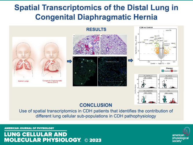
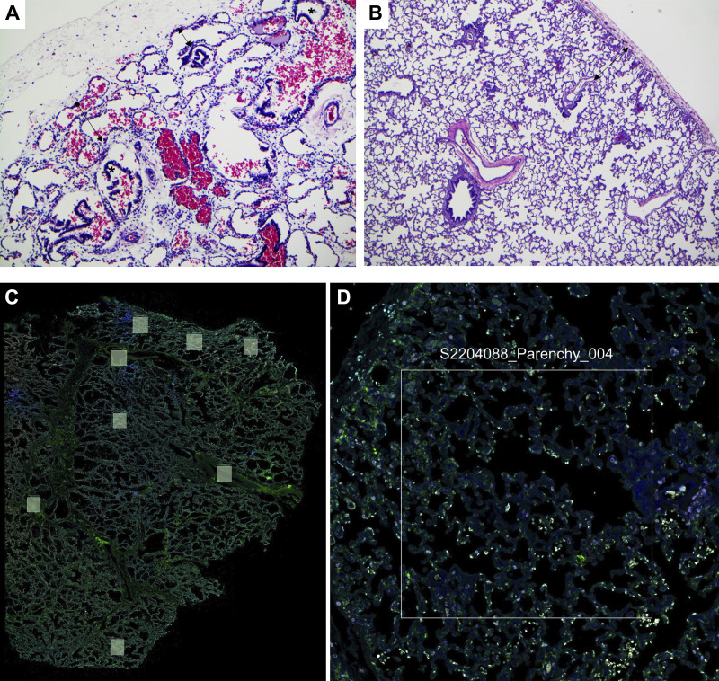
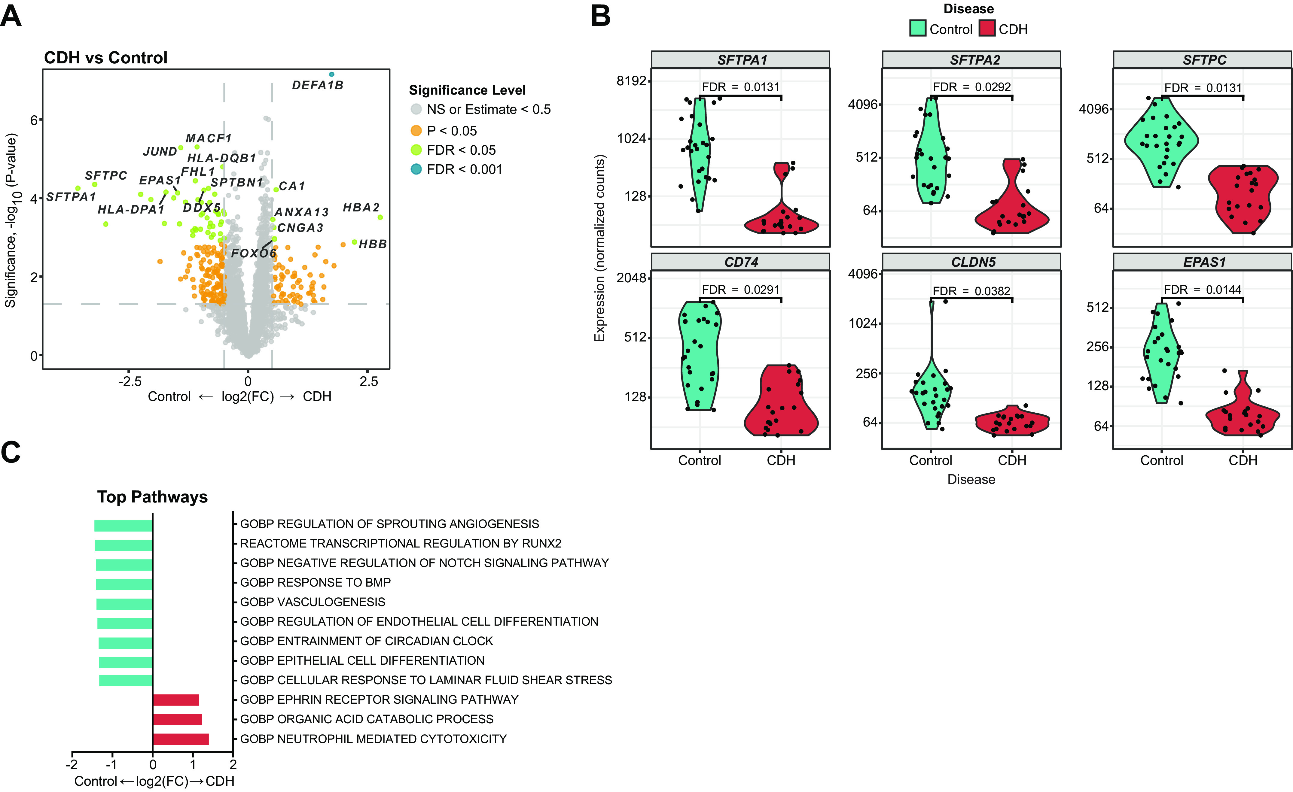
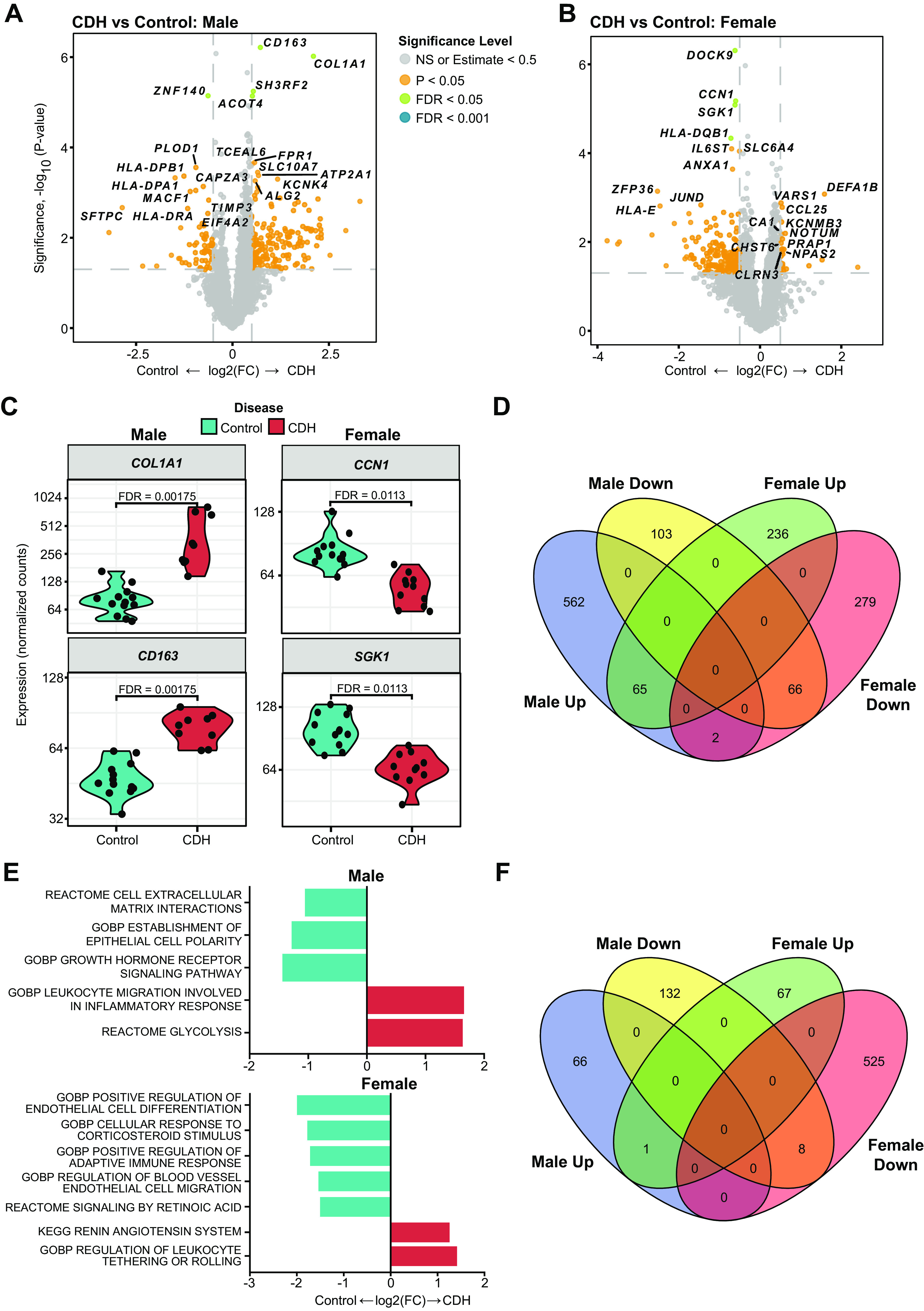
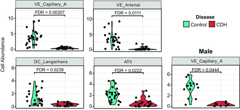
Abnormal pulmonary vascular development and function in congenital diaphragmatic hernia (CDH) is a significant factor leading to pulmonary hypertension. The lung is a very heterogenous organ and has marked cellular diversity that is differentially responsive to injury and therapeutic agents. Spatial transcriptomics provides the unmatched capability of discerning the differences in the transcriptional signature of these distinct cell subpopulations in the lung with regional specificity. We hypothesized that the distal lung parenchyma (selected as a region of interest) would show a distinct transcriptomic profile in the CDH lung compared with control (normal lung). We subjected lung sections obtained from male and female CDH and control neonates to spatial transcriptomics using the Nanostring GeoMx platform. Spatial transcriptomic analysis of the human CDH and control lung revealed key differences in the gene expression signature. Increased expression of alveolar epithelial-related genes (SFTPA1 and SFTPC) and angiogenesis-related genes (EPAS1 and FHL1) was seen in control lungs compared with CDH lungs. Response to vitamin A was enriched in the control lungs as opposed to abnormality of the coagulation cascade and TNF-alpha signaling via NF-kappa B in the CDH lung parenchyma. In male patients with CDH, higher expression of COL1A1 (ECM remodeling) and CD163 was seen. Increased type 2 alveolar epithelial cells (AT-2) and arterial and lung capillary endothelial cells were seen in control lung samples compared with CDH lung samples. To the best of our knowledge, this is the first use of spatial transcriptomics in patients with CDH that identifies the contribution of different lung cellular subpopulations in CDH pathophysiology and highlights sex-specific differences.NEW & NOTEWORTHY This is the first use of spatial transcriptomics in patients with congenital diaphragmatic hernia (CDH) that identifies the contribution of different lung cellular subpopulations in CDH pathophysiology and highlights sex-specific differences.
Keywords: congenital diaphragmatic hernia; pulmonary hypertension; spatial transcriptomics.
Conflict of interest statement
No conflicts of interest, financial or otherwise, are declared by the authors.
Figures
Similar articles
-
Endothelial-to-Mesenchymal Transition in Human and Murine Models of Congenital Diaphragmatic Hernia.Neonatology. 2024;121(4):512-518. doi: 10.1159/000537802. Epub 2024 Apr 8. Neonatology. 2024. PMID: 38588643
-
Decreased Endoglin expression in the pulmonary vasculature of nitrofen-induced congenital diaphragmatic hernia rat model.Pediatr Surg Int. 2017 Feb;33(2):263-268. doi: 10.1007/s00383-016-4004-0. Epub 2016 Nov 7. Pediatr Surg Int. 2017. PMID: 27822781
-
Increased c-kit and stem cell factor expression in the pulmonary vasculature of nitrofen-induced congenital diaphragmatic hernia.J Pediatr Surg. 2016 May;51(5):706-9. doi: 10.1016/j.jpedsurg.2016.02.007. Epub 2016 Feb 11. J Pediatr Surg. 2016. PMID: 26932254
-
Congenital diaphragmatic hernia-associated pulmonary hypertension.Semin Perinatol. 2020 Feb;44(1):151167. doi: 10.1053/j.semperi.2019.07.006. Epub 2019 Jul 30. Semin Perinatol. 2020. PMID: 31519366 Review.
-
Congenital diaphragmatic hernia. A cause of persistent pulmonary hypertension of the newborn which lacks an effective therapy.Biol Neonate. 1998 Nov;74(5):323-36. doi: 10.1159/000014050. Biol Neonate. 1998. PMID: 9742261 Review.
Cited by
-
Epithelial Dysfunction in Congenital Diaphragmatic Hernia: Mechanisms, Models and Emerging Therapies.Cells. 2025 May 9;14(10):687. doi: 10.3390/cells14100687. Cells. 2025. PMID: 40422190 Free PMC article. Review.
-
Fetal hypoplastic lungs have multilineage inflammation that is reversed by amniotic fluid stem cell extracellular vesicle treatment.Sci Adv. 2024 Jul 26;10(30):eadn5405. doi: 10.1126/sciadv.adn5405. Epub 2024 Jul 26. Sci Adv. 2024. PMID: 39058789 Free PMC article.
-
Fetal Tracheal Occlusion Correlates with Normalized YAP Expression and Alveolar Epithelial Differentiation in Congenital Diaphragmatic Hernia.Am J Respir Cell Mol Biol. 2025 Jun;72(6):688-697. doi: 10.1165/rcmb.2024-0323OC. Am J Respir Cell Mol Biol. 2025. PMID: 39661950 Free PMC article.
-
Sex-specific differences in the severity of pulmonary hypoplasia in experimental congenital diaphragmatic hernia and implications for extracellular vesicle-based therapy.Pediatr Surg Int. 2024 Oct 28;40(1):278. doi: 10.1007/s00383-024-05856-0. Pediatr Surg Int. 2024. PMID: 39467854
References
-
- Olutoye OO, Short WD, Gilley J, Hammond JD, Belfort MA, Lee TC, King A, Espinoza J, Joyeux L, Lingappan K, Gleghorn JP, Keswani SG. The cellular and molecular effects of fetoscopic endoluminal tracheal occlusion in congenital diaphragmatic hernia. Front Pediatr 10: 925106, 2022. doi:10.3389/FPED.2022.925106. - DOI - PMC - PubMed
Publication types
MeSH terms
Substances
Associated data
Grants and funding
LinkOut - more resources
Full Text Sources
Medical
Research Materials
Miscellaneous