

doi: 10.2340/actadv.v103.4500.
Unusually Aggressive Actinic Keratosis of the Eyelid and Conjunctiva
Affiliations
- PMID: 37605896
- PMCID: PMC10461307
- DOI: 10.2340/actadv.v103.4500
Item in Clipboard
Unusually Aggressive Actinic Keratosis of the Eyelid and Conjunctiva
Acta Derm Venereol.
.
No abstract available
Conflict of interest statement
Figures
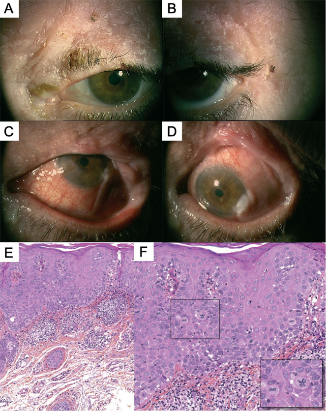
(a, b) Initial clinical presentation in 2010, with a scaly erythematous plaque, affecting the free border of the left eyelid and extending upward to the eyebrow, initially without conjunctival involvement. (c–d) Relapse in 2015, with conjunctival involvement: papillomatous hyperaemic plaques of the temporal bulbar conjunctiva, extending from the limbus to the lateral canthus and to the superior palpebral conjunctiva. (e, f) Histological analysis showed evidence of actinic keratosis without evidence of invasion (e: haematoxylin and eosin (H&E) ×4; f: H&E ×10, inset ×40).
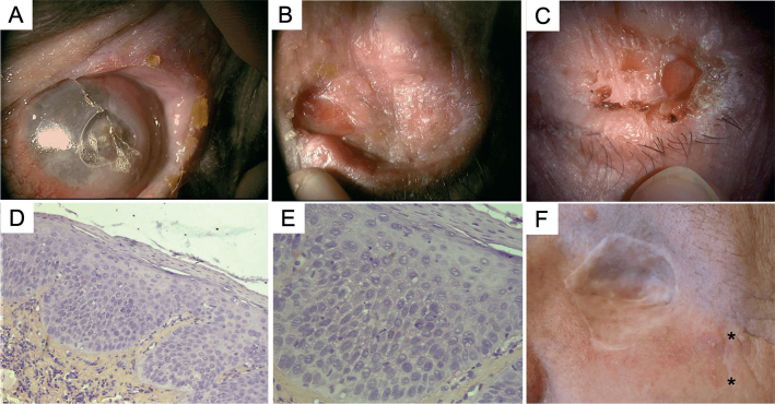
(a) Progressive disease despite topical treatment leading to a non-functional eye with excruciating pain. (d, e) Evisceration with reconstructive surgeries (using Cutler Beard flap) were performed in 2016, and histopathological analysis showed conjunctival leukokeratosis with few focal lesions of low-grade intra-epithelial neoplasia but without any evidence of invasion. (b, c) However, new actinic keratoses (AK) continued to appear over the remnant of the eyelid despite many topical treatments over 4 years. (f) Orbital exenteration and grafting were then done, but the patient was still developing cutaneous AK lesions in proximity to the skin graft even 6 months later. (d: H&E, ×4; e: H&E, ×10).
References
-
- Werner RN, Sammain A, Erdmann R, Hartmann V, Stockfleth E, Nast A. The natural history of actinic keratosis: a systematic review. Br J Dermatol 2013; 169: 502–518. - PubMed
-
- Richard MA, Amici JM, Basset-Seguin N, Claudel JP, Cribier B, Dreno B. Management of actinic keratosis at specific body sites in patients at high risk of carcinoma lesions: expert consensus from the AKTeam of expert clinicians. J Eur Acad Dermatol Venereol 2018; 32: 339–346. - PubMed
MeSH terms
LinkOut - more resources
Full Text Sources
