. 2023 Aug 22;8(16):e166386.
doi: 10.1172/jci.insight.166386.

Species-specific roles for the MAFA and MAFB transcription factors in regulating islet β cell identity

Affiliations

Species-specific roles for the MAFA and MAFB transcription factors in regulating islet β cell identity

Jeeyeon Cha et al. JCI Insight. .

Abstract

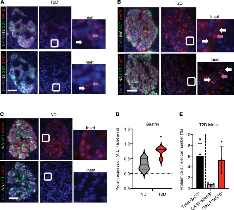
Type 2 diabetes (T2D) is associated with compromised identity of insulin-producing pancreatic islet β cells, characterized by inappropriate production of other islet cell-enriched hormones. Here, we examined how hormone misexpression was influenced by the MAFA and MAFB transcription factors, closely related proteins that maintain islet cell function. Mice specifically lacking MafA in β cells demonstrated broad, population-wide changes in hormone gene expression with an overall gene signature closely resembling islet gastrin+ (Gast+) cells generated under conditions of chronic hyperglycemia and obesity. A human β cell line deficient in MAFB, but not one lacking MAFA, also produced a GAST+ gene expression pattern. In addition, GAST was detected in human T2D β cells with low levels of MAFB. Moreover, evidence is provided that human MAFB can directly repress GAST gene transcription. These results support a potentially novel, species-specific role for MafA and MAFB in maintaining adult mouse and human β cell identity, respectively. Here, we discuss the possibility that induction of Gast/GAST and other non-β cell hormones, by reduction in the levels of these transcription factors, represents a dysfunctional β cell signature.

Keywords: Beta cells; Cell Biology; Diabetes; Endocrinology; Islet cells.


Figures

Figure 1
Figure 1. Non–β cell hormone expression is increased in MafA∆β islets.
(A and B) Analysis of MafA, Insulin 1 (Ins1), and non–β cell hormone mRNA levels in MafAWT and MafA∆β islets from 3-month-old adult male (A) and female (B) mice. Data are shown as mean ± SEM. n = 3–4 animals/group. *P < 0.05 by Student’s t test; n.d., not detectable. (C) Gast mRNA was induced more in male than in female MafA∆β islets. Data are shown as mean ± SEM. n = 3–4 animals/group. *P < 0.05 by Student’s t test. (D) Top panel: Stomach G cells from GASTWT and GASTKO mice served as positive and negative control for Gast antibody staining. n = 2–3 animals/group. Middle panels: Representative images for Gast (red) and Insulin (white) immunostaining in male MafAWT and MafA∆β islets. Insets for Gast staining are shown in right panels. Bottom left panel: Gastrin signal (A.U.) per mouse islet cell. Bottom right panel: Gast+ cells per islet (% of islet cells). Four to 6 islets per mouse. n = 3–4 animals/group were quantified using ImageJ. Scale bar, 50 μm. Data are shown as mean ± SEM. *P < 0.05 by Student’s t test.
Figure 2
Figure 2. Islet Gast+ cells produced in insulin-resistant mice share molecular signatures with the broader MafA∆β islet population.
(A) Administration of the S961 insulin receptor antagonist for 4 days elevated random blood glucose levels in relation to PBS-treated control mice. Data are shown as mean ± SEM. n = 2–3 animals/group. *P < 0.05 by Student’s t test. (B) MafA (red) protein levels were markedly reduced in S961-treated mice. Insulin (white). n = 2–4 animals/group. Scale bar, 50 μm. (C) UMAP analyses of the single-cell RNA-Seq data in male S961-treated islets. Left panel: Cluster annotation shows that ~80% of islet cells were Insulin+ β cells (left, circled in red). Right panels: Gast+ cells (blue dots) were enriched in the β cell population. (D) Heatmap showing the 20 upregulated genes found in S961 Gast+ cells by single-cell RNA sequencing (Supplemental Table 1) were also elevated in male MafA∆β islets. MafA∆β β cell RNA-Seq were was used in this analysis. n = 3 animals/experimental group. (E) Heatmap showing limited overlap between the upregulated genes found in S961 Gast+ cells and other exocrine and islet cell types.
Figure 3
Figure 3. MAFBKD, but not MAFAKD, induces non–β cell hormone expression in human β cells.
(AD) Analysis of MAFA/B protein (A and C) and hormone-related mRNA levels (B and D) in shControl (scrambled construct), shMAFA-, or shMAFB-treated EndoC-βH2 (i.e., only MAFB+) (A) and EndoC-βH1 (i.e., MAFA+MAFB+) cells (C). β-Actin served as the internal control and expression was normalized to shControl levels. MAFA knockdown in shMAFA-treated cells was reduced by ~55% of the control level, and shMAFB-treated cells decreased by ~71%. Data are shown as mean ± SEM. *P < 0.05, **P < 0.005 by Student’s t test in B. *P < 0.05 by 2-way ANOVA in D. n = 2–3 replicates/experiment, and experiments were repeated 4 times. (E) Immunostaining for SST (white) and GAST (green) in MAFBKD EndoC-βH2 cells. These proteins were undetectable in shControl-treated cells. Magnification, 20×. Left: Green arrows denote GAST+ SST cells, while red arrows denote GAST+ SST+ cells. Right: Quantification of hormone+ cells shown in MAFBKD EndoC-βH2 cells relative to the total β cell number compared with shControl. Data are shown as mean ± SEM. n = 2–3 replicates/experiment, and experiments were repeated 4 times.
Figure 4
Figure 4. Unique and common gene signatures are produced in the Gast+ cells of mouse S961-treated β cells, hESC-derived MAFBKO β-like cells, and bona fide stomach G cells.
(A) UMAP of the single-cell RNA-Seq data from hESC-derived MAFBWT and MAFBKO cells from Russell et al. (8). Left panel: Cluster annotation (as determined by markers outlined in Supplemental Tables 7 and 8) identifies cell fates derived from this differentiation protocol. Right panel: Enrichment of GAST+ cells (encircled) and compromised INS+ cell numbers in the MAFBKO cell population relative to MAFBWT cells. (B) Top 10 most influenced GO biological processes based on the 61 upregulated genes in GAST+ MAFBKO cells compared with GAST MAFBKO cells. (C and D) Venn diagrams of DEGs comparing single-cell data from GAST+ cells from hESC-derived MAFBKO β-like cells and Gast+ β cells of mouse S961-treated islets (C) as well as human stomach G cells (D). Gene products common to both conditions are listed on the right. Bolded gene products indicate those common to all 3 data sets.
Figure 5
Figure 5. GAST+ MAFBKO cell genes upregulated in MAFBKD EndoC-βH2 cells.
(A) Venn diagram of DEGs showing that 18 upregulated genes are shared between MAFBKD EndoC-βH2 cells and the GAST+ MAFBKO hESC–derived β-like cells. (B) Heatmap of these 18 upregulated shared genes in shMAFB- and shControl-treated EndoC-βH2 cells. n = 3 replicates/group. (C) qPCR analysis of a subset of the shared genes in shMAFB- and shControl-treated EndoC-βH2 cells. β-Actin served as the internal control and expression normalized to levels of shControl. Data are shown as mean ± SEM. *P < 0.05 by Student’s t test. n = 2 replicates/experiment, and experiments were repeated 3 times. (D) Venn diagram of DEGs of GAST+ MAFBKO β-like cells, MAFBKD EndoC-βH2 cells, and human stomach G cells. The 5 genes common to all 3 data sets are listed on the right.
Figure 6
Figure 6. GAST is produced in MAFB-deficient T2D islet cells.
(AC) Representative images of immunostaining performed on serial sections from male age-matched diabetic (A and B) and nondiabetic (ND) (C) pancreata. MAFB, red in top panels; GAST, red in bottom panels; SST, green; INS, white; and nuclei, blue. GAST+ cells were not detected in healthy donor islets by immunofluorescence analysis (C). However, GAST production showed interislet variability and was associated with lower MAFB levels in T2D islet cells (A and B). Insets show magnified view of representative GASTMAFB+ (purple arrows) and rare GAST+MAFB (white arrows) cells. n = 4 age- and sex-matched donors/condition. Scale bar, 100 μm. (D) Quantification of GAST signal (A.U.) per islet between ND and T2D male donors. Data are shown as mean ± SEM. *P < 0.05 by Student’s t test. n = 4 age- and sex-matched donors/condition, 4–6 islets per donor. (E) Quantification of GAST and MAFB colocalization in T2D male donor islets (% islet cells). Data are shown as mean ± SEM. n = 4 age- and sex-matched donors/condition, 4–6 islets per donor. Donor information in Supplemental Table 3.
Figure 7
Figure 7. MAFB binds within the human GAST 5′-flanking region and suppresses gene expression in β cells.
(A) ChIP-Seq data derived from whole islets by Pasquali et al. (36) illustrating islet-enriched PDX1, NKX2.2, FOXA2, NKX6.1, and MAFB TF binding sites (TFBS) within approximately 1.5 kbp of the human islet GAST gene transcription start site. (B) Gel shift analysis of MAFB (left) or MAFA (right) protein binding to a biotin-labeled human INSULIN (INS) probe to a MAFA/B binding site starting at position –135 in the presence of human INS and human GAST –1,825/–1,795, –1,525/–1,495, or –1,411/–1,381 bp unlabeled competitors. While all 3 GAST sites competed as effectively as the INS element, only the GAST –1,525/–1,495 element is conserved in mouse and also binds MAFA (right panel). n = 3 experimental replicates. (C) Representative MAFA/B binding site mutation in the –1,525/–1,495 element stimulated –1.68 kbp GAST-driven luciferase reporter activity in human EndoC-βH1 cells. pGL3 Basic-LUC represents the control vector without insert. Data are shown as mean ± SEM. *P < 0.05 by 2-way ANOVA. n = 3 experimental replicates, and experiments were repeated 3 times.

References

    1. Hang Y, Stein R. MafA and MafB activity in pancreatic β cells. Trends Endocrinol Metab. 2011;22(9):364–373. doi: 10.1016/j.tem.2011.05.003. - DOI - PMC - PubMed
    1. Pan FC, Wright C. Pancreas organogenesis: from bud to plexus to gland. Dev Dyn. 2011;240(3):530–565. doi: 10.1002/dvdy.22584. - DOI - PubMed
    1. Artner I, et al. MafA and MafB regulate genes critical to beta-cells in a unique temporal manner. Diabetes. 2010;59(10):2530–2539. doi: 10.2337/db10-0190. - DOI - PMC - PubMed
    1. Cyphert HA, et al. Examining how the MAFB transcription factor affects islet β-cell function postnatally. Diabetes. 2019;68(2):337–348. doi: 10.2337/db18-0903. - DOI - PMC - PubMed
    1. Banerjee RR, et al. Gestational diabetes mellitus from inactivation of prolactin receptor and MafB in islet β-cells. Diabetes. 2016;65(8):2331–2341. doi: 10.2337/db15-1527. - DOI - PMC - PubMed

Publication types