Review
. 2023 Jul 31;12(3):50.
doi: 10.3390/antib12030050.

Diagnosis and Treatment of Paraneoplastic Neurologic Syndromes

Affiliations
Review

Diagnosis and Treatment of Paraneoplastic Neurologic Syndromes

Daniel Chiu et al. Antibodies (Basel). .

Abstract

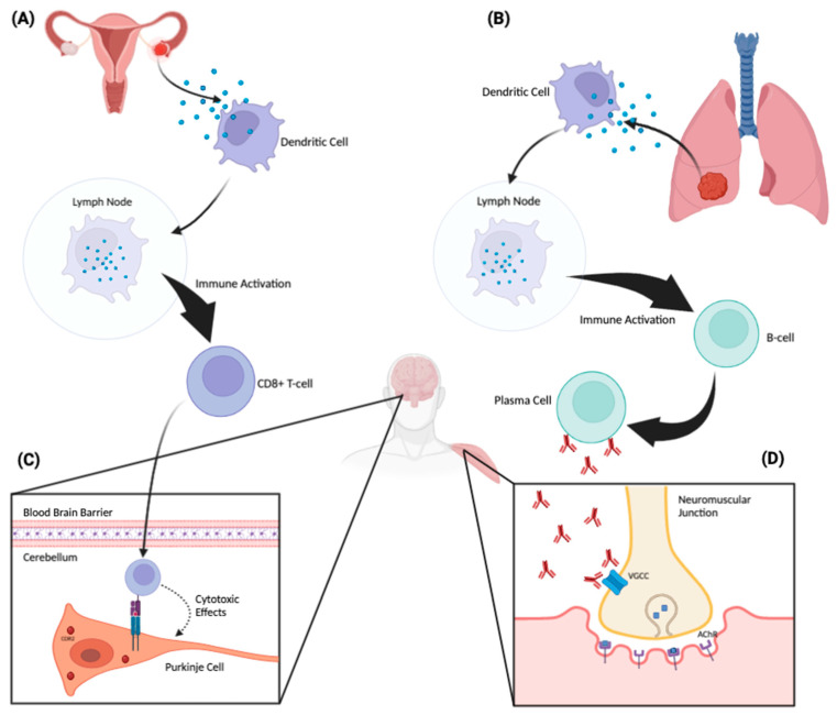
Paraneoplastic antibody syndromes result from the anti-tumor antibody response against normal antigens ectopically expressed by tumor cells. Although this antibody response plays an important role in helping clear a nascent or established tumor, the engagement of antigens expressed in healthy tissues can lead to complex clinical syndromes with challenging diagnosis and management. The majority of known paraneoplastic antibody syndromes have been found to affect the central and peripheral nervous system. The present review provides an update on the pathophysiology of paraneoplastic neurologic syndromes, as well as recommendations for their diagnosis and treatment.

Keywords: autoimmune; onconeural antibodies; paraneoplastic neurologic syndrome.


Conflict of interest statement

The authors declare no conflict of interest.

Figures

Figure 1
Figure 1
Proposed pathophysiologic mechanism of paraneoplastic neurologic disorders. A tumor aberrantly expresses a neuronal protein (antigen) that the immune system recognizes as non-self. An ovarian tumor (A) and a lung tumor (B) are depicted as examples. These tumor-associated proteins are phagocytosed by dendritic cells and subsequently presented to lymphocytes in regional lymph nodes. There, they activate antigen-specific CD8+ cytotoxic T cells and antibody-producing B cells. These antibodies and cytotoxic T cells can then trigger an antigen-specific PNS affecting the peripheral or central nervous system. When the corresponding neuronal antigen is located intracellularly, CD8+ cytotoxic T cells can recognize and bind to these antigens when they are presented on the cell surface by MHC-1 molecules (C). Through this binding, the T cell can then exert cytotoxic effects on the target neuronal cell. Panel C demonstrates a CDR2-specific T-cell response against a Purkinje cell in the cerebellum. On the other hand, when the corresponding neuronal antigen is located on the cell surface, the antibodies themselves can be pathogenic (D). Panel (D) depicts antibodies acting directly against voltage-gated calcium channels located on the surface of a presynaptic nerve terminal at the neuromuscular junction. This inhibits the influx of calcium into the nerve, which, in turn, attenuates the release of acetylcholine into the neuromuscular junction, producing the Lambert–Eaton myasthenic syndrome.

Similar articles

Cited by

References

    1. Oppenheim H. Über Hirnsymptome bei Carcinomatose ohne nachweisbare Veränderungen im Gehirn. Charité-Annalen. 1888;13:335–344.
    1. Schulz P., Prüss H. “Hirnsymptome bei Carcinomatose”—Hermann Oppenheim an Early Description of a Paraneoplastic Neurological Syndrome. J. Hist. Neurosci. 2015;24:371–377. doi: 10.1080/0964704X.2015.1021120. - DOI - PMC - PubMed
    1. Auché M. Des névrites périphériques chez les cancéreux. Rev. Méd. 1890;10:785–807.
    1. Denny-Brown D. Primary sensory neuropathy with muscular changes associated with carcinoma. J. Neurol. Neurosurg. Psychiatry. 1948;11:73–87. doi: 10.1136/jnnp.11.2.73. - DOI - PMC - PubMed
    1. Wilkinson P.C., Zeromski J. Immunofluorescent detection of antibodies against neurones in sensory carcinomatous neuropathy. Brain J. Neurol. 1965;88:529–583. doi: 10.1093/brain/88.3.529. - DOI - PubMed

LinkOut - more resources