

Immunolocalization of Some Epidermal Proteins and Glycoproteins in the Growing Skin of the Australian Lungfish (Neoceratodus forsteri)
- PMID: 37606491
- PMCID: PMC10443291
- DOI: 10.3390/jdb11030035
Immunolocalization of Some Epidermal Proteins and Glycoproteins in the Growing Skin of the Australian Lungfish (Neoceratodus forsteri)
Abstract
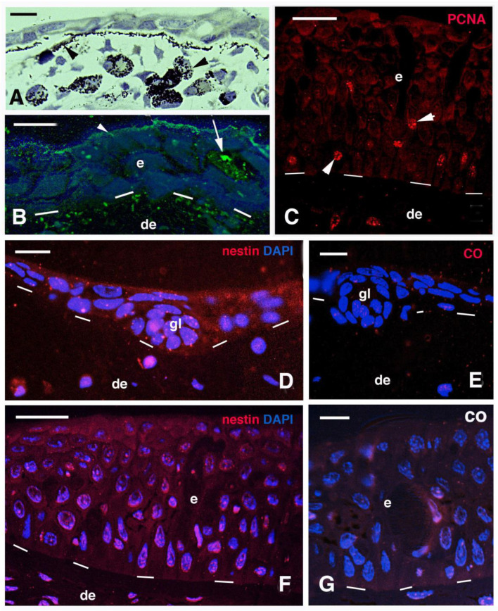
Here we report the immunolocalization of mucin, nestin, elastin and three glycoproteins involved in tissue mineralization in small and large juveniles of Neoceratodus forsteri. Both small and larger juvenile epidermis are mucogenic and contain a diffuse immunolabeling for nestin. Sparse PCNA-labeled cells, indicating proliferation, are found in basal and suprabasal epidermal layers. No scales are formed in small juveniles but are present in a 5 cm long juvenile and in larger juveniles. Elastin and a mineralizing matrix are localized underneath the basement membrane of the tail epidermis where lepidotriches are forming. The latter appears as "circular bodies" in cross sections and are made of elongated cells surrounding a central amorphous area containing collagen and elastin-like proteins that undergo calcification as evidenced using the von Kossa staining. However, the first calcification sites are the coniform teeth of the small juveniles of 2-3 cm in length. In the superficial dermis of juveniles (16-26 cm in length) where scales are formed, the spinulated outer bony layer (squamulin) of the elasmoid scales contains osteonectin, alkaline phosphatase, osteopontin, and calcium deposits that are instead absent in the underlying layer of elasmodin. In particular, these glycoproteins are localized along the scale margin in juveniles where scales grow, as indicated by the presence of PCNA-labeled cells (proliferating). These observations suggest a continuous deposition of new bone during the growth of the scales, possibly under the action of these mineralizing glycoproteins, like in the endoskeleton of terrestrial vertebrates.
Keywords: evolution; immunolabeling; lungfish; skin; structure; ultrastructure.
Conflict of interest statement
The author declares no conflict of interest. This study was self-supported, working on biological material prepared under a J. Joss permit (Australian Research Council 40%; Refs. [22,30]). The antibodies purchased from DSHB were produced and maintained by the facilities of the Hybridoma Bank, University of Iowa, IA, USA, supported by the US NIH.
Figures


References
-
- Whitear M. The skin of fishes including cyclostomes. Epidermis. In: Bereither-Hahn J., Matoltsy G.A., Sylvia-Richards K., editors. Biology of the Integument. Volume 2. Springer; Berlin, Germany: 1986. pp. 8–38. Vertebrates.
-
- Whitear M. The skin of fishes including cyclostomes. Epidermis. In: Bereither-Hahn J., Matoltsy G.A., Sylvia-Richards K., editors. Biology of the Integument. Volume 2. Springer; Berlin, Germany: 1986. pp. 39–55. Vertebrates.
LinkOut - more resources
Full Text Sources
Research Materials
Miscellaneous
