. 2023 Dec;40(23-24):2500-2521.
doi: 10.1089/neu.2023.0117. Epub 2023 Oct 11.

Recovery of Forearm and Fine Digit Function After Chronic Spinal Cord Injury by Simultaneous Blockade of Inhibitory Matrix Chondroitin Sulfate Proteoglycan Production and the Receptor PTPσ

Affiliations

Recovery of Forearm and Fine Digit Function After Chronic Spinal Cord Injury by Simultaneous Blockade of Inhibitory Matrix Chondroitin Sulfate Proteoglycan Production and the Receptor PTPσ

Adrianna J Milton et al. J Neurotrauma. 2023 Dec.

Abstract

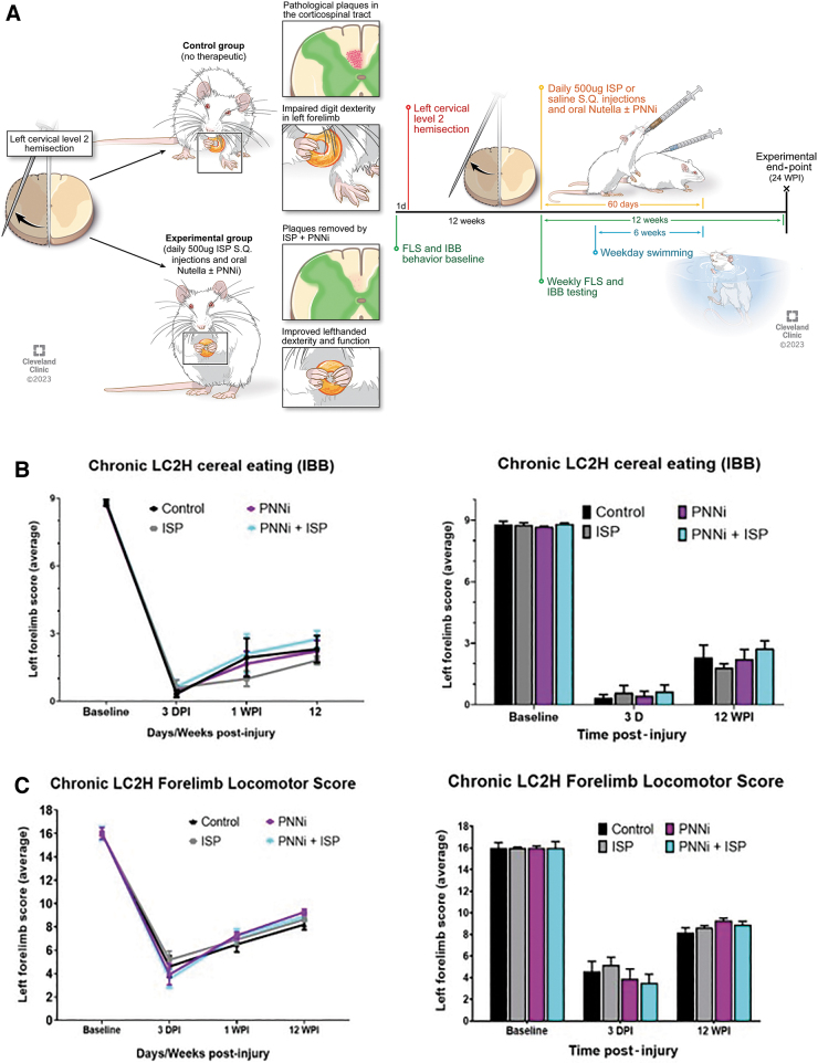
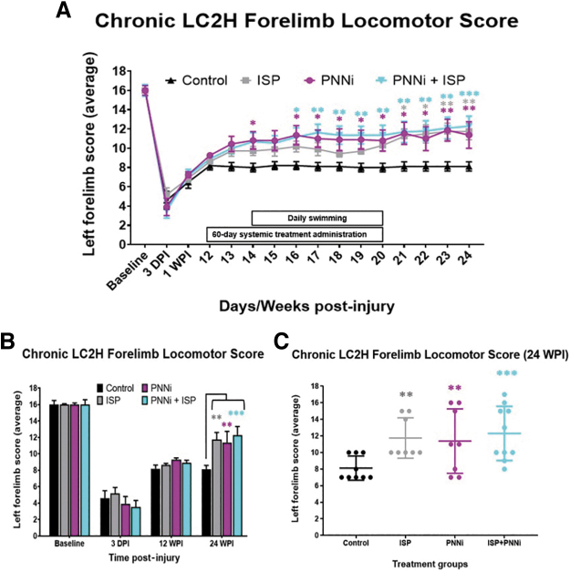
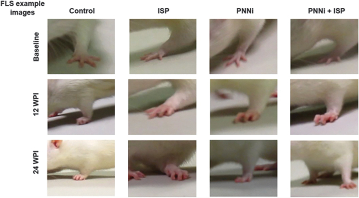
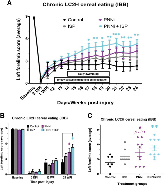
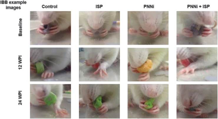
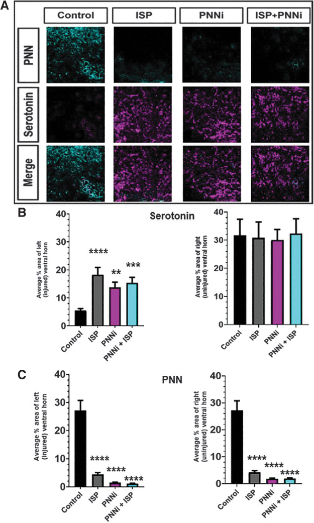
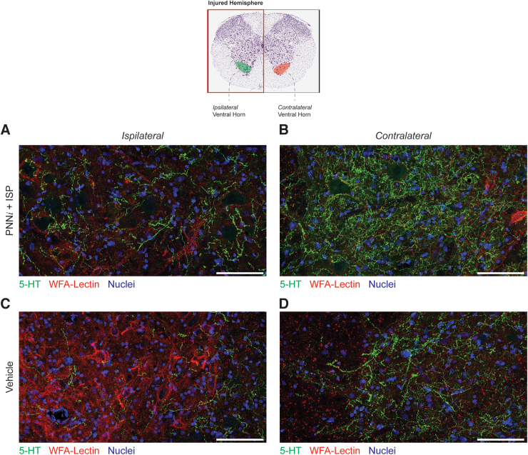
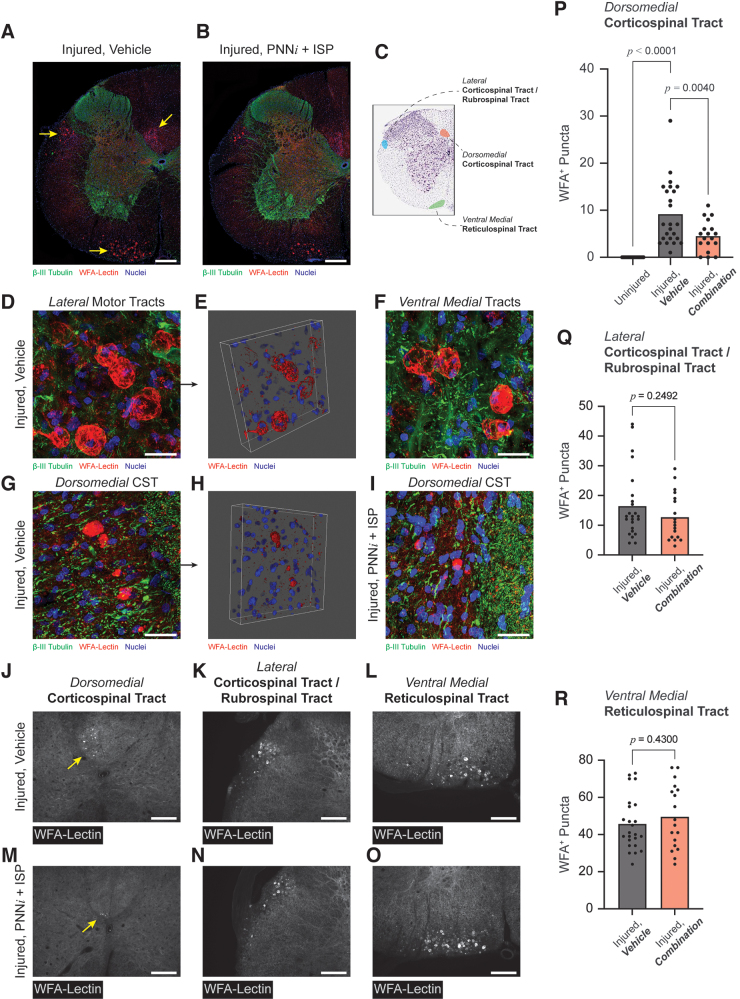
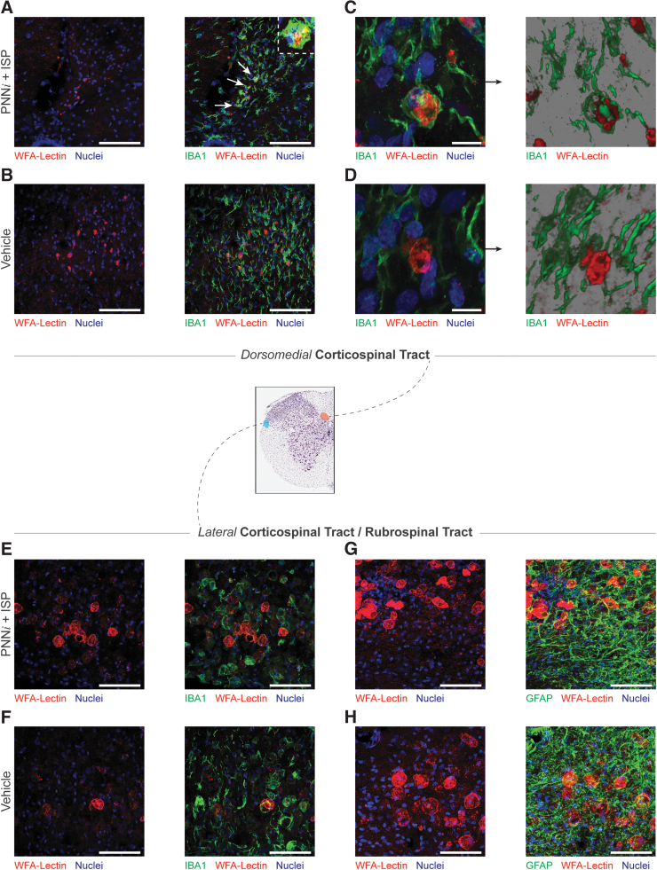
Spinal cord injuries (SCI), for which there are limited effective treatments, result in enduring paralysis and hypoesthesia, in part because of the inhibitory microenvironment that develops and limits regeneration/sprouting, especially during chronic stages. Recently, we discovered that targeted enzymatic removal of the inhibitory chondroitin sulfate proteoglycan (CSPG) component of the extracellular and perineuronal net (PNN) matrix via Chondroitinase ABC (ChABC) rapidly restored robust respiratory function to the previously paralyzed hemi-diaphragm after remarkably long times post-injury (up to 1.5 years) following a cervical level 2 lateral hemi-transection. Importantly, ChABC treatment at cervical level 4 in this chronic model also elicited improvements in gross upper arm function. In the present study, we focused on arm and hand function, seeking to highlight and optimize crude as well as fine motor control of the forearm and digits at lengthy chronic stages post-injury. However, instead of using ChABC, we utilized a novel and more clinically relevant systemic combinatorial treatment strategy designed to simultaneously reduce and overcome inhibitory CSPGs. Following a 3-month upper cervical spinal hemi-lesion using adult female Sprague Dawley rats, we show that the combined treatment had a profound effect on functional recovery of the chronically paralyzed forelimb and paw, as well as on precision movements of the digits. The regenerative and immune system related events that we describe deepen our basic understanding of the crucial role of CSPG-mediated inhibition via the PTPσ receptor in constraining functional synaptic plasticity at lengthy time points following SCI, hopefully leading to clinically relevant translational benefits.

Keywords: CSPGs; axon regeneration; axon sprouting; perineuronal net; receptor PTPσ; spinal cord injury.


Conflict of interest statement

No competing financial interests exist.

Figures

FIG. 1.
FIG. 1.
(A–D) Incomplete cervical spinal cord injury (SCI) severely impairs forelimb function during walking and eating behavior at chronic stages. (A) Timeline of the experimental protocol. Representative image depicting surgical SCI procedure at C2. All animals received a left hemi-lesion at C2 removing ipsilateral descending motor inputs. (B and C) Lateral cervical level two hemi-section (LC2H) severely impairs forelimb walking ability and cereal eating behavior at 3 days post-injury (DPI), 1 week post-injury (WPI), and 12 WPI. There were no significant differences across the experimental treatment groups compared with controls. Color image is available online.
FIG. 2.
FIG. 2.
(A–C) The Forelimb Locomotor Score (FLS) is significantly improved following systemic treatment to remove chondroitin sulfate proteoglycans (CSPGs) in the perineuronal net (PNN) and/or modulate receptor PTPσ. (A) Chronic spinal cord injury (SCI) rats were measured for forelimb locomotor function before and after lateral cervical level two hemi-section (LC2H) and systemic intracellular sigma peptide (ISP) ± perineuronal net inhibitor (PNNi). Both ISP ± PNNi significantly improved weight-bearing stepping after chronic PTPσ-PNN binding perturbation (average FLS score ISP analysis of variance [ANOVA], p = 0.037). (B, C) At 24 weeks post-injury (WPI), animals receiving either systemic drug showed an average improvement in walking behavior, with combinatory recovery showing the greatest significance and rate of recovery (p* < 0.05, p** < 0.005, p*** < 0.0005). Bonferonni post-hoc analysis, data are reported as average ± standard error of the mean (SEM). Color image is available online.
FIG. 3.
FIG. 3.
Forelimb Locomotor Scale (FLS) walking assessment. Example still-frame images from actively walking rats at baseline, 12 weeks post-injury (WPI), and 24 WPI (4 weeks after systemic treatment ended) during FLS analysis (refer to Fig. 2 and Supplementary Videos 1–5). Color image is available online.
FIG. 4.
FIG. 4.
(A–C) Cereal eating ability assessed using the Irvine, Beattie, and Breshnahan (IBB) rating scale is significantly improved following combined systemic treatment to both remove chondroitin sulfate proteoglycans (CSPGs) in the perineural net (PNN) and modulate receptor PTPσ. (A) Chronic spinal cord injury (SCI) rats were measured for forepaw cereal manipulability before and after lateral cervical level two hemi-section (LC2H) and systemic intracellular sigma peptide (ISP) ± PNN inhibitor (PNNi). Only animals receiving combinatory therapy showed significant improvement as early as 17 weeks post-injury (WPI). (B,C) Only chronic injured animals receiving ISP + PNNi improved significantly in cereal eating behavior by 24 WPI (panel C). Rats that received PNNi alone demonstrated a trend toward significance 4 weeks after the termination of oral administration (p* < 0.05, # = 0.1601). Bonferonni post-hoc analysis, data are reported as average ± standard error of the mean (SEM). Color image is available online.
FIG. 5.
FIG. 5.
Irvine, Beattie, and Breshnahan (IBB) cereal eating assessment of fine digit and wrist functional recovery. Example still-frame images of animals eating cereal at baseline, 12 weeks post-injury (WPI), and 24 WPI (4 weeks after systemic treatment ended) during IBB analysis (refer to Fig. 4 and Supplementary Videos 6 and 7). Color image is available online.
FIG. 6.
FIG. 6.
(A–C) Perineural nets (PNNs) are decreased and serotonergic fibers are significantly increased in the cervical enlargement gray matter. (A) Example immunofluorescent image of sprouted ventral horn serotonergic fibers (magenta) immediately caudal to the lateral cervical level two hemi-section (LC2H) in an animal treated with combined intracellular sigma peptide (ISP) ± PNN inhibitor (PNNi). (B, C) Quanitifications of analyzed images shown in (A). Serotonergic fibers are significantly increased, while PNNs demonstrate an inversely correlated decrease within the extracellular gray matter of the cervical enlargement. These results are not observed when chronic cervical spinal cord injury (SCI) rats receive saline + Nutella as control (analysis of variance [ANOVA] serotonin [5-HT]: ISP ipsilateral p < 0.0001, contralateral p = 0.991; PNNi ipsilateral p < 0.0033, contralateral p = 0.9898; ISP+PNNi ipsilateral p < 0.0004, ISP+PNNi contralateral p = 0.9993); (ANOVA Wisteria floribunda agglutinin [WFA], ISP ipsilateral p < 0.0001, contralateral p < 0.0001; PNNi ipsilateral p < 0.0001, contralateral p < 0.0001; ISP+PNNi ipsilateral p < 0.0001, contralateral p < 0.0001) Bonferonni post-hoc analysis, data are reported as average ± standard error of the mean (SEM). Color image is available online.
FIG. 7.
FIG. 7.
High-magnification histology confirms that perineural net inhibitor (PNNi) + intracellular sigma peptide (ISP) enhances serotonergic innervation. This analysis verified decreased chondroitin sulfate proteoglycans (CSPGs) and increased serotonergic sprouting within the injured ventral horn resulting from daily treatment with PNNi + ISP when compared with the injured, ipsilateral hemisphere (A, C). Representative immuno-micrographs demonstrate protection or possible enhancement of serotonergic, axon innervation contralateral to the injury (B, D). Scale bar = 100 μm. Color image is available online.
FIG. 8.
FIG. 8.
Clearing of chondroitin sulfate proteoglycans (CSPG) aggregates from the dorsomedial corticospinal tract (CST) is associated with behavioral recovery. At 24 weeks post-injury (WPI), low-magnification immuno-microscopy (A, B) reveals that CSPG aggregates are isolated to three descending motor pathways: (1) the dorsomedial CST, (2) the rubrospinal tract or possibly the lateral CST, and (3) the ventral medial reticulospinal tract (C). Aggregates with the lateral and ventral medial pathways were large diameter, globular structures (D–F). In contrast, aggregates within the dorsomedial CST were smaller in diameter and presented ellipsoid or spherical morphologies (G–H). Quantification of CSPG aggregates from these three anatomical sites (J–O) revealed that treatment with perineuronal net inhibitor (PNNi) + intracellular sigma peptide (ISP) associated with substantial clearing of these plaque-like structures from the dorsomedial CST (P). Aggregates within the lateral and ventral medial pathways remained intact (Q, R). A, B scale bar = 250 μm; D–I scale bar = 25 μm; J–O scale bar = 100 μm. P value for panel P was determined based on one-way analysis of variance (ANOVA); p values for panels Q and R were determined by unpaired t test. Color image is available online.
FIG. 9.
FIG. 9.
Microglia may clear chondroitin sulfate proteoglycan (CSPG) aggregates from the dorsomedial corticospinal tract (CST). Histological analysis of the cervical cord (C5–C9; 24 weeks post-injury) revealed that IBA1+ microglia internalize CSPG aggregates within the CST of combination-treated animals (A–D). Internalized aggregates are identified with white arrows in panel A. The larger diameter aggregates in the lateral motor tracts are proximal to microglia but were neither internalized nor cleared in either condition (E, F). CSPGs accumulated within fields of reactive astrocytes (G, H), especially in lateral motor tracts; however, it was not clear whether reactive astroglia were engaged with the generation or clearance of these structures. Scale bar = 100 μm. Color image is available online.

Similar articles

Cited by

References

    1. Sekhon LHS, Fehlings MG. Epidemiology, demographics, and pathophysiology of acute spinal cord injury. Spine (Phila Pa 1976) 2001;26(Supplement):S2–S12; doi: 10.1097/00007632-200112151-00002 - DOI - PubMed
    1. Kumar R, Lim J, Mekary RA, et al. . Traumatic spinal injury: global epidemiology and worldwide volume. World Neurosurg 2018;113:e345–e363; doi: 10.1016/j.wneu.2018.02.033 - DOI - PubMed
    1. Anderson KD. Targeting recovery: priorities of the spinal cord-injured population. J Neurotrauma 2004;21(10):1371–1383; doi: 10.1089/neu.2004.21.1371 - DOI - PubMed
    1. Snow DM, Steindler DA, Silver J. Molecular and cellular characterization of the glial roof plate of the spinal cord and optic tectum: a possible role for a proteoglycan in the development of an axon barrier. Dev Biol 1990;138(2):359–376; doi: 10.1016/0012-1606(90)90203-U - DOI - PubMed
    1. Brittis PA, Canning DR, Silver J. Chondroitin sulfate as a regulator of neuronal patterning in the retina. Science (1979) 1992;255(5045):733–736; doi: 10.1126/science.1738848 - DOI - PubMed

Publication types

Substances