. 2023 Sep 28;97(9):e0079023.
doi: 10.1128/jvi.00790-23. Epub 2023 Aug 21.

Characterization of a mouse-adapted strain of bat severe acute respiratory syndrome-related coronavirus

Affiliations

Characterization of a mouse-adapted strain of bat severe acute respiratory syndrome-related coronavirus

Hao-Feng Lin et al. J Virol. .

Abstract

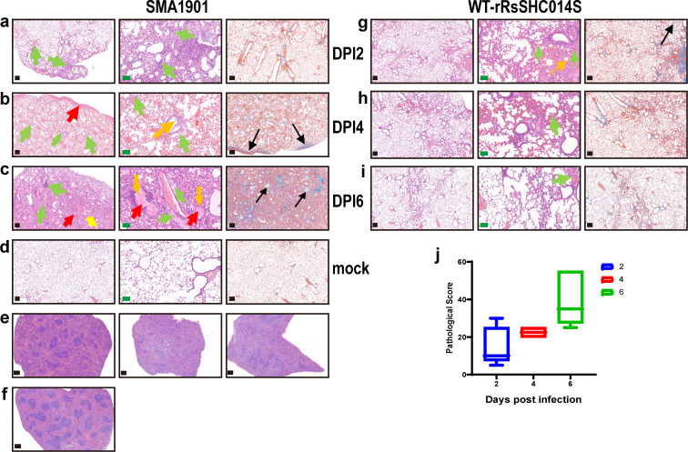
Bats carry genetically diverse severe acute respiratory syndrome-related coronaviruses (SARSr-CoVs). Some of them utilize human angiotensin-converting enzyme 2 (hACE2) as a receptor and cannot efficiently replicate in wild-type mice. Our previous study demonstrated that the bat SARSr-CoV rRsSHC014S induces respiratory infection and lung damage in hACE2 transgenic mice but not wild-type mice. In this study, we generated a mouse-adapted strain of rRsSHC014S, which we named SMA1901, by serial passaging of wild-type virus in BALB/c mice. SMA1901 showed increased infectivity in mouse lungs and induced interstitial lung pneumonia in both young and aged mice after intranasal inoculation. Genome sequencing revealed mutations in not only the spike protein but the whole genome, which may be responsible for the enhanced pathogenicity of SMA1901 in wild-type BALB/c mice. SMA1901 induced age-related mortality similar to that observed in SARS and COVID-19. Drug testing using antibodies and antiviral molecules indicated that this mouse-adapted virus strain can be used to test prophylactic and therapeutic drug candidates against SARSr-CoVs. IMPORTANCE The genetic diversity of SARSr-CoVs in wildlife and their potential risk of cross-species infection highlights the importance of developing a powerful animal model to evaluate the antibodies and antiviral drugs. We acquired the mouse-adapted strain of a bat-origin coronavirus named SMA1901 by natural serial passaging of rRsSHC014S in BALB/c mice. The SMA1901 infection caused interstitial pneumonia and inflammatory immune responses in both young and aged BALB/c mice after intranasal inoculation. Our model exhibited age-related mortality similar to SARS and COVID-19. Therefore, our model will be of high value for investigating the pathogenesis of bat SARSr-CoVs and could serve as a prospective test platform for prophylactic and therapeutic candidates.

Keywords: animal model; bat SARS-related coronavirus; mouse-adapted strain; pathogenicity; pneumonia.


Conflict of interest statement

The authors declare no conflict of interest.

Figures

Fig 1
Fig 1
Characterization of a mouse-adapted strain of the recombinant virus rRsSHC014S. (a) Body weight loss of mice during the passaging process. Three 6-week-old female or male BALB/c mice were infected intranasally with the supernatant of lung homogenate from the mice infected with former passaged viruses. Body weight changes after 3–5 DPI was recorded as compared with the initial weight (BALB/c mice as a standardized experimental mice, weighing about 20 g). (b and c) Weight loss and hematoxylin and eosin (H&E) staining of lung sections of the mice after being infected with the lung homogenate supernatant of passage 19. Virus-infected mice show interstitial pneumonia with thickened alveolar septa and damaged alveoli, and exudation of fibrin can be seen in some alveoli. (d and e) Schematic of rRsSHC014S genome and all the adapted mutations. Amino acid mutations of rRsSHC014S were generated during passaging and the nucleotide changes in the 19th passage. * indicates mutation of the stop codon nucleotide.
Fig 2
Fig 2
Infectivity of SMA1901 and WT-rRsSHC014S in vitro. Comparison of the host range and replication efficiency of SMA1901 and WT-rRsSHC014S in different cells and human airway organoid at an MOI = 1.0. Virus replication was detected by immunofluorescence assay with rabbit serum against the SARSr-CoV-Rp3 Np followed by Cy3-conjugated goat anti-rabbit IgG and nuclei were stained with DAPI in virus-infected cells at 48 h post-infection. Scale bars: the scale bar is 400 (white) or 58 (red) μm. Viral genome copies were determined by quantitative reverse transcription (RT)-PCR in supernatants of virus-infected cells. Error bars indicate the standard error. Statistical significance was measured by two-way analysis of variance (ANOVA) between the SMA1901 and WT-rRsSHC014S infected. *P < 0.05
Fig 3
Fig 3
Pathogenicity of SMA1901 in 6-week-old mice. Mice were divided into two groups, one for monitoring weight change and the survival rate and one for tissue sampling at various time points after infection. (a) In the tissue sampling group, 6-week-old mice were mock-infected (n = 6) or intranasally infected (n = 24) with 1 × 10⁵ TCID50 of SMA1901 and the group of SMA1901-infected was sacrificed for tissue collection at 2, 4, and 6 DPI. (b and c) In the survival monitoring groups, mouse body weight and survival were monitored until 6 DPI upon challenge with DMEM (n = 8) or SMA1901 (n = 8). (d and e) Infectious viral titers in lung and brain tissues in mice infected with SMA1901 were detected by plaque formation assay. (f) The SMA1901-infected mice (n = 8) were sacrificed for tissue collection (heart, liver, spleen, lung, kidney, intestine, brain, trachea, and turbinate) at 2, 4, and 6 DPI, respectively; the copy numbers of viral RNA were measured using RT-qPCR. (g) The SMA1901-infected mice (n = 8) were sacrificed to examine the pathological changes in the lungs at 2, 4, and 6 DPI compared to the mock-infected group. Yellow arrow indicates thickened alveolar septa. Blue arrows indicate inflammatory cell infiltration. Green arrows indicate increased numbers of peri-bronchial and peri-vascular lymphocytes. Red arrow indicates fibrin leakage. Black arrow indicates the exudation of fibroblast hyperplasia. Error bars indicate the standard error. Images were collected using a Pannoramic MIDI system. The scale bar is 50 (green) or 200 (black) μm. Dash line in Fig. 3f represents the limit of detection.
Fig 4
Fig 4
Pathogenicity of SMA1901 in 10-month-old aged mice. Ten-month-old mice were mock-infected or intranasally infected with 1 × 10⁵ TCID50 of SMA1901 or WT-rRsSHC014S and all were sacrificed for tissue collection at 2, 4, and 6 DPI. (a) Mouse body weight was monitored until 7 DPI. (b) Survival curve of BALB/c mice upon challenge with DMEM (MOCK), SMA1901, or WT-rRsSHC014S. (c–e, h, and i) Mice infected with SMA1901 or WT-rRsSHC014S (n ≥ 5) were sacrificed for tissue collection (heart, liver, spleen, lung, kidney, intestine, brain, trachea, and turbinate) at 2, 4, and 6 DPI, and the copy numbers of viral RNA were measured using RT-qPCR. (f and g) Infectious viral titers in lung and brain tissues in mice infected with SMA1901 were detected by plaque formation assay. (j) Lung cytokine and chemokine heatmap in SMA1901-infected or WT-rRsSHC014S-infected mice compared with mock-infected mice at 2 DPI. Error bars indicate the standard error. Statistical significance was measured by two-way ANOVA between the SMA1901 and WT-rRsSHC014S infected. *P < 0.05, **P < 0.01, ***P < 0.001. Dash line in Fig. 4a represents the ethical death line of the animal, other dash lines represent the limit of detection.
Fig 5
Fig 5
Histopathological changes and pathological scoring in 10-month-old aged mice. Mice were sacrificed to examine the pathological changes in the lungs and spleen at 2, 4, and 6 DPI. (a) SMA1901-infected mouse lung at 2 DPI showed minor thickened alveolar septa with inflammatory cell (green arrow) infiltration. (b) Moderate interstitial pneumonia was observed at 4 DPI with inflammatory cell (green arrow) infiltration and exfoliation of bronchial epithelial cells (orange arrow). Eosinophilic mucus acid (red arrow) was seen at the margins of the tissue, and exudation of fibroblast hyperplasia (black arrow) was found. (c) Severe pneumonia was observed in the majority of infected mice at 6 DPI. The histopathological changes included massive peri-bronchial and peri-vascular inflammatory cell infiltration (green arrow), fibroplasia (black arrow), exudation of eosinophilic mucus acid (red arrow), edema with the hyaline membrane (yellow arrow) formation, and dissolving and exfoliation of bronchial epithelial cells (orange arrow). (d) Mock-infected BALB/c mouse lung. (e and f) H&E staining of spleen sections infected with SMA1901 (n = 5) compared with the mock-infected mice. (G–I) The mild lung pathology of WT-rRsSHC014S-infected aged mice showed the exfoliation of bronchial epithelial cells (orange arrow), infiltration of inflammatory cell (green arrow), and exudation of fibroblast hyperplasia (black arrow). (j) Semiquantitative analysis of the pathology of the H&E-stained lung section from SMA1901-infected mice 2 (n = 5), 4 (n = 5), and 6 (n = 5) DPI. The scores were determined based on the percentage of inflammation in the pulmonary section by using the following 0–70 point scoring system: 0, no inflammation; 15, affected area (≤1%); 30, affected area (>1%, ≤10%); 45, affected area (>10%, ≤50%); 60, affected area (>50%). Additional 10 points were added when pulmonary edema, inflammatory cell infiltration, fibroplasia, and/or hyaline membrane formation were observed. Error bars indicate the standard error. Images were collected using a Pannoramic MIDI system. The scale bar is 50 (green) or 200 (black) μm.
Fig 6
Fig 6
SRB12 inhibits SMA1901 in 6-month-old mice. (a) Six-month-old BALB/c mice were intraperitoneally injected with SRB12 with a dose of 200 µg/mouse (n = 8) or PBS (n = 6) followed by intranasal infection with the 1 × 10⁵ TCID50 of SMA1901 24 h later. (b) Mouse body weight was monitored until 5 DPI. (c) Mice were sacrificed for tissue collection (lung, trachea, and turbinate) at 5 DPI, and the copy numbers of viral RNA were measured using RT-qPCR. (d) Survival curves of mice in the SRB12 and mock treatment groups. (e) H&E staining of lung sections from the SRB1 prophylactic group and PBS group. The PBS group showed inflammatory lung injury, thickened alveolar septa (green arrow), eosinophilic mucus acid exudation (red arrow), hyaline membrane formation (yellow arrow), and inflammatory cell infiltration (blue arrow). Error bars indicate the standard error. Statistical significance was measured by two-way ANOVA between the PBS and prophylactic groups. *P < 0.05, **P < 0.01, ***P < 0.001. Images were collected using a Pannoramic MIDI system. The scale bar is 50 (green) or 200 (black) μm. Dash line in Fig. 6c represents the limit of detection.
Fig 7
Fig 7
Molnupiravir inhibits SMA1901 in 10-month-old Mice. (a) Antiviral efficacy of molnupiravir in Vero E6 cells against viruses (rWIV1, rRsSHC014S, and SMA1901). Vero E6 cells were pre-treated with the drug at different dilutions for 2 h, and the virus (MOI = 0.1) was added to allow attachment for 1 h after the drugs was removed. Then the virus was removed and the cells were cultured with the drug-containing medium until the end of the experiment. After 48 h, the virus RNA was measured via RT-qPCR. (b) Ten-month-old BALB/c mice were orally administered with molnupiravir with a dose of 250 mg/kg (n ≥ 6) or vehicle (n ≥ 6) followed by intranasal infecton with 1 × 10⁵ TCID50 of SMA1901 2 h later. (c) Mouse body weight was monitored until 7 DPI. (d) Survival curves of mice in molnupiravir and vehicle treatment groups. (e and f) Mice were sacrificed for tissue collection (lung, trachea, and turbinate) at 5 DPI, and the copy numbers of viral RNA were measured using RT-qPCR. Infectious viral titers in lung tissues in mice were detected by plaque formation assay. (g and h) Scores and H&E staining of lung sections from the prophylactic or PBS group. Error bars indicate the standard error. Statistical significance was measured by two-way ANOVA between the PBS and prophylactic groups. *P < 0.05, **P < 0.01, ***P < 0.001. Images were collected using a Pannoramic MIDI system. The scale bar is 50 (green) or 200 (black)μm. Dash line in Fig. 7c represents the ethical death line of the animal, and Fig. 7e represents the limit of detection.
Fig 8
Fig 8
The RsSHC014 S protein and its mutants efficiently bind and utilize mouse ACE2. (a) The 293T/17 cells transfected with hACE2- or mACE2-expressing plasmids were infected by WT-rRsSHC014S (MOI = 1.0), and then the ratio of the infected cells was detected by flow cytometry. (b and d) The RsSHC014-S1 and its mutants were expressed and purified. hACE2- or mACE2-expressing plasmids were transfected in HEK 293T/17 cells, incubated with the protein, and then the S1-ACE2 interaction was detected by flow cytometry. (c) HEK 293T/17 cells expressing human/mouse ACE2 were infected with the RsSHC014 and its mutant spike-pseudotyped viruses. The lysates of infected cell were analyzed by measuring luciferase activities. All experiments were repeated independently for three times. Error bars indicate the standard error. Statistical significance was measured by two-way ANOVA, ***P < 0.001.

Similar articles

Cited by

References

    1. Drosten C, Günther S, Preiser W, van der Werf S, Brodt H-R, Becker S, Rabenau H, Panning M, Kolesnikova L, Fouchier RAM, Berger A, Burguière A-M, Cinatl J, Eickmann M, Escriou N, Grywna K, Kramme S, Manuguerra J-C, Müller S, Rickerts V, Stürmer M, Vieth S, Klenk H-D, Osterhaus ADME, Schmitz H, Doerr HW. 2003. Identification of a novel coronavirus in patients with severe acute respiratory syndrome. N Engl J Med 348:1967–1976. doi:10.1056/NEJMoa030747 - DOI - PubMed
    1. Zaki AM, van Boheemen S, Bestebroer TM, Osterhaus ADME, Fouchier RAM. 2012. Isolation of a novel coronavirus from a man with pneumonia in Saudi Arabia. N Engl J Med 367:1814–1820. doi:10.1056/NEJMoa1211721 - DOI - PubMed
    1. Zhou P, Yang X-L, Wang X-G, Hu B, Zhang L, Zhang W, Si H-R, Zhu Y, Li B, Huang C-L, Chen H-D, Chen J, Luo Y, Guo H, Jiang R-D, Liu M-Q, Chen Y, Shen X-R, Wang X, Zheng X-S, Zhao K, Chen Q-J, Deng F, Liu L-L, Yan B, Zhan F-X, Wang Y-Y, Xiao G-F, Shi Z-L. 2020. A pneumonia outbreak associated with a new coronavirus of probable bat origin. Nature 588:E6. doi:10.1038/s41586-020-2951-z - DOI - PMC - PubMed
    1. Yang XL, Hu B, Wang B, Wang MN, Zhang Q, Zhang W, Wu LJ, Ge XY, Zhang YZ, Daszak P, Wang LF, Shi ZL. 2015. Isolation and characterization of a novel bat coronavirus closely related to the direct progenitor of severe acute respiratory syndrome coronavirus. J Virol 90:3253–3256. doi:10.1128/JVI.02582-15 - DOI - PMC - PubMed
    1. Hu B, Zeng L-P, Yang X-L, Ge X-Y, Zhang W, Li B, Xie J-Z, Shen X-R, Zhang Y-Z, Wang N, Luo D-S, Zheng X-S, Wang M-N, Daszak P, Wang L-F, Cui J, Shi Z-L, Drosten C. 2017. Discovery of a rich gene pool of bat SARS-related coronaviruses provides new insights into the origin of SARS coronavirus. PLoS Pathog 13:e1006698. doi:10.1371/journal.ppat.1006698 - DOI - PMC - PubMed

Publication types

MeSH terms

LinkOut - more resources