. 2023 Dec 31;24(1):2246206.
doi: 10.1080/15384047.2023.2246206.

Transcription factor 3 promotes migration and invasion potential and maintains cancer stemness by activating ID1 expression in esophageal squamous cell carcinoma

Affiliations

Transcription factor 3 promotes migration and invasion potential and maintains cancer stemness by activating ID1 expression in esophageal squamous cell carcinoma

Zhao-Xing Li et al. Cancer Biol Ther. .

Abstract

Transcription factor 3 (TCF3) is a member of the basic Helix - Loop - Helix (bHLH) transcription factor (TF) family and is encoded by the TCF3 gene (also known as E2A). It has been shown that TCF3 functions as a key transcription factor in the pathogenesis of several human cancers and plays an important role in stem cell maintenance and carcinogenesis. However, the effect of TCF3 in the progression of esophageal squamous cell carcinoma (ESCC) is poorly known. In our study, TCF3 was found to express highly and correlated with cancer stage and prognosis. TCF3 was shown to promote ESCC invasion, migration, and drug resistance both from the results of in vivo and in vitro assays. Moreover, further studies suggested that TCF3 played these roles through transcriptionally regulating Inhibitor of DNA binding 1(ID1). Notably, we also found that TCF3 or ID1 was associated with ESCC stemness. Furthermore, TCF3 was correlated with the expression of cancer stemness markers CD44 and CD133. Therefore, maintaining cancer stemness might be the underlying mechanism that TCF3 transcriptionally regulated ID1 and further promoted ESCC progression and drug resistance.

Keywords: ID1; TCF3; cancer stem cell; cancer stemness; esophageal squamous cell carcinoma.


Conflict of interest statement

No potential conflict of interest was reported by the author(s).

Figures

Figure 1.
Figure 1.
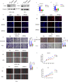
TCF3 is highly expressed in esophageal squamous cell carcinoma and correlates with patient prognosis. a. 6 pairs of esophageal squamous cell carcinoma tissue microarray shows that TCF3 is highly expressed in esophageal carcinoma compared to normal tissues and the expression increases with increasing malignancy. b. TCGA database shows that TCF3 expression in ESCC is highly expressed compared to other genes. c. Esophageal squamous cell carcinoma tissue microarray from 79 patients shows that TCF3 is highly expressed in ESCC tissues compared to normal tissues. d-e. The expression level of TCF3 is corresponding to ESCC progression. f. High TCF3 expression correlates with the prognosis of ESCC.g.TCF3 protein is highly expressed in ESCC tissues of five randomly selected patients. h. TCF3 is higher in ESCC cell lines especially in KYSE-150, TE1, and KYSE-410 than in normal esophageal epithelial cell line HET-1A.
Figure 2.
Figure 2.
TCF3 promotes proliferation, migration, and invasion of ESCC cells. a-d. KYSE-150 and TE-1 have decreased TCF3 protein expression after siRnas targeting TCF3 could significantly knockdown the expression of TCF3 protein and TCF3 mRNA. E-F. The fluorescence intensity of TCF3 was reduced in KYSE-150 and TE-1 after si-TCF3 was transfected. G-H. The migration and invasion abilities of KYSE-150 and TE-1 were decreased when TCF3 was knocked down. I&J. The wound healing rates of KYSE-150 and TE-1 were decreased when TCF3 was knocked down. K&L. Cell proliferation is affected by KYSE-150 and TE-1 after the siRNA knockdown of TCF3.
Figure 3.
Figure 3.
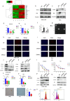
TCF3 regulates the expression of cancer stem markers CD44 and CD133 by transcriptionally regulating ID1. a. RNA-seq results show significantly upregulated and downregulated gene signatures. b-c. In KYSE-150 and TE-1, with the knockdown of TCF3 the protein expression level of ID1 is subsequently reduced. d-e. In KYSE-150 and TE-1, with the knockdown of TCF3, the mRNA expression of ID1 is subsequently reduced. f-g. CHIP and Dual luciferase reporter assay suggested TCF3 could transcriptionally regulate ID1.h-i. With knockdown of TCF3 in KYSE-150 and TE-1 CD44, fluorescence intensity decreased. J-K. The protein expression of CD44 and CD133 was reduced with the knockdown of TCF3 in KYSE-150 and TE-1. l -m. With the knockdown of TCF3 in KYSE-150 and TE-1 knockdown increased sensitivity to the chemotherapeutic drug cisplatin. n-o. mRNA expression of ID1 was reduced in KYSE-150 and TE-1 after siRNA knockdown. p-q. The protein expression level of CD44 and CD133 was subsequently reduced in KYSE-150 and TE-1 when ID1 was knocked down. I. KYSE-150 has a significant decrease in the sphere formation efficiency after si-TCF3 was transfected. s. with knockdown of TCF3, CD44+ ESCC cells number were decreased.
Figure 4.
Figure 4.
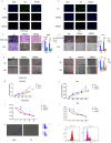
ID1 promotes proliferation, migration, invasion, and drug resistance of ESCC cells. a-b. The fluorescence intensity of ID1 was reduced in KYSE-150 and TE-1 after ID1 was knocked down. c-d. The migration and invasion abilities of KYSE-150 and TE-1 were decreased when ID1 was knocked down. e -f. The wound healing rates of KYSE-150 and TE-1 were decreased when ID1 was knocked down. g-h. Cell proliferation is affected in KYSE-150 and TE-1 after the siRNA knockdown of ID1.I&J. Increased sensitivity to the chemotherapeutic drug cisplatin with knockdown of TCF3 in KYSE-150 and TE-1. k. KYSE150 has a significant decrease in the sphere formation efficiency after si-ID1 was transfected. l. with knockdown of ID1, CD44+ ESCC cells number were decreased.
Figure 5.
Figure 5.
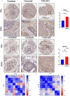
The expressions of cancer stem markers CD44 and CD133 were correlated with the progression of ESCC. a. The expression of CD44 is corresponding to the tumor staging of ESCC. b. The expression of CD133 is corresponding to the tumor staging of ESCC. c. The expression of CD44 positively correlates with TCF3 expression.d.CD133 positively correlates with the expression of TCF3. *p < .05, **p < .01. ***p < .001.
Figure 6.
Figure 6.
TCF3 promotes the progression of ESCC in vivo. a-b. Tumor appearance in mice after 1 month of subcutaneous tumor formation. c. Smaller tumor volume in the knockdown group compared to the NC group. D. Smaller tumor weight in the knockdown group compared to the NC group. e-f. Reduced TCF3 expression in the knockdown group compared to the NC group.

Similar articles

Cited by

References

    1. Chen T, Qin WZ, Yao LQ, Zhong YS, Zhang YQ, Chen WF, Hu JW, Ooi M, Chen LL, Hou YY, et al. Long-term outcomes of endoscopic submucosal dissection for high-grade dysplasia and early-stage carcinoma in the colorectum. Cancer Commun. 2018;38(1):3. doi:10.1186/s40880-018-0273-4. - DOI - PMC - PubMed
    1. Lambert AW, Pattabiraman DR, Weinberg RA.. Emerging biological principles of metastasis. Cell. 2017;168(4):670–11. doi:10.1016/j.cell.2016.11.037. - DOI - PMC - PubMed
    1. Celia-Terrassa T, Kang Y. Distinctive properties of metastasis-initiating cells. Genes Dev. 2016;30(8):892–908. doi:10.1101/gad.277681.116. - DOI - PMC - PubMed
    1. Slyper M, Shahar A, Bar-Ziv A, Granit RZ, Hamburger T, Maly B, Peretz T, Ben-Porath I. Control of breast cancer growth and initiation by the stem cell-associated transcription factor TCF3. Cancer Res. 2012;72(21):5613–5624. doi:10.1158/0008-5472.CAN-12-0119. - DOI - PubMed
    1. Pu XY, Zheng DF, Lv T, Zhou YJ, Yang JY, Jiang L. Overexpression of transcription factor 3 drives hepatocarcinoma development by enhancing cell proliferation via activating Wnt signaling pathway. Hepatobiliary Pancreat Dis Int. 2022;21(4):378–386. doi:10.1016/j.hbpd.2022.01.003. - DOI - PubMed

Publication types

MeSH terms

Substances