. 2023 Aug 29;120(35):e2302083120.
doi: 10.1073/pnas.2302083120. Epub 2023 Aug 22.

SARS-CoV-2 Mac1 is required for IFN antagonism and efficient virus replication in cell culture and in mice

Affiliations

SARS-CoV-2 Mac1 is required for IFN antagonism and efficient virus replication in cell culture and in mice

Yousef M Alhammad et al. Proc Natl Acad Sci U S A. .

Abstract

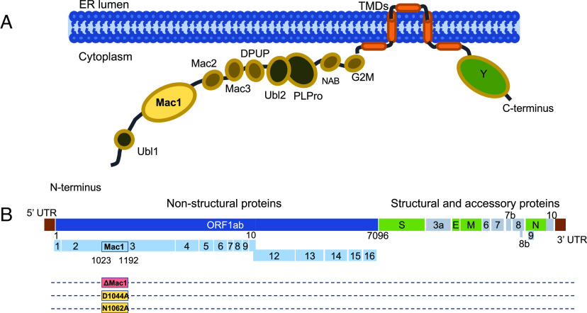
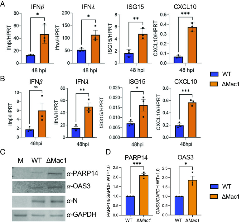
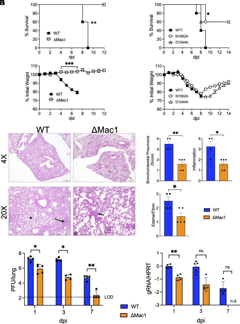
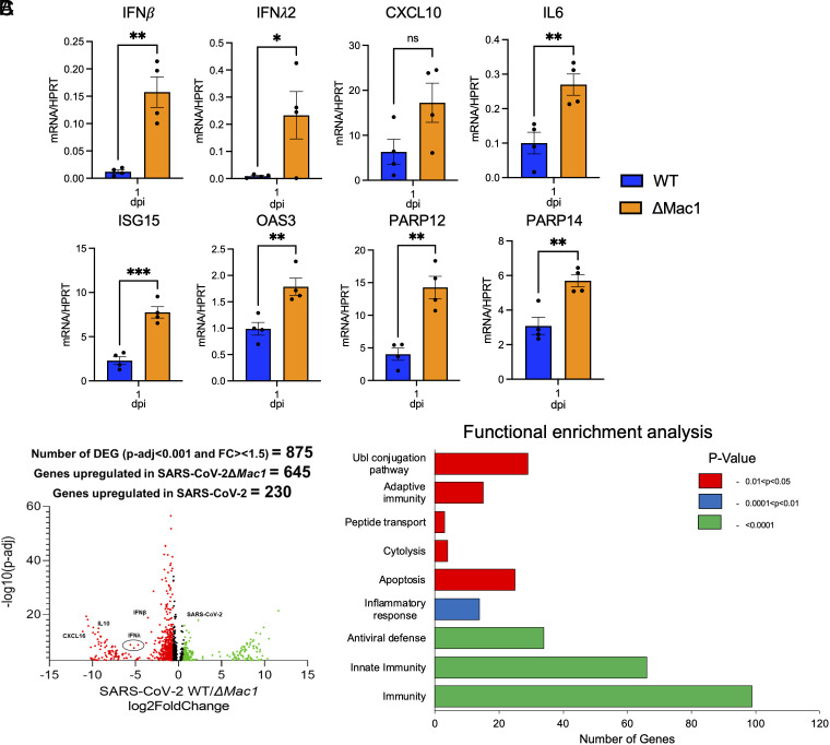
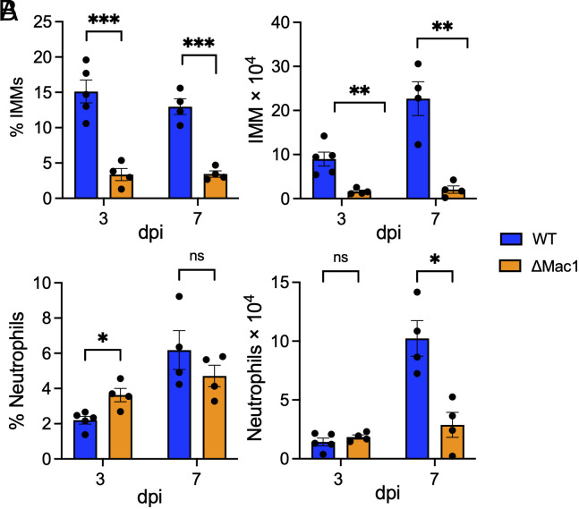
Several coronavirus (CoV) encoded proteins are being evaluated as targets for antiviral therapies for COVID-19. Included in these drug targets is the conserved macrodomain, or Mac1, an ADP-ribosylhydrolase and ADP-ribose binding protein encoded as a small domain at the N terminus of nonstructural protein 3. Utilizing point mutant recombinant viruses, Mac1 was shown to be critical for both murine hepatitis virus (MHV) and severe acute respiratory syndrome (SARS)-CoV virulence. However, as a potential drug target, it is imperative to understand how a complete Mac1 deletion impacts the replication and pathogenesis of different CoVs. To this end, we created recombinant bacterial artificial chromosomes (BACs) containing complete Mac1 deletions (ΔMac1) in MHV, MERS-CoV, and SARS-CoV-2. While we were unable to recover infectious virus from MHV or MERS-CoV ΔMac1 BACs, SARS-CoV-2 ΔMac1 was readily recovered from BAC transfection, indicating a stark difference in the requirement for Mac1 between different CoVs. Furthermore, SARS-CoV-2 ΔMac1 replicated at or near wild-type levels in multiple cell lines susceptible to infection. However, in a mouse model of severe infection, ΔMac1 was quickly cleared causing minimal pathology without any morbidity. ΔMac1 SARS-CoV-2 induced increased levels of interferon (IFN) and IFN-stimulated gene expression in cell culture and mice, indicating that Mac1 blocks IFN responses which may contribute to its attenuation. ΔMac1 infection also led to a stark reduction in inflammatory monocytes and neutrophils. These results demonstrate that Mac1 only minimally impacts SARS-CoV-2 replication, unlike MHV and MERS-CoV, but is required for SARS-CoV-2 pathogenesis and is a unique antiviral drug target.

Keywords: ADP-ribosylation; SARS-CoV-2; coronavirus; interferon; macrodomain.


Conflict of interest statement

A.R.F. and R.C. were named as inventors on a patent filed by the University of Kansas.

Figures

Fig. 1.
Fig. 1.
SARS-CoV-2 nonstructural protein three domains and location of Mac1 in SARS-CoV-2 genome. (A) Cartoon diagram illustrating the different domains of SARS-CoV-2 nsp3. (B) Schematic of the SARS-CoV-2 genome with location of Mac1, indicating the area of the Mac1 deletion and the point mutations that were engineered.
Fig. 2.
Fig. 2.
SARS-CoV-2 ΔMac1 replicates normally in Vero E6 and A549 cells but has a mild replication defect in Calu-3 and primary epithelial cells. (A and B) Vero E6 (A) and A549-ACE2 (B) cells were infected with SARS-CoV-2 WT and ΔMac1 at an MOI of 0.1 PFU/cell. Both cell-associated and cell-free virus was collected at indicated time points and virus titers were determined by plaque assays. Data shown are one experiment representative of three independent experiments. n = 3 per group for each experiment. The data in A and B are from one experiment representative of at least three independent experiments. n = 3 per group. (C and D) Calu-3 cells were infected with SARS-CoV-2 WT and ΔMac1 viruses at low MOI. Both cell-associated and cell-free virus was collected at indicated times, and virus titers were determined by the plaque assay. The data in C are from one experiment representative of at least three independent experiments. n = 3 per group. (D) The results of all combined experiments from Calu-3 cells (C), where the average WT values from each experiment were normalized to 1.0 at 24 and 48 hpi are shown. Each point represents a separate biological replicate. (E and F) Calu-3 cells were infected at an MOI of 1 PFU/cell, and cell lysates were collected, and viral protein levels were determined by immunoblotting (E) or cells were fixed at 24 hpi were costained with DAPI and either anti-nsp3 or anti-N and then analyzed by confocal microscopy at 20× magnification (F). The data in E and F show data from one representative experiment of two independent experiments. (G) Primary human bronchial epithelial cells grown at air–liquid interface were infected apically with SARS-CoV-2 WT and ΔMac1 at an MOI of 0.2 (Donor 1) or 0.02 (Donor 2) PFU/cell. Cell-free virus was collected from the apical side at indicated time points and virus titers were determined by plaque assays. n = 2 to 3 per group for each experiment.
Fig. 3.
Fig. 3.
IFN-γ, but not IFN-β, pretreatment enhances replication defect of ΔMac1 and Mac1 point mutant viruses in Calu-3 cells. Calu-3 cells were pretreated for 18 h with increasing concentrations (0, 5, 50, and 500 units) of IFN-β (A) and IFN-γ (B), then infected with either SARS-CoV-2 WT or ∆Mac1 at an MOI of 0.1 PFU/cell. Cells were collected at 48 hpi, and titers were determined by the plaque assay. Fold differences between WT and ΔMac1 are indicated at each amount of IFN. The data shown are of one experiment representative of two (A) and three (B) independent experiments with n = 3 for each group. (C) Calu-3 cells were pretreated with 500 units of IFN-γ for 18 h and infected with SARS-CoV-2 WT, N1062A, and D1044A at an MOI of 0.1 PFU/cell. Cells were collected at 48 hpi, and titers were determined by the plaque assay. The data shown are of one experiment representative of three independent experiments with n = 3 for each group. (D) Fold-reduction of virus titer for each of the indicated viruses as compared to WT virus was determined. These data are the combined results of three independent experiments.
Fig. 4.
Fig. 4.
ΔMac1 induces increased IFN-I, IFN-III, and cytokine responses compared to WT SARS-CoV-2 in cell culture. Calu-3 (A) and A549-ACE2 (B) cells were infected with SARS-CoV-2 WT and ΔMac1 at an MOI of 0.1 PFU/cell, and total RNA was collected 48 hpi. IFN-β, IFN-λ, ISG15, and CXCL10 levels were determined by qPCR using the ΔCt method with primers listed in SI Appendix, Table S2 and normalized to HPRT mRNA levels. The data show one experiment representative of three independent experiments with n = 3 for each experiment. (C) Calu-3 cells were infected as described above, and cell lysates were collected at 48 hpi and protein levels of PARP14 and OAS3 were determined by immunoblotting. The data show one experiment representative of three independent experiments. (D) Quantification of immunoblots shown in C. The data are the combined results of three independent experiments.
Fig. 5.
Fig. 5.
ΔMac1 is highly attenuated in K18-ACE2 mice. (A and B) K18-ACE2 C57BL/6 mice were infected with 2.5 × 10⁴ PFU of WT or ΔMac1 SARS-CoV-2 and survival (A) and weight loss (B) were measured over 12 d. (C and D) K18-ACE2 C57BL/6 mice were infected with 2.5 × 10⁴ PFU of WT, N1062, and D1044A and survival (C) and weight loss (D) were measured over 14 d. (AD) n = 5 for each group. (E) Photomicrographs (hematoxylin and eosin stain) of lungs from WT and ΔMac1-infected mice at 7 dpi demonstrating bronchointerstitial pneumonia (black arrow) and edema and fibrin (black asterisk). (F) Mice were scored for bronchointerstitial pneumonia, inflammation, and edema/fibrin deposition. (G and H) WT n = 4; ΔMac1 n = 5. (G and H) K18-ACE2 C57BL/6 mice were infected as described above and lung titers (G) and gRNA levels (H) were determined by the plaque assay and RT-qPCR with primers specific for nsp12 and normalized to HPRT, respectively. Results are from one experiment representative of two independent experiments with n = 4 to 10 mice per group.
Fig. 6.
Fig. 6.
ΔMac1 induces a robust innate immune response in the lungs following infection. (A) K18-ACE2 C57BL/6 mice were infected with 2.5 × 10⁴ PFU of indicated viruses and lungs were harvested at 1 dpi and total RNA was isolated. The relative levels of indicated transcripts were determined by qPCR using the ΔCt method with primers listed in SI Appendix, Table S2 normalized to HPRT mRNA levels. The results are from one experiment representative of two independent experiments with n = 4 to 8 mice per group. (BC) The total RNA from the samples in A was analyzed by RNAseq to determine the full transcriptome in the lung following infection. (B) Volcano plot indicating differentially expressed genes (DEGs) between WT and ΔMac1-infected mice. (C) Functional enrichment analysis of biological processes enriched in the transcriptome of mice infected with ΔMac1 performed using DAVID functional annotation tool.
Fig. 7.
Fig. 7.
ΔMac1 infection results in reduced inflammatory monocytes and neutrophils. (A and B) K18-ACE2 C57BL/6 mice were infected as described above, and lungs were harvested at the indicated days post infection, and the percentages and total numbers of infiltrating inflammatory monocytes (A) and neutrophils (B) were determined by flow cytometry. Data are derived from the results of one experiment representative of two independent experiments performed with n = 4 to 5 mice/group/experiment.

Update of

References

    1. Wang Y., Grunewald M., Perlman S., Coronaviruses: An updated overview of their replication and pathogenesis. Methods Mol. Biol. 2203, 1–29 (2020). - PMC - PubMed
    1. Blanco-Melo D., et al. , Imbalanced host response to SARS-CoV-2 drives development of COVID-19. Cell 181, 1036–1045.e1039 (2020). - PMC - PubMed
    1. Parker E. P. K., et al. , Response to additional COVID-19 vaccine doses in people who are immunocompromised: A rapid review. Lancet Glob. Health 10, e326–e328 (2022). - PMC - PubMed
    1. Beigel J. H., Tomashek K. M., Dodd L. E., Remdesivir for the treatment of covid-19 - preliminary report. N. Engl. J. Med. 383, 994 (2020). - PubMed
    1. Wahl A., et al. , SARS-CoV-2 infection is effectively treated and prevented by EIDD-2801. Nature 591, 451–457 (2021). - PMC - PubMed

Publication types

Substances