. 2023 Aug 29;120(35):e2306479120.
doi: 10.1073/pnas.2306479120. Epub 2023 Aug 22.

PI3K block restores age-dependent neurovascular coupling defects associated with cerebral small vessel disease

Affiliations

PI3K block restores age-dependent neurovascular coupling defects associated with cerebral small vessel disease

Pratish Thakore et al. Proc Natl Acad Sci U S A. .

Abstract

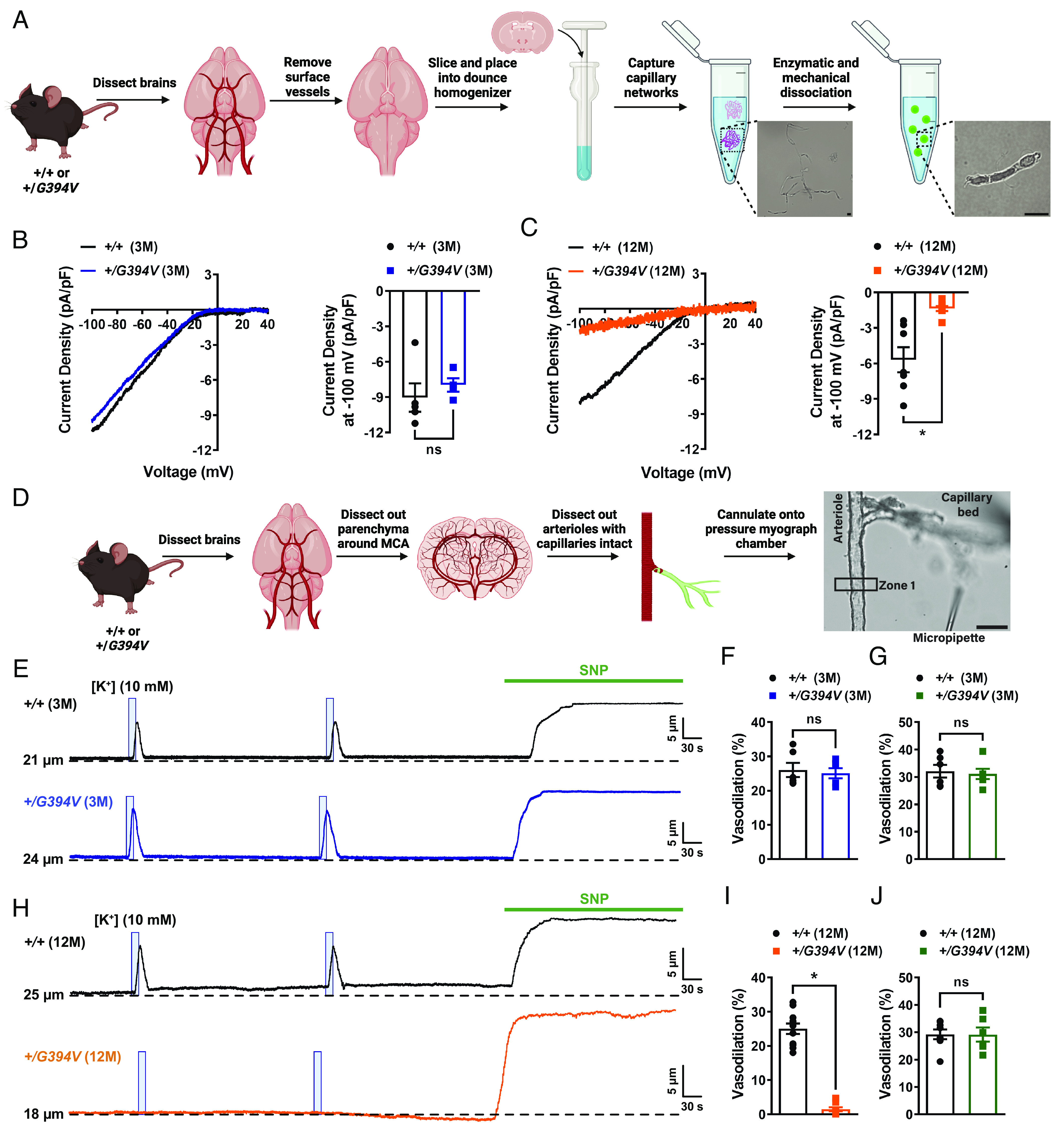
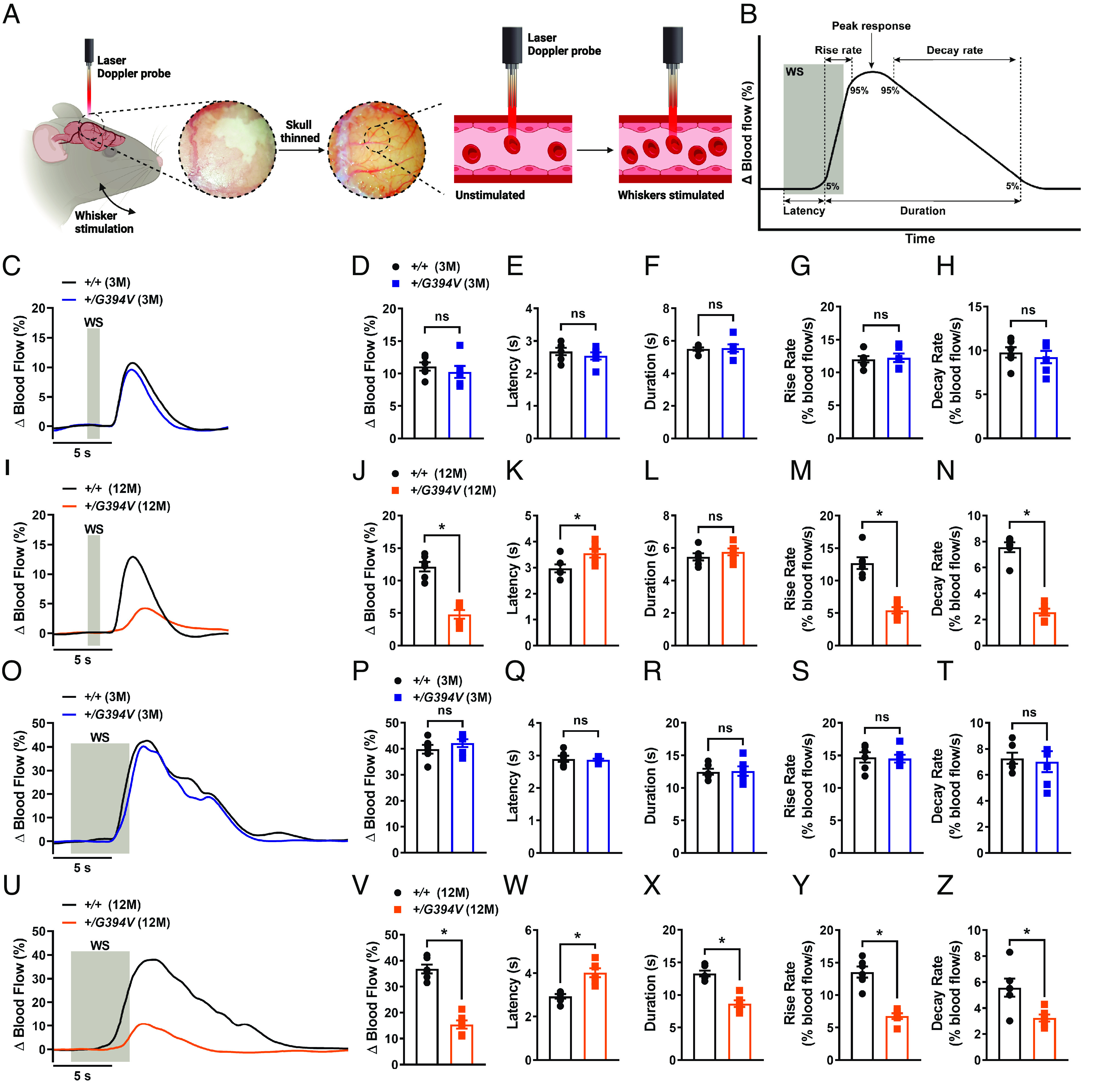
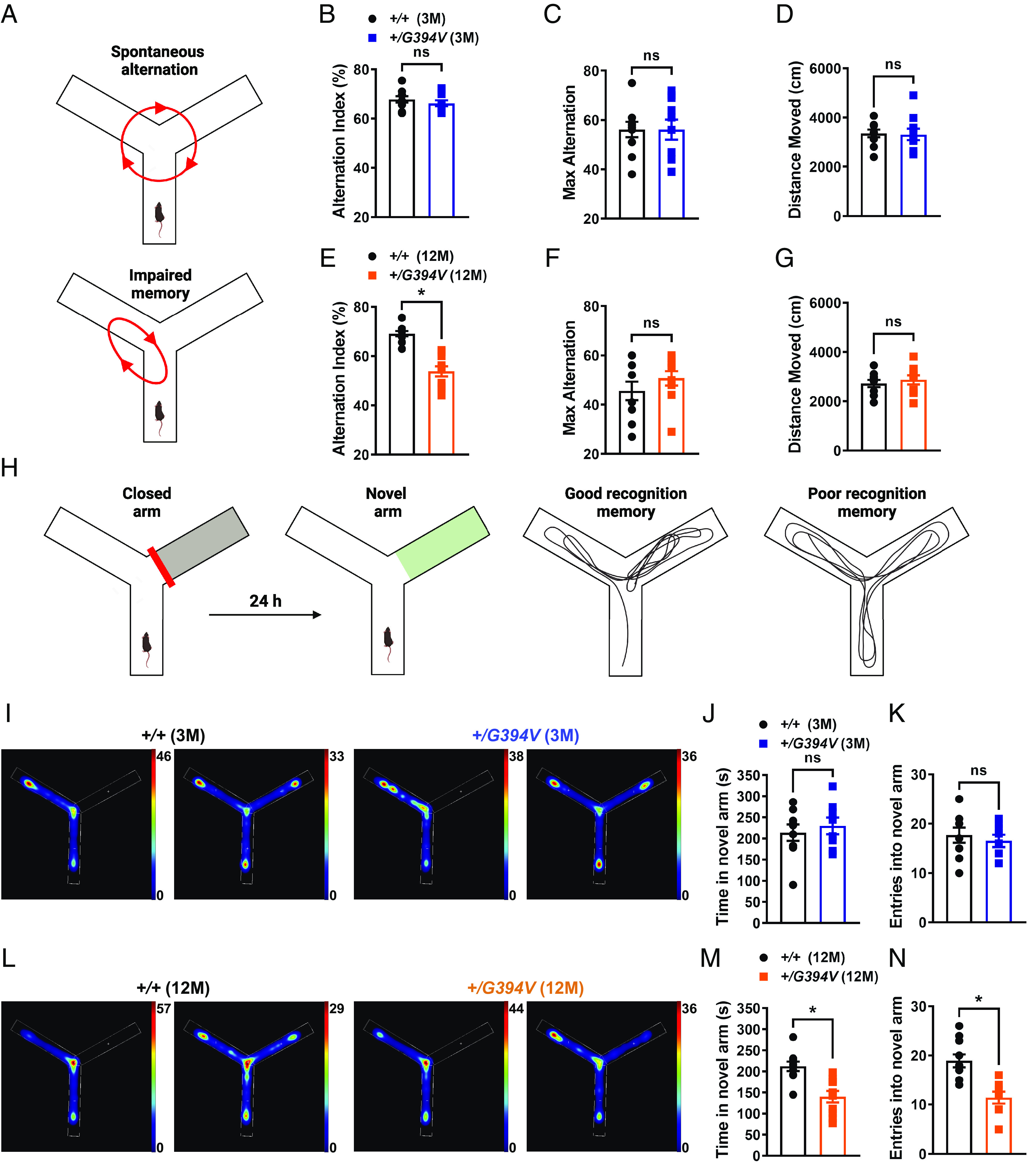
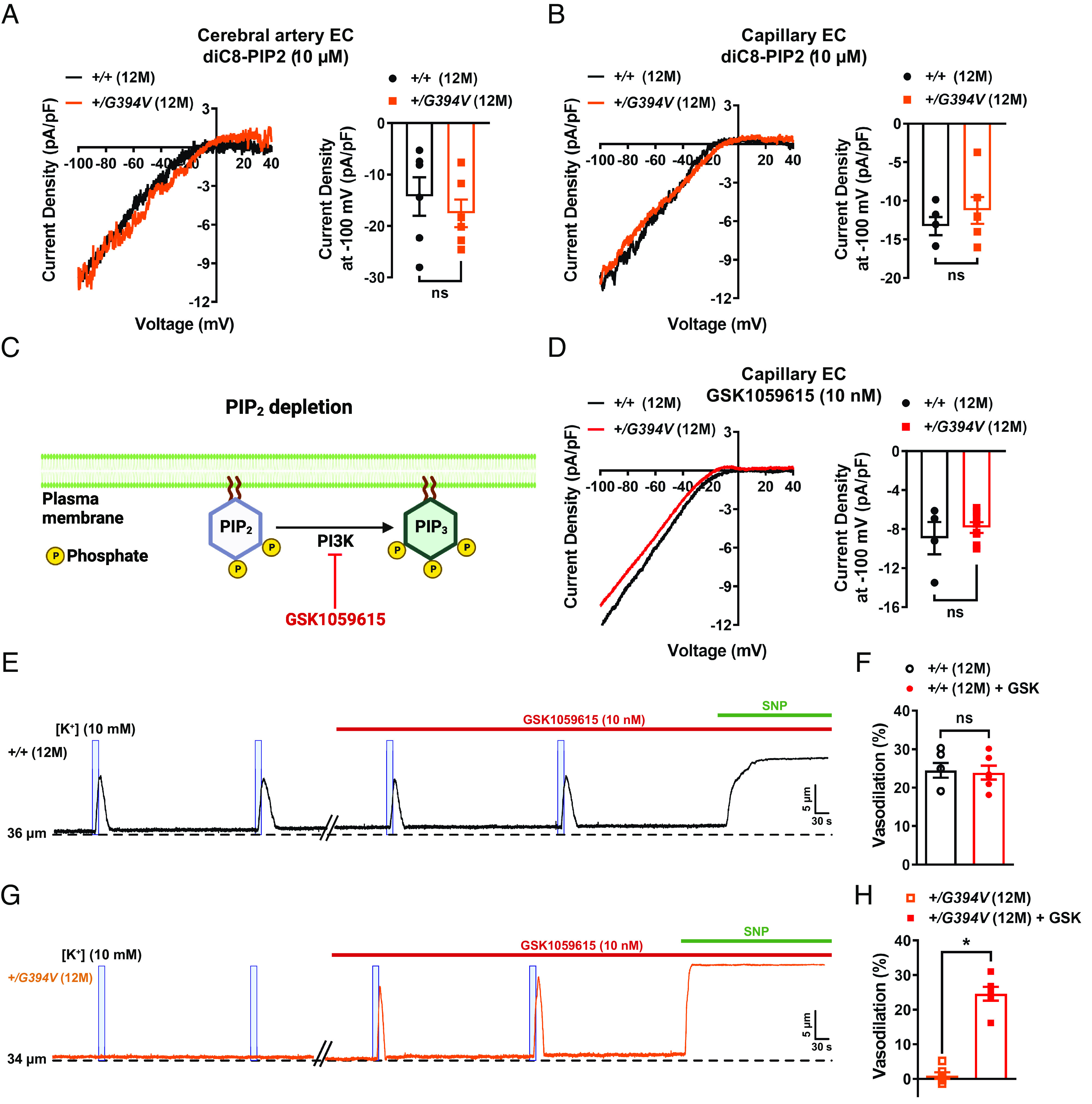
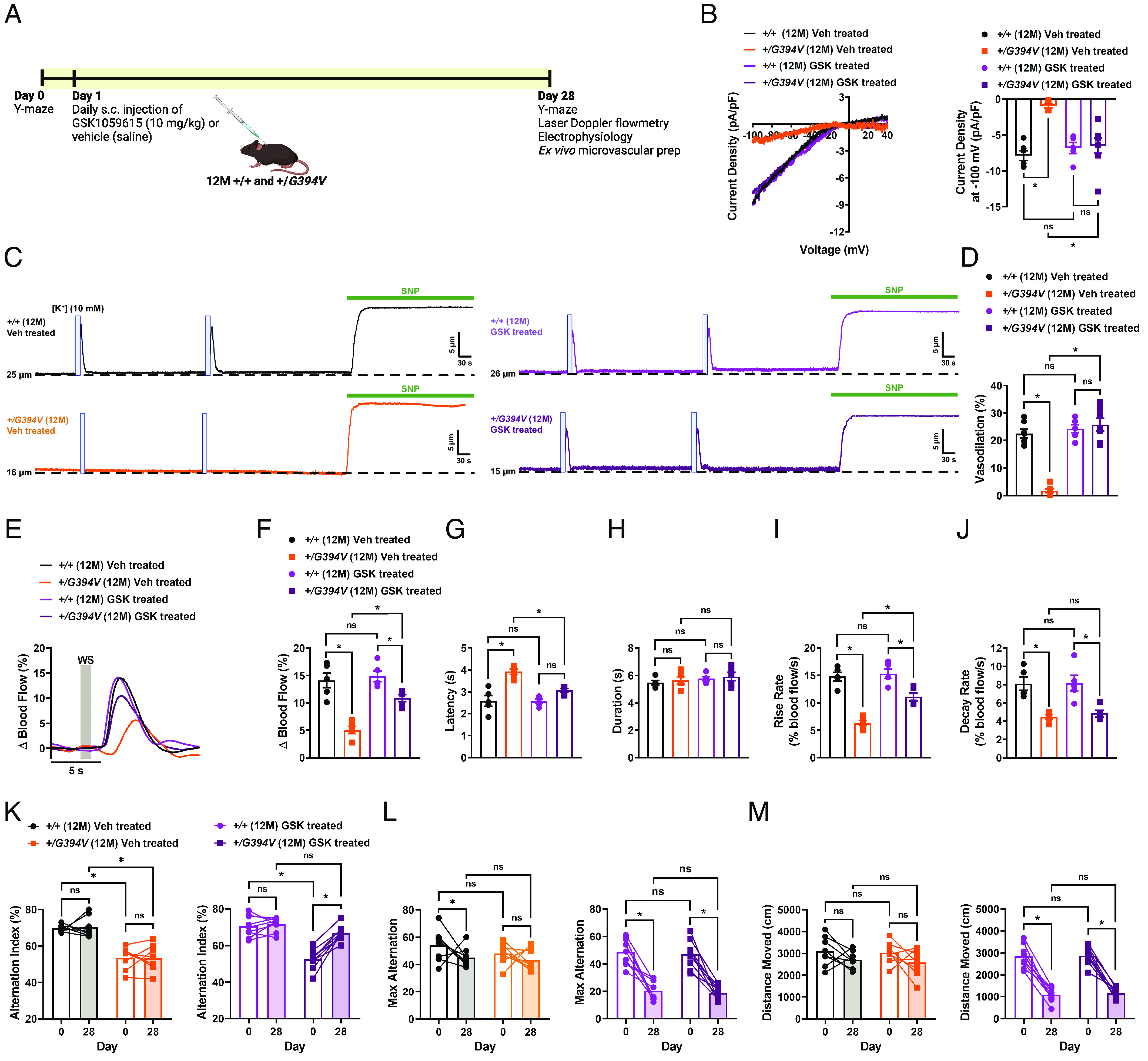
Neurovascular coupling (NVC), a vital physiological process that rapidly and precisely directs localized blood flow to the most active regions of the brain, is accomplished in part by the vast network of cerebral capillaries acting as a sensory web capable of detecting increases in neuronal activity and orchestrating the dilation of upstream parenchymal arterioles. Here, we report a Col4a1 mutant mouse model of cerebral small vessel disease (cSVD) with age-dependent defects in capillary-to-arteriole dilation, functional hyperemia in the brain, and memory. The fundamental defect in aged mutant animals was the depletion of the minor membrane phospholipid phosphatidylinositol 4,5 bisphosphate (PIP2) in brain capillary endothelial cells, leading to the loss of inwardly rectifying K+ (Kir2.1) channel activity. Blocking phosphatidylinositol-3-kinase (PI3K), an enzyme that diminishes the bioavailability of PIP2 by converting it to phosphatidylinositol (3, 4, 5)-trisphosphate (PIP3), restored Kir2.1 channel activity, capillary-to-arteriole dilation, and functional hyperemia. In longitudinal studies, chronic PI3K inhibition also improved the memory function of aged Col4a1 mutant mice. Our data suggest that PI3K inhibition is a viable therapeutic strategy for treating defective NVC and cognitive impairment associated with cSVD.

Keywords: COL4A1; Kir2.1 channels; cerebral small vessel disease; extracellular matrix; functional hyperemia.


Conflict of interest statement

S.E. and D.B.G. have filed a provisional patent for the use of PI3 kinase inhibitors to treat cSVDs.

Figures

Fig. 1.
Fig. 1.
Age-dependent loss of Kir2.1 channel activity and capillary-to-arteriole dilation in Col4a1+/G394V mice. (A) Illustration of the brain capillary EC isolation procedure. (Scale bar, 10 µm.) (B) Representative I-V traces and summary data showing Kir2.1 current densities in freshly isolated capillary ECs from 3-M-old Col4a1+/+ and Col4a1+/G394V mice (n = 4 to 5 cells from 4 to 5 animals per group, ns = not significant, unpaired t test). (C) Representative I-V traces and summary data showing Kir2.1 current densities in freshly isolated capillary ECs from 12-M-old Col4a1+/+ and Col4a1+/G394V mice (n = 7 to 8 cells from four animals per group; *P < 0.05, unpaired t test). (D) Illustration of the microvascular preparation. Parenchymal arterioles with intact capillaries were carefully dissected and cannulated onto a pressure myograph chamber, and compounds of interest were focally applied to capillary extremities. (Scale bar, 50 µm.) (E and F) Representative traces (E) and summary data (F) showing K+ (10 mM, blue box)-induced dilation of upstream arterioles in preparations from 3-M-old Col4a1+/+ and Col4a1+/G394V mice (n = 6 preparations from three animals per group, ns = not significant, unpaired t-test). (G) The dilation produced by superfusing SNP (10 µM) in preparations from 3-M-old Col4a1+/+ and Col4a1+/G394V mice (n = 6 preparations from three animals per group, ns = not significant, unpaired t-test). (H and I) Representative traces (H) and summary data (I) showing K+ (10 mM, blue box)-induced dilation of upstream arterioles in preparations from 12-M-old Col4a1+/+ and Col4a1+/G394V mice (n = 11 preparations from 6 to 7 animals per group, *P < 0.05, unpaired t-test). (J) The dilation produced by superfusing SNP (10 µM) in preparations from 12-M-old Col4a1+/+ and Col4a1+/G394V mice (n = 6 to 8 preparations from 3 to 5 animals per group, ns = not significant, unpaired t-test).
Fig. 2.
Fig. 2.
Age-dependent impairment of functional hyperemia in Col4a1+/G394V mice. (A) Illustration demonstrating the functional hyperemia assessment procedure in the mouse somatosensory cortex. (B) Illustration demonstrating the parameters that were analyzed. (C and D) Representative traces (C) and summary data (D) showing the increase in blood flow following 1-s contralateral whisker stimulation (WS) in 3-M-old Col4a1+/+ and Col4a1+/G394V mice (n = 6 animals per group, ns = not significant, unpaired t test). (EH) Latency (E), duration (F), rise rate (G), and decay rate (H) were also analyzed (n= 6 animals per group, ns = not significant, unpaired t test). (I and J) Representative traces (I) and summary data (J) showing the increase in blood flow following 1-s contralateral WS in 12-M-old Col4a1+/+ and Col4a1+/G394V mice (n = 6 animals per group, *P < 0.05, unpaired t test). (KN) Latency (K), duration (L), rise rate (M), and decay rate (N) were also analyzed (n = 6 animals per group, *P < 0.05, ns = not significant, unpaired t-test). (O and P) Representative traces (O) and summary data (P) showing the increase in blood flow following 5-s contralateral WS in 3-M-old Col4a1+/+ and Col4a1+/G394V mice (n = 6 animals per group, ns = not significant, unpaired t-test). (QT) Latency (Q), duration (R), rise rate (S), and decay rate (T) were also analyzed (n= 6 animals per group, ns = not significant, unpaired t-test). (U and V) Representative traces (U) and summary data (V) showing the increase in blood flow following 5-s contralateral WS in 12-M-old Col4a1+/+ and Col4a1+/G394V mice (n = 6 animals per group, *P < 0.05, unpaired t test). (WZ) Latency (W), duration (X), rise rate (Y), and decay rate (Z) were also analyzed (n = 6 animals per group, *P < 0.05, unpaired t test).
Fig. 3.
Fig. 3.
Age-dependent memory deficits in Col4a1+/G394V mice. (A) Illustration of the Y-maze spontaneous alternation behavior assay showing examples of a spontaneous (Top) and nonspontaneous (Bottom) alternation. (B) Summary data showing alternation index, an indicator spatial working memory, in 3-M-old Col4a1+/+ and Col4a1+/G394V mice (n = 10 animals per group, ns = not significant, unpaired t test). (C and D) Summary data showing max alternation (C) and distance moved (D), indicative of exploratory activity, in 3-M-old Col4a1+/+ and Col4a1+/G394V mice (n = 10 animals per group, ns = not significant, unpaired t test). (E) Summary data showing the alternation index of 12-M-old Col4a1+/+ and Col4a1+/G394V mice (n = 10 animals per group, *P < 0.05, unpaired t test). (F and G) Summary data showing max alternation (F) and distance moved (G) in 12-M-old Col4a1+/+ and Col4a1+/G394V mice (n = 10 animals per group, ns = not significant, unpaired t test). (H) Illustration demonstrating typical and impaired Y-maze novel arm behavior. (I) Representative heatmaps showing the time (s) 3-M-old Col4a1+/+ and Col4a1+/G394V mice spent in areas of the Y-maze during the novel arm test. (J and K) Summary data showing the time spent (J) and entries (K) into the novel arm (n = 8 to 9 animals per group, ns = not significant, unpaired t test). (L) Representative heatmaps showing the time (s) 12-M-old Col4a1+/+ and Col4a1+/G394V mice spent in areas of the Y-maze during the novel arm test. (M and N) Summary data showing the time spent (M) and entries (N) into the novel arm (n = 10 animals per group, *P < 0.05, unpaired t-test).
Fig. 4.
Fig. 4.
PIP2 depletion reduces Kir2.1 currents in 12-M-old Col4a1+/G394V mice. (A) Representative I-V traces and summary data showing Kir2.1 current densities in freshly isolated cerebral artery ECs from 12-M-old Col4a1+/+ and Col4a1+/G394V mice with internal solution supplemented with diC8-PIP2 (10 µM) (n = 6 cells from four animals per group, ns = not significant, unpaired t-test). (B) Representative I-V traces and summary data showing Kir2.1 current densities in freshly isolated brain capillary ECs from 12-M-old Col4a1+/+ and Col4a1+/G394V mice with internal solution supplemented with diC8-PIP2 (10 µM) (n = 5 to 6 cells from 4 to 5 animals per group, ns = not significant, unpaired t test). (C) PIP2 depletion pathway. (D) Representative I-V traces and summary data showing Kir2.1 current densities in freshly isolated brain capillary ECs from 12-M-old Col4a1+/+ and Col4a1+/G394V mice treated with the PI3K blocker GSK1059615 (10 nM) (n = 4 to 8 cells from 3 to 6 animals per group, ns = not significant, unpaired t test). (E and F) Representative trace (E) and summary data (F) showing K+ (10 mM, blue box)-induced dilation of upstream arterioles in preparations from 12-M-old Col4a1+/+ mice before and after superfusing the PI3K blocker GSK1059615 (10 nM, 30 min) (n = 6 preparations from five animals per group, ns = not significant, paired t test). (G and H) Representative trace (G) and summary data (H) showing K+ (10 mM, blue box)-induced dilation of upstream arterioles in preparations from 12-M-old Col4a1+/G394V mice before and after superfusing the PI3K blocker GSK1059615 (10 nM, 30 min) (n = 6 preparations from five animals per group, *P < 0.05, paired t test).
Fig. 5.
Fig. 5.
Chronic PI3K inhibition restores Kir2.1 currents, K+-induced dilation, functional hyperemia, and memory deficits in 12-M-old Col4a1+/G394V mice. (A) Illustration showing GSK1059615 treatment plan. (B) Representative I-V traces and summary data showing Kir2.1 current densities in freshly isolated capillary ECs from 12-M-old Col4a1+/+ and Col4a1+/G394V mice treated with vehicle (saline) or GSK1059615 (10 mg/kg) for 28 d, s.c. (n = 5 to 8 cells from 3 to 4 animals per group, *P < 0.05, ns = not significant, nonrepeated measures two-way ANOVA). (C and D) Representative traces (C) and summary data (D) showing K+ (10 mM, blue box)-induced dilation of upstream arterioles in preparations from 12-M-old Col4a1+/+ and Col4a1+/G394V mice treated with vehicle (saline) or GSK1059615 (10 mg/kg) for 28 d, s.c. (n = 6 to 7 preparations from three animals per group, *P < 0.05, ns = not significant, nonrepeated measures two-way ANOVA). (E and F) Representative traces (E) and summary data (F) showing the increase in blood flow following 1-s contralateral whisker stimulation (WS) in 12-M-old Col4a1+/+ and Col4a1+/G394V mice treated with vehicle (saline) or GSK1059615 (10 mg/kg) for 28 d, s.c. (n = 5 animals per group, *P < 0.05, ns = not significant, nonrepeated measures two-way ANOVA). (GJ) Latency (G), duration (H), rise rate (I), and decay rate (J) were also analyzed (n = 5 animals per group, *P < 0.05, ns = not significant, nonrepeated measures two-way ANOVA). (K) Summary data showing alternation index, an indicator spatial working memory, of 12-M-old Col4a1+/+ and Col4a1+/G394V mice before and after treatment with vehicle (saline) or GSK1059615 (10 mg/kg) for 28 d, s.c. (n = 8 to 9 animals per group, *P < 0.05, ns = not significant, repeated measures two-way ANOVA). (L and M) Summary data showing max alternation (L) and distance moved (M), indicative of exploratory activity, in 12-M-old Col4a1+/+ and Col4a1+/G394V mice before and after treatment with vehicle (saline) or GSK1059615 (10 mg/kg) for 28 d, s.c. (n = 8 to 9 animals per group, *P < 0.05, ns = not significant, repeated measures two-way ANOVA).

Update of

Similar articles

Cited by

References

    1. Corriveau R. A., et al. , The science of vascular contributions to cognitive impairment and dementia (VCID): A framework for advancing research priorities in the cerebrovascular biology of cognitive decline. Cell Mol. Neurobiol. 36, 281–288 (2016). - PMC - PubMed
    1. Wardlaw J. M., Smith C., Dichgans M., Mechanisms of sporadic cerebral small vessel disease: Insights from neuroimaging. Lancet Neurol. 12, 483–497 (2013). - PMC - PubMed
    1. Debette S., Schilling S., Duperron M. G., Larsson S. C., Markus H. S., Clinical significance of magnetic resonance imaging markers of vascular brain injury a systematic review and meta-analysis. Jama Neurol. 76, 81–94 (2019). - PMC - PubMed
    1. Dichgans M., Leys D., Vascular cognitive impairment. Circ. Res. 120, 573–591 (2017). - PubMed
    1. Shi Y., Wardlaw J. M., Update on cerebral small vessel disease: A dynamic whole-brain disease. Stroke Vasc. Neurol. 1, 83–92 (2016). - PMC - PubMed

Publication types

Substances