. 2023 Oct 23;58(20):2032-2047.e6.
doi: 10.1016/j.devcel.2023.07.020. Epub 2023 Aug 21.

Skin-type-dependent development of murine mechanosensory neurons

Affiliations

Skin-type-dependent development of murine mechanosensory neurons

Charalampia Koutsioumpa et al. Dev Cell. .

Abstract

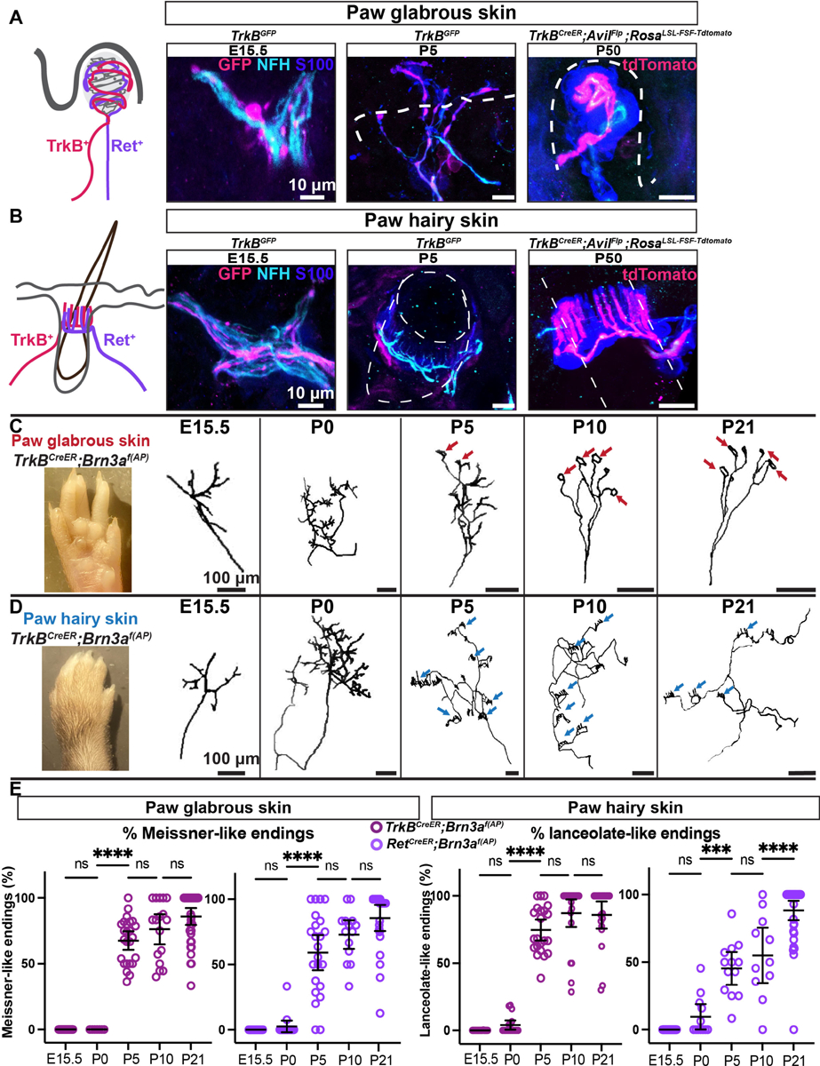
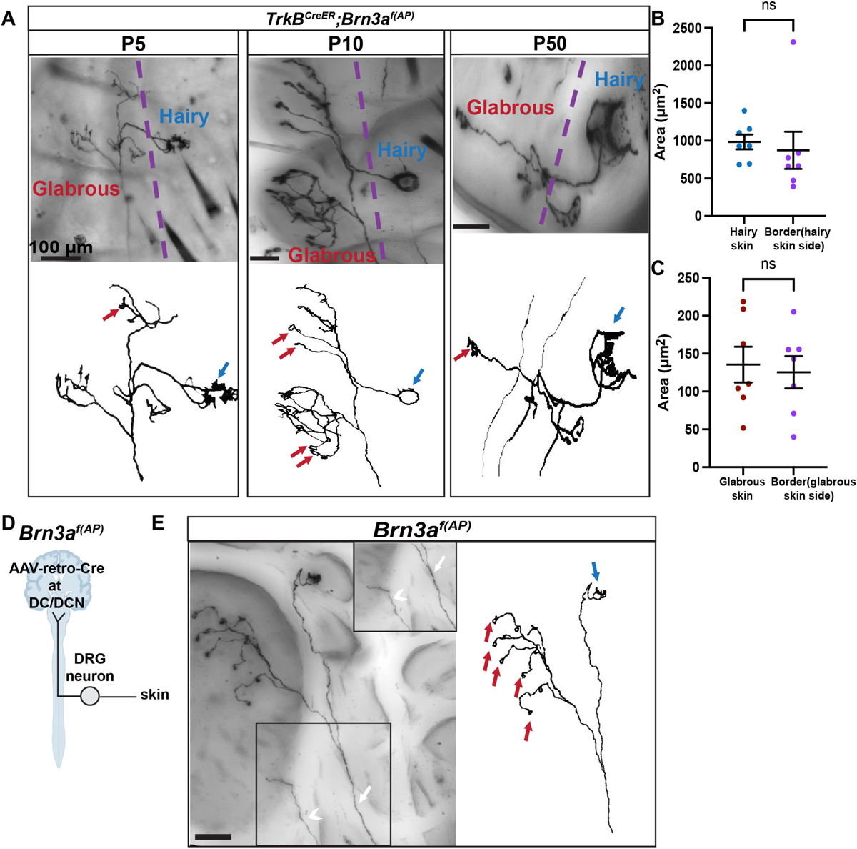
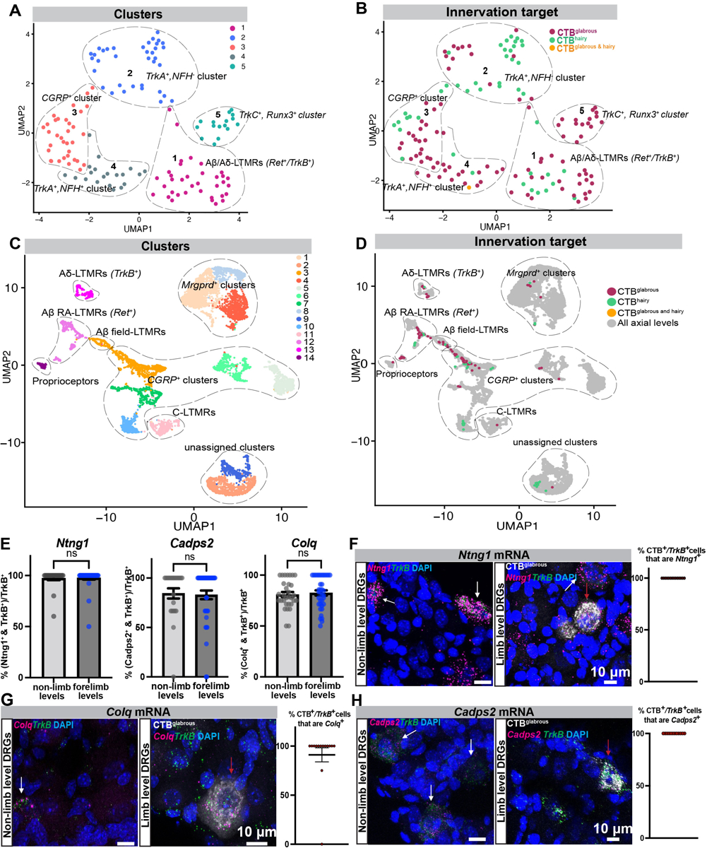
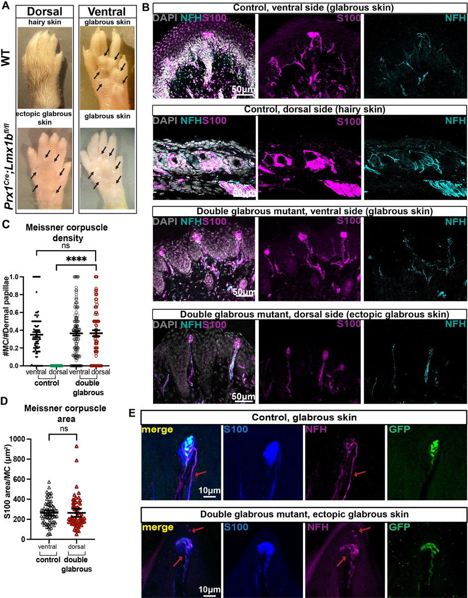
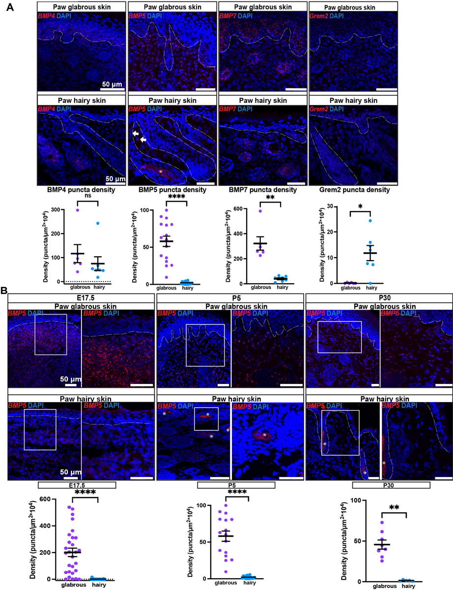
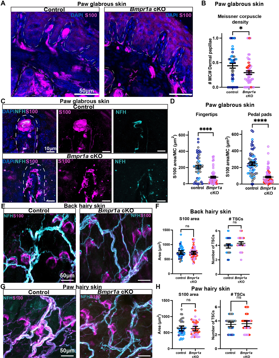
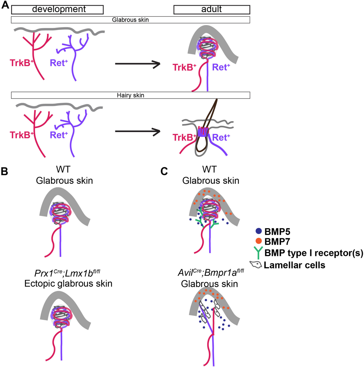
Mechanosensory neurons innervating the skin underlie our sense of touch. Fast-conducting, rapidly adapting mechanoreceptors innervating glabrous (non-hairy) skin form Meissner corpuscles, while in hairy skin, they associate with hair follicles, forming longitudinal lanceolate endings. How mechanoreceptors develop axonal endings appropriate for their skin targets is unknown. We report that mechanoreceptor morphologies across different skin regions are indistinguishable during early development but diverge post-natally, in parallel with skin maturation. Neurons terminating along the glabrous and hairy skin border exhibit hybrid morphologies, forming both Meissner corpuscles and lanceolate endings. Additionally, molecular profiles of neonatal glabrous and hairy skin-innervating neurons largely overlap. In mouse mutants with ectopic glabrous skin, mechanosensory neurons form end-organs appropriate for the altered skin type. Finally, BMP5 and BMP7 are enriched in glabrous skin, and signaling through type I bone morphogenetic protein (BMP) receptors in neurons is critical for Meissner corpuscle morphology. Thus, mechanoreceptor morphogenesis is flexibly instructed by target tissues.

Keywords: axonal development; mechanosensory neurons; morphogenesis; touch end-organs.


Conflict of interest statement

Declaration of interests The authors declare no competing interests.

Figures

Figure 1.
Figure 1.. LTMR axon terminals in glabrous and hairy skin are comparable embryonically but diverge in the first postnatal week
(A and B) Example images of TrkB+ endings (TrkBGFP or TrkBCreER; AvilFlp; RosaLSL-FSF-Tdtomato) in paw glabrous skin (A) and paw hairy skin (B) at E15.5, P5, and P50. The sections were stained with S100, NFH and GFP or tdTomato. The white dotted lines represent the dermal-epidermal junction in glabrous skin and the hair follicle outline in hairy skin. TrkB+ and Ret+ endings in glabrous skin innervate Meissner corpuscles in dermal papillae. TrkB+ and Ret+ endings in hairy skin form longitudinal lanceolate endings around hair follicles (Ret+ endings are present around guard and awl/auchene hair follicles, while TrkB+ endings around awl/auchene and zigzag hair follicles). (C and D) Example reconstructions of sparsely labeled TrkBCreER; Brn3af(AP) endings in paw glabrous skin (C) and paw hairy skin (D) at different developmental ages. Red arrows point to developing Meissner corpuscles while blue arrows to developing lanceolate endings. (E) Quantification of percentages of Meissner-like endings (left panel) in paw glabrous skin of TrkBCreER; Brn3af(AP) and RetCreER; Brn3af(AP) across development. Each datapoint represents the quantification of endings from a single neuron. Quantification of percentages of lanceolate-like endings (right panel) in paw hairy skin of TrkBCreER; Brn3af(AP) and RetCreER; Brn3af(AP) sensory neurons across development (n=3–5 mice per timepoint and per genotype). For Meissner ending analysis: TrkBCreER; Brn3af(AP) one-way-ANOVA p<0.0001, R2=0.8, RetCreER;Brn3af(AP) one-way-ANOVA p<0.0001, R2=0.7. Pairwise comparisons for TrkBCreER; Brn3af(AP) were performed: E15.5 vs P0 p>0.99, P0 vs P5 p<0.0001, P5 vs P10 p=0.43 and P10 vs P21 p=0.25. Pairwise comparisons for RetCreER; Brn3af(AP) were performed: E15.5 vs P0 p=0.99, P0 vs P5 p<0.0001, P5 vs P10 p=0.29 and P10 vs P21 p=0.37. For lanceolate-ending analysis: TrkBCreER; Brn3af(AP) one-way-ANOVA p<0.0001, R2=0.86, RetCreER;Brn3af(AP) one-way-ANOVA p<0.0001, R2=0.79. Pairwise comparisons for TrkBCreER; Brn3af(AP) were performed: E15.5 vs P0 p=0.93, P0 vs P5 p<0.0001, P5 vs P10 p=0.09 and P10 vs P21 p=0.99. Pairwise comparisons for RetCreER; Brn3af(AP) were performed: E15.5 vs P0 p= 0.65, P0 vs P5 p=0.0001, P5 vs P10 p=0.73 and P10 vs P21 p<0.0001. See also Figure S1, S2.
Figure 2.
Figure 2.. Individual neurons with branches that extend into both glabrous and hairy skin form ending types appropriate for both skin regions
(A) Representative example images (top) and reconstructions (bottom) of P5, P10 and P50 “border neurons” from TrkBCreER; Brn3af(AP) mice. These neurons terminate in the finger of the paws and exhibit branches that form both lanceolate endings in hairy skin (blue arrows) and Meissner corpuscle endings in glabrous skin (red arrows). The purple dotted line indicates the approximate border of glabrous and hairy skin. (B) End-organ area quantifications from TrkBCreER; Brn3af(AP) endings terminating in hairy paw skin. Lanceolate endings that terminate only in hairy skin and endings from border neuron branches in hairy skin have similar areas. Each datapoint represents the area of a single lanceolate ending. N=3 mice per group. Mann Whitney test p=0.09, U=11. (C) End-organ area quantifications from TrkBCreER; Brn3af(AP) Meissner corpuscle endings terminating only in paw glabrous skin and endings from border neuron branches in glabrous skin. Each datapoint represents a single Meissner corpuscle. N=3 mice per group. Mann Whitney test p=0.7, U=21. (D) Schematic of brainstem viral labeling approach. AAV-retro-Cre virus was injected in the dorsal column (DC) or dorsal column nuclei (DCN) of Brn3af(AP) mice. (E) Representative example image (left) and reconstruction (right) of an identified border neuron. The neuron terminates in the ventral hindpaw innervating the hair follicles (blue arrows) present in the middle of the paw on one side and the glabrous pedal pads on the other (red arrows). The imaging approach distinguishes single axons, the white arrow indicates the border neuron axon in the skin while the white arrowhead points to a neighboring axon, also shown in the inset (top right corner). See also Figure S3 and Supplementary Videos 1–3.
Figure 3.
Figure 3.. Transcriptional signatures of glabrous and hairy skin innervating neuronal classes
(A) UMAP plot with the clusters identified from the P5 sensory neuron cell picking experiment: cluster 1, 36 neurons (10 hairy skin innervating, 26 glabrous skin innervating neurons); cluster 2, 36 neurons (22 hairy skin innervating, 14 glabrous skin innervating); cluster 3, 34 neurons (10 hairy skin innervating, 24 glabrous skin innervating); cluster 4, 20 neurons (2 hairy skin innervating, 1 glabrous and hairy skin innervating, 17 glabrous skin innervating); and cluster 5, 16 neurons (16 glabrous skin innervating). (B) UMAP plot with color-coding according to the innervation target (glabrous skin innervation, hairy skin innervation, or for one neuron both glabrous and hairy skin). (C) UMAP plot and clusters of the integration analysis from the P5 cell picking and P5 scRNA seq dataset. (D) UMAP plot of the integration analysis with color-coding according to the innervation target (E) Quantification of the percentages of cells double-positive for TrkB+ and Ntng1/Cadps2/Colq in nonlimb and forelimb (C5–C8) DRGs, n=3 mice per condition. All comparisons were done using the Mann Whitney test. For Ntng1: p>0.9, U=402, 35 C5–C8 and 23 non-limb cells quantified, Cadps2: p=0.98, U=447, 39 C5–C8 and 23 non-limb cells quantified, Colq: p =0.58, U=529 37 C5–C8 and 31 non-limb cells quantified. (F) Example images (left panels) of RNAscope for Ntng1 and TrkB mRNA. Nonlimb and limb level DRG sections of P5 mice injected with CTB in forepaw glabrous skin (CTBglabrous) were examined. White arrows point to Ntng1 and TrkB double positive and red arrows to CTB, TrkB and Ntng1 triple positive cells. Quantifications (right panel) of the percentages of neurons that were triple positive for CTB, TrkB and Ntng1 (14 cells analyzed from 3 mice). (G) Example images (left panels) of RNAscope for Colq and TrkB mRNA. Nonlimb and limb level DRG sections of P5 mice injected with CTB into forepaw glabrous skin (CTBglabrous) were examined. White arrows point to TrkB and Colq double positive and red arrows to CTB, TrkB and Colq triple positive cells. Quantifications (right panel) of the percentages of neurons that were triple positive for CTB, TrkB and Colq (14 cells analyzed from 3 mice). (H) Example images (left panels) of RNAscope for Cadps2 and TrkB mRNA. Nonlimb and limb level DRG sections of P5 mice injected with CTB in forepaw glabrous skin (CTBglabrous) were examined. White arrows point to TrkB and Cadps2 double positive and red arrows to CTB, TrkB and Cadps2 triple positive cells. Quantifications (right panel) of the percentages of neurons that were triple positive for CTB, TrkB and Cadps2 (12 cells analyzed from 3 mice). See also Figure S4.
Figure 4.
Figure 4.. Ectopic glabrous skin in double glabrous mutant mice becomes innervated and displays Meissner corpuscle-like structures
(A) Example images of the dorsal and ventral surfaces of wild type and Prx1Cre; Lmx1bfl/fl mice, also referred to as double glabrous mutants. The majority of the dorsal surface of the mutants is covered by glabrous skin, with areas of hairy skin interspersed. Black arrows point to the pedal pads. A small amount of hair removal (Nair) was applied to all surfaces. (B) Sections of P21 control and double glabrous forepaws with NFH, S100, and DAPI. Control ventral and dorsal skin regions (top two rows) have Meissner corpuscles on the ventral side and lanceolate endings on the dorsal side. Double glabrous mutant ventral and dorsal skin (bottom two rows) is predominantly glabrous, and these mutants contain Meissner corpuscles on both the ventral and dorsal sides of the paw. Hair follicles innervated with lanceolate endings are found in some of the mutant’s dorsal surfaces. (C) Quantifications of Meissner corpuscles density (Meissner corpuscles divided by the number of dermal papillae) in control and double glabrous mice demonstrate that they are similar in control ventral, double glabrous dorsal, and double glabrous ventral skin but distinct from control dorsal skin. Each datapoint represents a forepaw/hindpaw skin section. N=3–4 mice were analyzed per genotype, 85 sections for control ventral, 19 for control dorsal, 127 for double glabrous ventral, and 78 for dorsal, One-way Kruskal Wallis test p<0.0001, Kruskal Wallis statistic 43.21. Additional Mann Whitney tests between two conditions: ns, and <0.0001, Mann Whitney U=209. (D) Lamellar cell area of Meissner corpuscles quantified in skin sections of forepaw and hindpaw skin in control and double glabrous mice. Each datapoint represents the average S100+ area of Meissner corpuscles in a skin section. N=3 mice per condition, 72 control and 59 double glabrous sections quantified (Mann Whitney test p=0.4, U=1951). (E) Forelimb pedal pad sections of control Npy2r-GFP glabrous skin (top) and Prx1Cre; Lmx1bfl/fl; Npy2r-GFP double glabrous mutant, ectopic glabrous skin (bottom). The sections are stained using S100, GFP and NFH. Red arrows point to the GFP axon (presumably Ret+). N=3 mice per condition. See also Figure S5.
Figure 5.
Figure 5.. Characterization of BMP ligand expression patterns in paw glabrous and hairy skin over development
(A) Example forepaw glabrous and hairy skin images visualizing BMP4, BMP5, BMP7 and Grem2 puncta at P5 (top panels) and puncta density quantifications (bottom panels). The asterisk indicates background signal in the hair shaft, and the arrows point to the mRNA signal. The dotted lines outline the dermo-epidermal junction in all panels. N=3 mice per group. The puncta density of BMP4, BMP5, BMP7 and Grem2 at P5 control glabrous and hairy skin are shown in the bottom panels. Mann Whitney test from left to right: p=0.13, p<0.0001, p= 0.001 and p=0.01. N=3 mice per condition. (B) BMP5 mRNA in skin sections of forepaw glabrous skin and forepaw hairy skin (top panels) and puncta density quantifications (bottom panels). A lower (left panel) and higher magnification (right panel, outlined in a white rectangle) are presented. The skin was examined at E17.5, P5 and P30 using in situ RNAscope. BMP5 puncta are enriched in the glabrous skin at all time points. The asterisk indicates background signal in the hair shaft. N=3 mice per group. The bottom panels include quantifications of BMP5 puncta density at E17.5, P5 and P30 control glabrous and hairy skin. Mann Whitney test from left to right: p<0.0001, p<0.0001 and p=0.001. N=3 mice per condition. See also Figure S6.
Figure 6.
Figure 6.. Conditional deletion of genes encoding BMP type I receptors in somatosensory neurons leads to dysmorphic Meissner corpuscles
(A) Sections of forepaw pedal pad glabrous skin of AvilCre; Bmpr1afl/fl and control littermate P30 mice. The dotted lines outline the dermal-epidermal junction and sweat glands. S100 was used to investigate the presence of Meissner corpuscles and DAPI the overall skin composition. (B) Quantifications of Meissner corpuscle density in forepaw fingertips in control and mutant mice (P28-P35). Bmpr1a cKO had comparable densities, which were decreased compared to control littermates. Each datapoint represents the Meissner corpuscle density quantified in a skin section; 46 control and 58 mutant sections. Mann Whitney test p=0.02, U=981. N=3 controls; AvilCre/+ (blue dots), Bmpr1afl/fl (light blue dots), and Bmpr1afl/fl;Actvr1fl/+ mice (grey dots). N=3 mutants; two AvilCre; Bmpr1afl/fl; Acvr1fl/+ mice (pink dots) and one AvilCre; Bmpr1afl/fl mouse (red dots). Mutant mice were born in sub-Mendelian ratios. (C) Sections of forepaw pedal pad skin and example images of Meissner corpuscles in control and mutant mice. The dotted line outlines a dermal papilla. The sections were stained with S100 to visualize the lamellar cell core, NFH for the myelinated axons and DAPI. (D) Quantifications of the lamellar S100+ area per Meissner corpuscle across forepaw fingertip (left panel) and forepaw pedal pads (right panel) in control and mutant mice. Each datapoint represents the S100+ area per MC quantified in a section of skin (for fingertips 49 control and 59 mutant sections, Mann Whitney test p<0.0001, U=799, for pedal pads 62 control and 80 mutant sections, Mann Whitney test p<0.0001, U=758.5). N=3 controls including AvilCre/+ (blue dots), Bmpr1afl/fl (light blue dots), Bmpr1afl/fl;Actvr1fl/+ mice (grey dots), n=3 mutants including two AvilCre; Bmpr1afl/fl; Acvr1fl/+mice (pink dots) and one AvilCre; Bmpr1afl/fl mouse (red dots). (E) Example images of back hairy skin wholemount staining of control and mutant mice. Overall innervation and lanceolate ending morphology are preserved in the mutants. (F) Quantifications of the terminal Schwann cell number and S100 area of lanceolate endings in back hairy skin. Each datapoint represents the Schwann cell S100 area or number of TSCs, respectively, for every non-guard hair follicle ending quantified. The lanceolate ending S100+ area (left panel) was similar in control (endings in 44 hair follicles quantified) and mutant mice (endings in 62 hair follicles quantified) n= 3 controls, n= 3 mutants, (two AvilCre;Bmpr1afl/fl;Acvr1fl/+ mice in pink, and one AvilCre;Bmpr1afl/fl in red, which were combined for statistical analysis). The number of TSCs (right panel) was similar in control (endings in 48 hair follicles quantified) and mutant mice (endings in 65 hair follicles quantified) (Mann Whitney p=0.82, U=1329 and p =0.053, U=1245 respectively). Controls genotypes are: AvilCre/+ (blue dots), Bmpr1afl/fl (light blue dots), and Bmpr1afl/fl;Actvr1fl/+ (grey dots). (G) Example images of paw hairy skin wholemount staining of control and mutant mice. Overall innervation and lanceolate ending morphology is preserved in the mutants. (H) Similar analysis as in (F) in the forepaw hairy skin of control and mutant mice. The lanceolate ending S100+ area (left panel) was similar in control (endings in 38 hair follicles quantified) and mutant mice (endings in 37 hair follicles quantified). The number of TSCs (right panel) was similar in control (endings in 40 hair follicles quantified) and mutant mice (endings in 38 hair follicles quantified) (Mann Whitney p=0.76, U=674 and p=0.9, U=750.5 respectively). Control genotypes are AvilCre/+ (blue dots), Bmpr1afl/fl (light blue dots), and Bmpr1afl/fl;Actvr1fl/+ (grey dots). See also Figure S7.
Figure 7.
Figure 7.. Model of LTMR development and its skin dependence
(A) Spatiotemporal development of TrkB+ and Ret+ Meissner corpuscle innervating and lanceolate ending forming neurons. (B) Dorsal-ventral skin alterations lead to ectopic Meissner corpuscle development. (C) Bmpr1a cKO mice exhibit highly aberrant Meissner development.

Comment in

Similar articles

Cited by

References

    1. Abraira VE, and Ginty DD (2013). The Sensory Neurons of Touch. Neuron 79, 618–639. 10.1016/j.neuron.2013.07.051. - DOI - PMC - PubMed
    1. Handler A, and Ginty DD (2021). The Mechanosensory Neurons of Touch and their Mechanisms of Activation. Nat Rev Neurosci 22, 521–537. 10.1038/s41583-021-00489-x. - DOI - PMC - PubMed
    1. McCray BA, and Scherer SS (2021). Axonal Charcot-Marie-Tooth Disease: from Common Pathogenic Mechanisms to Emerging Treatment Opportunities. Neurotherapeutics 18, 2269–2285. 10.1007/s13311-021-01099-2. - DOI - PMC - PubMed
    1. Orefice LL, Mosko JR, Morency DT, Wells MF, Tasnim A, Mozeika SM, Ye M, Chirila AM, Emanuel AJ, Rankin G, et al. (2019). Targeting Peripheral Somatosensory Neurons to Improve Tactile-Related Phenotypes in ASD Models. Cell 178, 867–886.e24. 10.1016/j.cell.2019.07.024. - DOI - PMC - PubMed
    1. Richards N, and McMahon SB (2013). Targeting novel peripheral mediators for the treatment of chronic pain. Br J Anaesth 111, 46–51. 10.1093/bja/aet216. - DOI - PubMed

Publication types