. 2023 Oct 15:336:199205.
doi: 10.1016/j.virusres.2023.199205. Epub 2023 Aug 25.

Chloroplast-related host proteins interact with NIb and NIa-Pro of soybeans mosaic virus and induce resistance in the susceptible cultivar

Affiliations

Chloroplast-related host proteins interact with NIb and NIa-Pro of soybeans mosaic virus and induce resistance in the susceptible cultivar

John Bwalya et al. Virus Res. .

Abstract

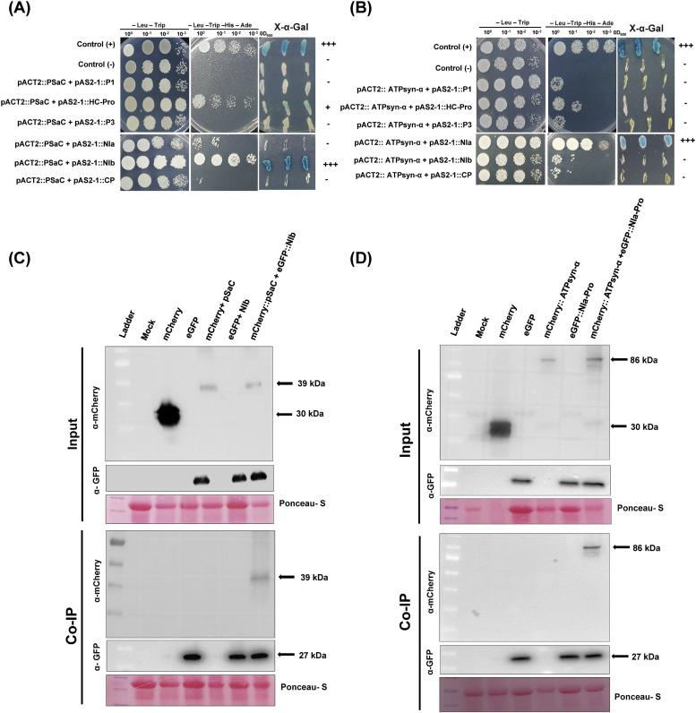
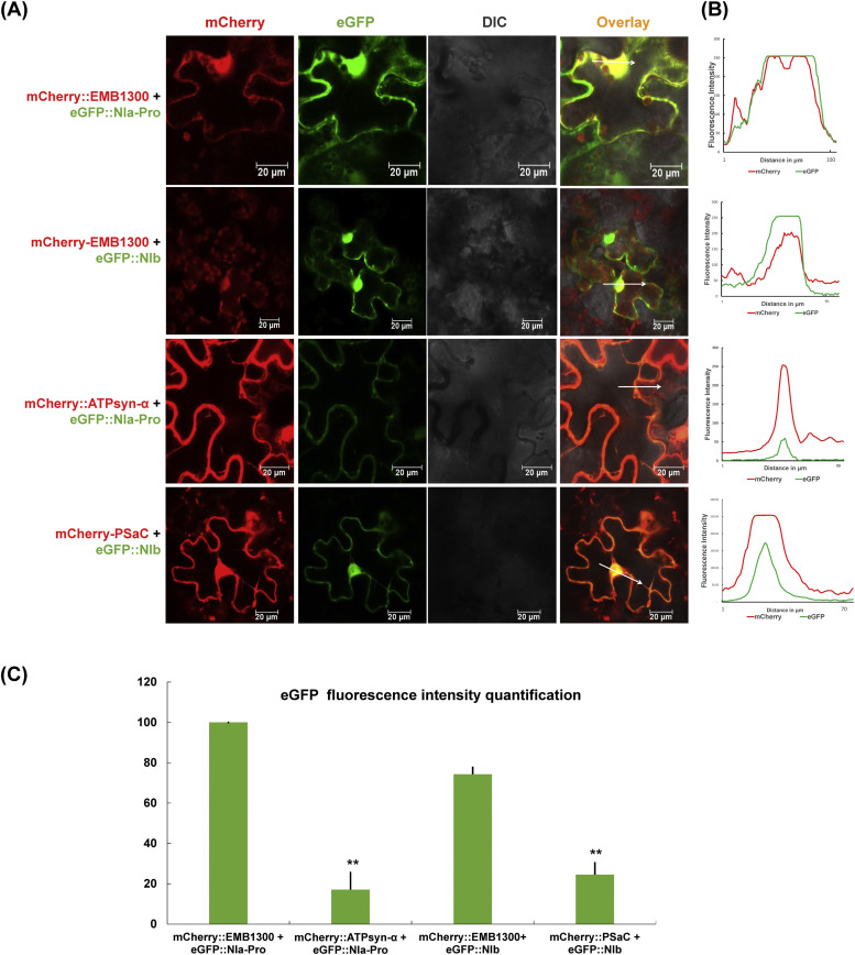
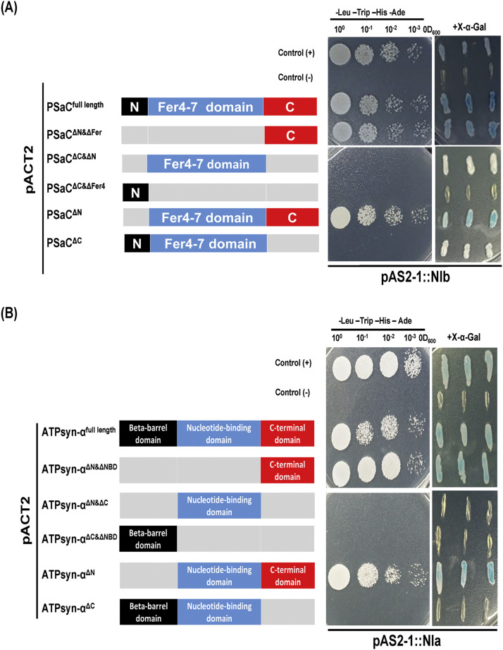
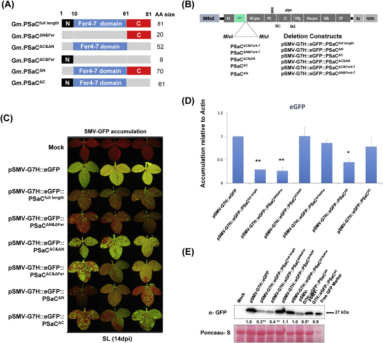
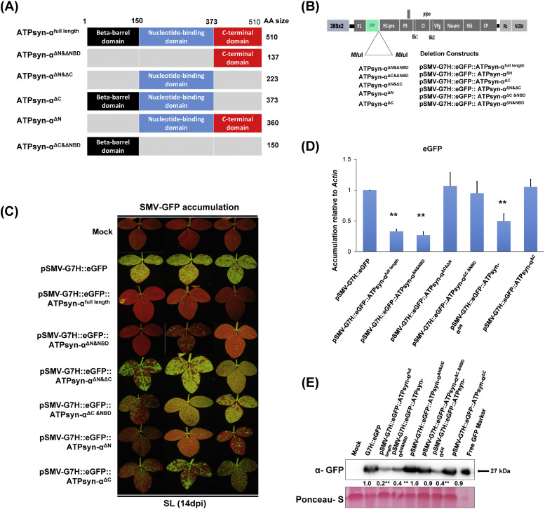
To gain a deeper understanding of the molecular mechanisms involved in viral infection and the corresponding plant resistance responses, it is essential to investigate the interactions between viral and host proteins. In the case of viral infections in plants, a significant portion of the affected gene products are closely associated with chloroplasts and photosynthesis. However, the molecular mechanisms underlying the interplay between the virus and host chloroplast proteins during replication remain poorly understood. In our previous study, we made an interesting discovery regarding soybean mosaic virus (SMV) infection in resistant and susceptible soybean cultivars. We found that the photosystem I (PSI) subunit (PSaC) and ATP synthase subunit α (ATPsyn-α) genes were up-regulated in the resistant cultivar following SMV-G7H and SMV-G5H infections compared to the susceptible cultivar. Overexpression of these two genes within the SMV-G7H genome in the susceptible cultivar Lee74 (rsv3-null) reduced SMV accumulation, whereas silencing of the PSaC and ATPsyn-α genes promoted SMV accumulation. We have also found that the PSaC and ATPsyn-α proteins are present in the chloroplast envelope, nucleus, and cytoplasm. Building on these findings, we now characterized protein-protein interactions between PSaC and ATPsyn-α with two viral proteins, NIb and NIa-Pro, respectively, of SMV. Through co-immunoprecipitation (Co-IP) experiments, we confirmed the interactions between these proteins. Moreover, when the C-terminal region of either PSaC or ATPsyn-α was overexpressed in the SMV-G7H genome, we observed a reduction in viral accumulation and systemic infection in the susceptible cultivar. Based on these results, we propose that the PSaC and ATPsyn-α genes play a modulatory role in conferring resistance to SMV infection by influencing the function of NIb and NIa-Pro-in SMV replication and movement. The identification of these photosynthesis-related genes as key players in the interplay between the virus and the host provides valuable insights for developing more targeted control strategies against SMV. Additionally, by utilizing these genes, it may be possible to genetically engineer plants with improved photosynthetic efficiency and enhanced resistance to SMV infection.

Keywords: Chloroplast-virus interplay; Plant defense; Soybean mosaic virus; Soybeans; Viral replication.


Conflict of interest statement

Declaration of Competing Interest The authors declare that they have no known competing financial interests or personal relationships that could have appeared to influence the work reported in this paper.

Figures

Fig 1
Fig. 1
Interactions between SMV viral proteins and soybean's chloroplast-related proteins. (A) Analysis of interactions between SMV proteins and GmPSaC in the yeast two-hybrid (Y2H) system. (B) Analysis of interactions between SMV proteins and GmATPsyn-α in the Y2H system. Dilutions (10°, 10−1,10−2, and 10−3) of yeast cultures at OD600 = 0.5 were spotted into -Leu -Trip -His -Ade deficient (left) or X-α-Gal -containing (right) plates and grown for 3 d at 28 °C. The symbol of "+” indicates the intensity of the interaction while “-" means no interaction. (C) Co-immunoprecipitation (Co-IP) analysis showing a direct interaction between SMV NIa-Pro-and ATPsyn-α. (D) Co-IP analysis showing a direct interaction between SMV NIb and PSaC.
Fig 2
Fig. 2
Co-expression of PSaC/ATPsyn-α with NIb or NIa-Pro-in the Nicotiana benthamiana cell. (A) Co-expression of PSaC or ATPsyn-α tagged with mCherry with NIb or NIa-Pro-tagged with a green fluorescent protein (GFP) in the N. benthamiana. The leaves were examined at 3 days post-co-infiltration, and fluorescence was assessed by confocal microscopy. (B) Pearson's coefficient of localization represents the degree of fluorescence coincidence. The intensity of eGFP fluorescence (green line) and mCherry fluorescence (red line) are on the right panels. (C) Statistical analysis was performed to compare eGFP expression levels, based on data from at least three biological replicates. The expression levels of eGFP with co-expression of mCherry::EMB1300 were used as a reference and compared using one-sided Student's t-tests. Symbols * and ** indicate a significant difference at p < 0.05 and p < 0.01, respectively.
Fig 3
Fig. 3
Analysis of SMV NIb/ NIa-Pro-interaction with GmPSaC/GmATPsyn-α deletions. (A) Schematic representation of GmPSaC and its deletion mutants used in Y2H assays are depicted in the left diagram and their interaction with NIb on the right. (B) Schematic representation of GmATPsyn-α and its deletion mutants used in Y2H assays are depicted in the left diagram and their interaction with NIa-Pro-on the right. Dilutions (10°, 10−1, 10−2, and 10−3) of yeast cultures at OD600 = 0.5 were spotted into -Leu -Trip -His -Ade deficient (left) or X-α-Gal -containing (right) plates and grown for 3 d at 28 °C. The symbol of "+” indicates the intensity of the interaction while “-" means no interaction.
Fig 4
Fig. 4
Experimental design, chimera constructions, and effect of GmPSaC full length and deletion mutants. (A) Scheme showing the protocol followed to generate deletion mutants. (B) A schematic diagram showing the genome organization of pSMV-G7H::eGFP on top and deletion constructs below. (C) Green fluorescent protein (GFP) visual levels in the systemically infected leaves (SL) of Lee74 plants. The first unifoliate leaves of 12-day-old seedlings were infected with pSMV-G7H::eGFP, pSMV-G7H::eGFP::PSaC, pSMV-G7H::eGFP::PSaCΔN&ΔFer, pSMV-G7H::eGFP::PSaCΔC&ΔN, pSMV-G7H::eGFP::PSaCΔC&ΔFer4, pSMV-G7H::eGFP::PSaCΔN and pSMV-G7H::eGFP::PSaCΔC. Fourteen days later, the SL from three plants (1, 2, and 3) were photographed under UV light. (D) Relative expression levels of GFP RNA in SL of Lee74 infected with constructs. Actin11 was used as the internal control. (E) Western protein blot for GFP levels in the systemic leaves the SL, and their quantified levels (lower panel). Ponceau S staining of RuBisCO was used on the loading control. Values are means of three biological replicates. Values were compared to that of the corresponding mock-treated plants (the bar on the left) with one-sided Student's t-tests. Symbols * and ** indicate a significant difference at p < 0.05 and p < 0.01, respectively.
Fig 5
Fig. 5
Experimental design, chimera constructions, and effect of GmATPsyn-α full length and deletion mutants. (A) Scheme showing the protocol followed to generate deletion mutants. (B) Schematic diagram showing the genome organization of pSMV-G7H::eGFP on top and deletion constructs below. (C) GFP visual levels in the systemically infected leaves (SL) of Lee74 plants. The first unifoliate leaves of 12-day-old seedlings were infected with pSMV-G7H::eGFP, pSMV-G7H::eGFP::ATPsyn-α, pSMV-G7H::eGFP::ATPsyn-αΔN&NBD, pSMV-G7H::eGFP::ATPsyn-αΔN&ΔC, pSMV-G7H::eGFP::ATPsyn-αΔC, pSMV-G7H::eGFP::ATPsyn-αΔN and pSMV-G7H::eGFP::ATPsyn-αΔC&NBD. Fourteen days later, the SL from three plants (1, 2, and 3) were photographed under UV light. (D) Relative expression levels of GFP RNA in SL of Lee74 infected with constructs. Actin11 was used as the internal control. (E) Western protein blot for GFP levels in the systemic leaves the SL, and their quantified levels (lower panel). Ponceau S staining of RuBisCO was used on the loading control. Values are means of three biological replicates. Values were compared to that of the corresponding mock-treated plants (the bar on the left) with one-sided Student's t-tests. Symbols * and ** indicate a significant difference at p < 0.05 and p < 0.01, respectively.

Similar articles

References

    1. Alazem M., Bwalya J., Pai H., Yu J., Cam H.C., Burch-Smith T., Kim K.H. Viral synergism suppresses R gene-mediated resistance by impairing downstream defense mechanisms in soybean. Plant Physiol. 2023:kiad255. - PMC - PubMed
    1. Alazem M., He M.H., Chang C.H., Cheng N., Lin N.S. Disrupting the homeostasis of high mobility group protein promotes the systemic movement of bamboo mosaic virus. Front. Plant Sci. 2020;11 - PMC - PubMed
    1. Bhattacharyya D., Chakraborty S. Chloroplast: the Trojan horse in plant–virus interaction. Mol. Plant Pathol. 2018;19:504–518. - PMC - PubMed
    1. Bobik K., Burch-Smith T.M. Chloroplast signaling within, between and beyond cells. Front. Plant Sci. 2015;6:781. - PMC - PubMed
    1. Broder Y.C., Katz S., Aronheim A. The ras recruitment system, a novel approach to the study of protein-protein interactions. Curr. Biol. 1998;8:1121–1124. - PubMed

Publication types

Substances

Supplementary concepts

LinkOut - more resources