. 2023 Oct;114(10):3972-3983.
doi: 10.1111/cas.15934. Epub 2023 Aug 22.

Promotion of squamous cell carcinoma tumorigenesis by oncogene-mediated THG-1/TSC22D4 phosphorylation

Affiliations

Promotion of squamous cell carcinoma tumorigenesis by oncogene-mediated THG-1/TSC22D4 phosphorylation

Nohara Goto et al. Cancer Sci. 2023 Oct.

Abstract

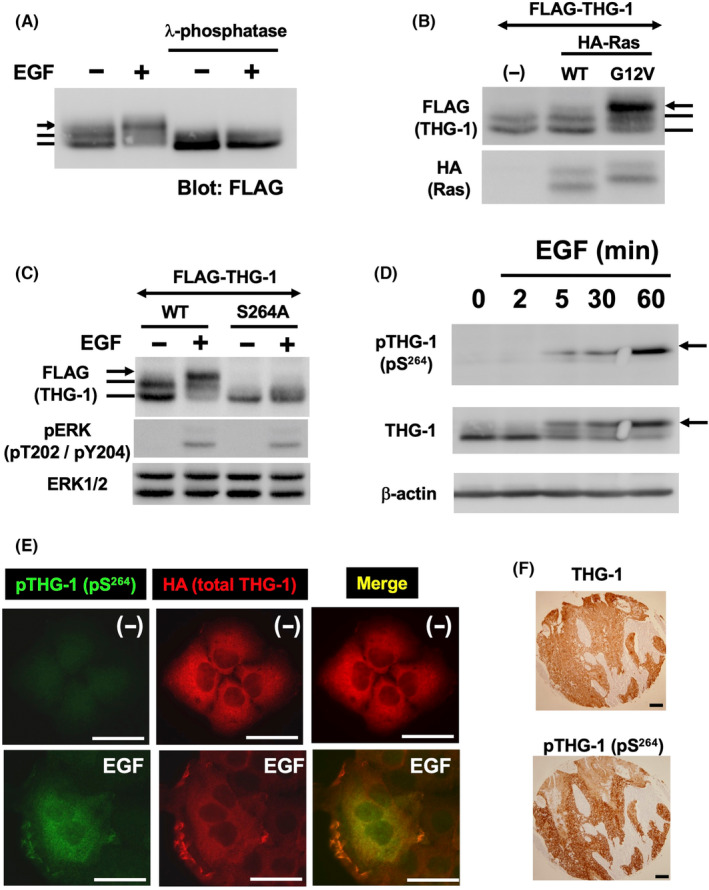
Carcinoma cells possess high proliferative and invasive potentials and exhibit a resilience against stresses, metabolic disorder, and therapeutic efforts. These properties are mainly acquired by genetic alterations including driver gene mutations. However, the detailed molecular mechanisms have not been fully elucidated. Here, we provide a novel mechanism connecting oncogenic signaling and the tumorigenic properties by a transforming growth factor-β1-stimulated clone 22 (TSC-22) family protein, THG-1 (also called as TSC22D4). THG-1 is localized at the basal layer of normal squamous epithelium and overexpressed in squamous cell carcinomas (SCCs). THG-1 knockdown suppressed SCC cell proliferation, invasiveness, and xenograft tumor formation. In contrast, THG-1 overexpression promoted the EGF-induced proliferation and stratified epithelium formation. Furthermore, THG-1 is phosphorylated by the receptor tyrosine kinase (RTK)-RAS-ERK pathway, which promoted the oncogene-mediated tumorigenesis. Moreover, THG-1 involves in the alternative splicing of CD44 variants, a regulator of invasiveness, stemness, and oxidative stress resistance under the RTK pathway. These findings highlight the pivotal roles of THG-1 as a novel effector of SCC tumorigenesis, and the detection of THG-1 phosphorylation by our established specific antibody could contribute to cancer diagnosis and therapy.

Keywords: CD44; THG-1; TSC22D4; monoclonal antibody; phosphorylation; receptor tyrosine kinase; squamous cell carcinoma.


Conflict of interest statement

The authors have no conflict of interest. Dr. Mitsuyasu Kato is an associate editor of Cancer Science.

Figures

FIGURE 1
FIGURE 1
Distribution of THG‐1 in normal stratified epithelium and squamous cell carcinomas (SCCs). (A) Immunohistochemistry of THG‐1 in normal esophagus, esophageal SCC, normal cervix uteri, and cervical SCC. (B) Immunohistochemistry of THG‐1 in normal lung and lung SCC, adenocarcinoma, large cell carcinoma, and small cell carcinoma. (C) Immunohistochemistry of THG‐1 in gastric SCC and adenocarcinoma. Scale bars: 100 μm.
FIGURE 2
FIGURE 2
Functions of THG‐1 in cell invasiveness and tumorigenicity. (A) Anti‐THG‐1, E‐Cadherin, and β‐Actin immunoblot analyses of TE13 cells expressing shRNAs targeting THG‐1 (shTHG‐1#2 and shTHG‐1#3) or control shRNA. (B) Effect of THG‐1 knockdown on cell proliferation. Error bars represent means ± SDs. (C, D) Effect of THG‐1 knockdown on cell invasion into the collagen gels in 3D culture assay. Scale bars: 50 μm. Error bars represent means ± SDs. **p < 0.01. (E, F) Effect of THG‐1 knockdown on tumor formation in NOD‐Scid mice (scale bar: 1 cm). Error bars represent means ± SDs. **p < 0.01. Immunohistochemistry of THG‐1 in xenograft tumors of TE13 cells (control, shTHG‐1#2, and shTHG‐1#3) (scale bars: 50 μm).
FIGURE 3
FIGURE 3
Effect of THG‐1 in epidermal growth factor (EGF)‐induced proliferation and stratified epithelium formation. (A) Anti‐THG‐1, FLAG, and β‐Actin immunoblot analyses of HaCaT cells expressing FLAG‐THG‐1 (clone 3 and clone 7) or control (mock). (B) Effect of THG‐1 on cellular proliferation and morphology in the presence of 50 ng/mL of EGF (in 0.1% FBS). Error bars represent means ± SDs. (C) Effect of THG‐1 on the formation of stratified epithelium on collagen gels in 3D culture assay. Morphology of stratified epithelium in control and THG‐1‐transfected HaCaT cells. Sections were stained with anti‐Ki67 antibody, and Ki67‐positive cells were counted (D). **p < 0.01. Error bars represent means ± SDs. Scale bars: 50 μm.
FIGURE 4
FIGURE 4
Phosphorylation of THG‐1 by the epidermal growth factor (EGF)‐RAS‐ERK pathway. (A) Phosphorylation of THG‐1 by EGF. HaCaT‐FLAG‐THG‐1 cells were treated with EGF. Immunoprecipitated THG‐1 was incubated with or without lambda protein phosphatase. Immunoblot analysis was performed using anti‐FLAG antibody. The arrow indicates the phosphorylated THG‐1. (B) THG‐1 is phosphorylated by constitutively active HRasG12V, but not by wild type (WT). HEK293T cells were transfected with FLAG‐THG‐1 with HRasWT and HRasG12V. Immunoblot analysis was performed using anti‐FLAG and HA antibodies. (C) S264 is an essential phosphorylation site of THG‐1. HEK293T cells were transfected with FLAG‐THG‐1 (WT and S264A). The cells were treated with 50 ng/mL of EGF, and immunoblot analysis was performed using anti‐FLAG, pERK, and ERK1/2 antibodies. (D) Using anti‐phospho‐THG‐1(pS264) mAb (clone 18), phosphorylated THG‐1 was detected by immunoblot analysis in TE13 cells. (E) The phosphorylation of THG‐1 was detected by immunofluorescence in HaCaT‐HA‐THG‐1 cells (scale bars: 20 μm) using anti‐phospho‐THG‐1(pS264) mAb (clone 21). Anti‐HA antibody detected HA‐THG‐1. (F) The phosphorylation of THG‐1 was detected by immunohistochemistry in esophageal SCC using anti‐phospho‐THG‐1(pS264) mAb (clone 21) and anti‐THG‐1 antibody. Scale bars: 100 μm.
FIGURE 5
FIGURE 5
Requirement of THG‐1 phosphorylation in Ras‐mediated tumorigenesis. (A) Anti‐phospho‐THG‐1(pS264) mAb (clone 18), FLAG, HA, and β‐Actin immunoblot analyses of HaCaT cells expressing HRasG12V, HRasG12V‐THG‐1 (WT), and HRasG12V‐THG‐1 (S264A) (B) Tumor formation in nude mice. Error bars represent means ± SDs. **p < 0.01. (C) The sections were stained with anti‐THG‐1 and Ki67 antibodies. Scale bars: 100 μm.
FIGURE 6
FIGURE 6
Regulation of epidermal growth factor (EGF)‐induced CD44 variant splicing by THG‐1. (A) Flow cytometry using anti‐Pan‐CD44 antibody conjugated with PE in TE13 cells expressing shRNAs targeting THG‐1 (shTHG‐1#2 and shTHG‐1#3) or control shRNA. (B) Schematic illustration of CD44s and CD44v. Primers to amplify CD44s and CD44v by semiquantitative RT‐PCR are indicated (arrows). (C) Semiquantitative RT‐PCR analysis of CD44s and CD44v in control and THG‐1‐knockdown cells in 10% FBS condition. (D) Semiquantitative RT‐PCR analysis of CD44s and CD44v in the presence or absence of 50 ng/mL of EGF (in 0.1% FBS) for 48 h. The indicated bands were identified as CD44v3–10, v6–10, and v8–10 by sequence analysis. (E) Cells were cultured in the presence or absence of 50 ng/mL of EGF (in 0.1% FBS) for 48 h. Flow cytometry was performed by anti‐CD44v9 (RV3) antibody. (F) The mock and CD44v3–10‐trasfected THG‐1‐knockdown cells (TE13 shTHG‐1 #2) were treated with anti‐CD44v3 (C44Mab‐6) and anti‐CD44v9 (C44Mab‐1) antibodies and analyzed using flow cytometry. (G) The sphere number (left) was counted, and the viability (right) was determined using the CellTiter‐Glo® 3D Cell Viability Assay in mock and CD44v3–10‐trasfected THG‐1‐knockdown cells. **p < 0.01. Error bars represent means ± SDs.
FIGURE 7
FIGURE 7
Regulation of CD44 variant splicing in THG‐1‐knockdown and ‐overexpressed tumors. (A) Decreased CD44v transcription in tumor sphere formation of THG‐1‐knockdown cells. (B) The xenograft tumor sections in Figure 2E were stained by anti‐CD44v9 (RV3) antibody. Scale bars: 100 μm. (C) Increased CD44v transcription in HaCaT‐HRasG12V‐THG‐1 (WT) cells compared with HRasG12V‐THG‐1 (S264A). (D) The xenograft tumor sections in Figure 5B were stained by anti‐CD44v9 (RV3) antibody. Scale bars: 100 μm.

Similar articles

Cited by

References

    1. Blanpain C, Fuchs E. Epidermal homeostasis: a balancing act of stem cells in the skin. Nat Rev Mol Cell Biol. 2009;10:207‐217. - PMC - PubMed
    1. Knoblich JA. Asymmetric cell division: recent developments and their implications for tumour biology. Nat Rev Mol Cell Biol. 2010;11:849‐860. - PMC - PubMed
    1. Alam M, Ratner D. Cutaneous squamous‐cell carcinoma. N Engl J Med. 2001;344:975‐983. - PubMed
    1. Rustgi AK, El‐Serag HB. Esophageal carcinoma. N Engl J Med. 2014;371:2499‐2509. - PubMed
    1. Bray F, Loos AH, McCarron P, et al. Trends in cervical squamous cell carcinoma incidence in 13 European countries: changing risk and the effects of screening. Cancer Epidemiol Biomarkers Prev. 2005;14:677‐686. - PubMed

Substances