. 2023 Sep 20;43(38):6479-6494.
doi: 10.1523/JNEUROSCI.0980-23.2023. Epub 2023 Aug 22.

KCNQ2/3 Gain-of-Function Variants and Cell Excitability: Differential Effects in CA1 versus L2/3 Pyramidal Neurons

Affiliations

KCNQ2/3 Gain-of-Function Variants and Cell Excitability: Differential Effects in CA1 versus L2/3 Pyramidal Neurons

Nissi Varghese et al. J Neurosci. .

Abstract

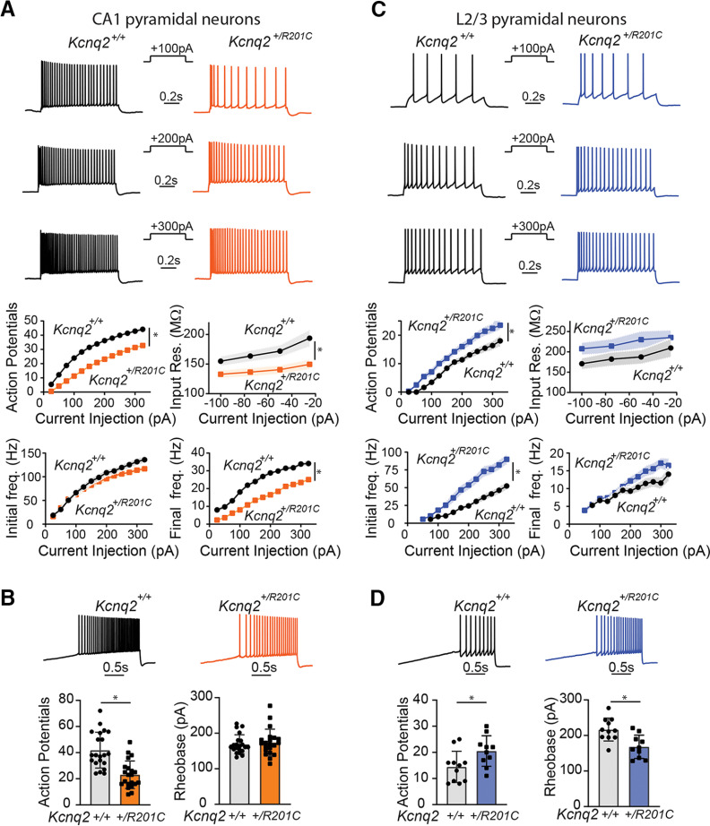
Gain-of-function (GOF) pathogenic variants in the potassium channels KCNQ2 and KCNQ3 lead to hyperexcitability disorders such as epilepsy and autism spectrum disorders. However, the underlying cellular mechanisms of how these variants impair forebrain function are unclear. Here, we show that the R201C variant in KCNQ2 has opposite effects on the excitability of two types of mouse pyramidal neurons of either sex, causing hyperexcitability in layer 2/3 (L2/3) pyramidal neurons and hypoexcitability in CA1 pyramidal neurons. Similarly, the homologous R231C variant in KCNQ3 leads to hyperexcitability in L2/3 pyramidal neurons and hypoexcitability in CA1 pyramidal neurons. However, the effects of KCNQ3 gain-of-function on excitability are specific to superficial CA1 pyramidal neurons. These findings reveal a new level of complexity in the function of KCNQ2 and KCNQ3 channels in the forebrain and provide a framework for understanding the effects of gain-of-function variants and potassium channels in the brain.SIGNIFICANCE STATEMENT KCNQ2/3 gain-of-function (GOF) variants lead to severe forms of neurodevelopmental disorders, but the mechanisms by which these channels affect neuronal activity are poorly understood. In this study, using a series of transgenic mice we demonstrate that the same KCNQ2/3 GOF variants can lead to either hyperexcitability or hypoexcitability in different types of pyramidal neurons [CA1 vs layer (L)2/3]. Additionally, we show that expression of the recurrent KCNQ2 GOF variant R201C in forebrain pyramidal neurons could lead to seizures and SUDEP. Our data suggest that the effects of KCNQ2/3 GOF variants depend on specific cell types and brain regions, possibly accounting for the diverse range of phenotypes observed in individuals with KCNQ2/3 GOF variants.

Keywords: KCNQ2; KCNQ3; gain-of-function; hippocampus; neurological disorders; potassium channels.


Figures

Figure 1.
Figure 1.
Expression of KCNQ2 R201C GOF variant in forebrain excitatory neurons lead to premature lethality, tonic-clonic seizures, and SUDEP. A, Illustration of the design of the Kcnq2+/cR201C mice and the effect of expression of Cre recombinase. B, Sequencing validation of expression of Kcnq2R201C transcript in Emx1cre::Kcnq2+/cR201C mice and survival curves of Emx1cre/+::Kcnq2+/+ (black, n = 10) and Emx1cre/+::Kcnq2+/cR201C (red, n = 16; Kaplan Meier p < 0.0001). We observed that mice expressing one copy of Kcnq2R201C die prematurely. C, Video-electrocorticography (ECoG) recordings of Kcnq2R201C GOF mice show presence of spontaneous tonic-clonic seizures, starting with behavioral arrest (i), followed by a tonic phase (ii), and culminating in a clonic phase with tonic hindlimb extension (iii). Simultaneous ECoG shows intermittent generalized spikes (i) followed by low-voltage fast activity with a direct current [DC] shift (ii) followed by rhythmic generalized spikes before seizure termination (iii). LF = left frontal, RF = right frontal, LP = left parietal, RP = right parietal. Scale bar = 100 μV and 5 s. D, Sudden unexpected death in epilepsy (SUDEP) event captured with video-EEG monitoring with brain death following a tonic clonic seizure, including (i) behavioral arrest with low-voltage fast activity, (ii) a tonic phase, and then (iii) a clonic phase with generalized spikes before seizure termination and death (horizontal scale = 12 s, 1 s in inset; vertical scale = 200 μV, 100 μV in inset). E, Global expression of KCNQ2R201C GOF variant leads to premature lethality. Left, survival curves of UBCcre-ERT2::Kcnq2+/cR201C and UBCcre-ERT2::Kcnq2+/+. We found that mice expressing one copy of Kcnq2R201C die prematurely two weeks after tamoxifen injection. Right, sanger sequencing validation of expression of Kcnq2R201C in UBCcre-ERT2::Kcnq2+/cR201C mice 12 d after tamoxifen injection.
Figure 2.
Figure 2.
KCNQ2R201C leads to hypoexcitability of CA1 pyramidal neurons and hyperexcitability of L2/3 pyramidal neurons. A, Top, Representative voltage responses in response to different depolarizing current injections (1 s) in CA1 pyramidal neurons. Bottom, Summary graphs showing the effects of one copy of Kcnq2R201C variant to CA1 pyramidal neurons action potential count, initial frequency and final frequency, and input resistance (Kcnq2+/+: n = 21, N = 9; Kcnq2+/R201C: n = 23, N = 8). B, Representative voltage responses to +120-pA/s ramp protocol from Kcnq2+/+ (black) and Kcnq2+/R201C (orange) CA1 pyramidal neurons. Summary graphs showing the effects on action potential number and rheobase (Kcnq2+/+: n = 21, N = 9; Kcnq2+/R201C: n = 21, N = 8). C, Top, Representative voltage responses in response to different depolarizing current injections (1 s) in L2/3 pyramidal neuron. Bottom, Summary graphs showing the effects of one copy of Kcnq2R201C variant to L2/3 pyramidal neurons action potential count, initial and final frequency, as well as input resistance (Kcnq2+/+: n = 11, N = 3; Kcnq2+/R201C: n = 10, N = 3). D, Representative voltage responses to +120-pA/s ramp protocol from Kcnq2+/+ (black) and Kcnq2+/R201C (blue) L2/3 pyramidal neurons. Summary graphs showing the effects on action potential number, and rheobase (Kcnq2+/+: n = 11, N = 3; Kcnq2+/R201C: n = 10, N = 3). Summary graphs show mean and SEM. * indicates p < 0.05. Detailed statistics are found in Table 2.
Figure 3.
Figure 3.
KCNQ2R201C leads to hypoexcitability of CA1 pyramidal neurons and hyperexcitability of L2/3 pyramidal neurons independent of the protocol. A, Representative voltage responses to the first wave of the four sinusoidal cycles (+100 pA, top, and +200 pA, bottom) for Kcnq2+/+ (black) and Kcnq2+/R201C mice in CA1 and L2/3 pyramidal neurons (orange and blue). B, Summary graphs of action potential count for the first and fourth wave of CA1 pyramidal neurons (Kcnq2+/+: n = 10, N = 3; Kcnq2+/R201C: n = 12, N = 4) and L2/3 pyramidal neurons (Kcnq2+/+: n = 11, N = 3; Kcnq2+/R201C: n = 10, N = 3) as well as the corresponding rheobase currents. Summary graphs show mean and SEM. * indicates p < 0.05. Detailed statistics are found in Table 2.
Figure 4.
Figure 4.
KCNQ2R201C-expressing L2/3 pyramidal neurons have greater M-current. A, Panel shows the M-current protocol and representative XE991-sensitive KCNQ-current traces in L2/3 pyramidal neurons (Kcnq2+/+: n = 8, N = 3; Kcnq2+/R201C: n = 11, N = 5). B, Summary graphs for XE991 sensitive currents, XE991 current and M-current density (deactivating current) were obtained using the traditional protocol in L2/3 pyramidal neurons of Kcnq2+/+ (black) and Kcnq2+/R201C (blue) mice. Summary graphs show mean and SEM. * indicates p < 0.05. Detailed statistics are found in Table 2.
Figure 5.
Figure 5.
Seizure activity captured with simultaneous neocortical (epidural) and hippocampal (depth) electrode recordings. Mice were implanted with bilateral frontal (epidural) and hippocampal (depth) electrodes. A, Recording from a P33 Kcnq2+/R201C mouse with frequent 1- to 2-s absence seizures (*) more prominently expressed in neocortical leads compared with hippocampal leads, associated with behavioral arrest. These are consistent with seizures in typical absence seizures, which engage neocortical more so than hippocampal circuitry. B, A recording from a P40 Kcnq2+/R201C mouse with convulsive seizure activity (filtered with a 2-Hz high pass filter to better illustrate electrographic onset) associated with repetitive tonic posturing, followed by clonic convulsive activity ∼12 s after electrographic seizure onset. Note the near-simultaneous onset of seizures in both neocortical and hippocampal leads; horizontal bar = 5 s, vertical bar = 250 μV.
Figure 6.
Figure 6.
Superficial and Deep CA1 pyramidal neurons have distinct firing properties. A, Representative traces of a voltage response to step current injections to +100 and +300 pA in superficial (top) and deep (bottom) dorsal CA1a pyramidal neurons. B, Summary graphs comparing action potential count, initial and final frequency and input resistance between deep and superficial CA1a pyramidal neurons. C, Top, Representative traces of voltage responses of deep (left) and superficial (right) CA1a pyramidal neurons to slow ramps (+120 pA/s). Bottom, Summary graphs of ramp action potential count and rheobase (Deep: n = 13, N = 7; Superficial: n = 14, N = 8). Summary graphs show mean and SEM. * indicates p < 0.05. Detailed statistics are found in Table 2.
Figure 7.
Figure 7.
KCNQ3 channels preferentially control the firing properties of Superficial CA1 pyramidal neurons. A, Representative traces of step current injections of +100, +200, and +300 pA in CA1a superficial pyramidal neurons of the hippocampus from Kcnq3+/+, constitutive Kcnq3 knock-out (Kcnq3−/−), and constitutive Kcnq3R231C (Kcnq3+/R231C) knock-in mice. Summary graphs show the action potential count, initial and final frequency between these genotypes (Kcnq3+/+: n = 27, N = 13; Kcnq3−/−: n = 16, N = 6; Kcnq3+/R231C: n = 17, N = 7). Gray filled circles indicate recordings from Kcnq3+/+ mice. B, Representative traces showing deep CA1a pyramidal neuron voltage responses in response to +100-, +200-, or +300-pA current injections from Kcnq3+/+, Kcnq3−/−, and Kcnq3+/R231C mice (Kcnq3+/+: n = 25, N = 14; Kcnq3−/−: n = 15, N = 6; Kcnq3+/R231C: n = 16, N = 8). Gray filled circles indicate recordings from Kcnq3+/+ mice. Summary graphs show the action potential count, initial and final frequency between these genotypes. Summary graphs show mean and SEM. * indicates p < 0.05. Detailed statistics are found in Table 2.
Figure 8.
Figure 8.
Expression of Kcnq2R201C dampens excitability of CA1 pyramidal neurons independent of radial layer. A, Representative traces of voltage responses to +100- and +300-pA step current injections in CA1a superficial pyramidal neurons (left) and CA1a deep pyramidal neurons (right) of the hippocampus. B, Left, Summary graphs of action potential count, initial, final frequency and input resistance of CA1a superficial pyramidal neurons. Right, summary graphs of action potential count, initial frequency, final frequency, and input resistance of CA1 deep neurons (Deep: Kcnq2+/+: n = 10, N = 5; Kcnq2+/R201C: n = 10, N = 4; Superficial: Kcnq2+/+: n = 11, N = 4; Kcnq2+/R201C: n = 13, N = 4). Summary graphs show mean and SEM. * indicates p < 0.05. Detailed statistics are found in Table 2.
Figure 9.
Figure 9.
KCNQ3R231C increases the excitability of L2/3 pyramidal neurons. A, Left, Representative traces of voltage responses to +200-pA step current injection from layer 2/3 pyramidal neurons of the somatosensory cortex. Right, Summary graph of action potential count. B, Left, Representative traces of voltage responses to ramp current protocol (+120 pA/s). Right, summary graphs of action potential count and rheobase current (Kcnq3+/+: n = 11, N = 3; Kcnq3+/R231C: n = 12, N = 3). Summary graphs show mean and SEM. * indicates p < 0.05. Detailed statistics are found in Table 2.
Figure 10.
Figure 10.
KCNQ2/3 gain-of-function increased neuronal excitability of pyramidal neurons in silico. A, Response of the L2/3 pyramidal neuron model to a +200-pA step current to the soma in case of WT (control case, black lines) and KCNQ2/3 GOF (red lines). In this model, KCNQ2/3 GOF changes only occur at soma and AIS. Top panel, Transmembrane voltage at the soma in response to the current step. Bottom panel, Somatic M-current density in response to the current step. B, Action potential-to-current curve estimated for the neuron model in the control WT case (black line) and KCNQ2/3 GOF. KCNQ2/3 GOF is alternatively paired with increased conductance gmax (uniform values) at the soma (red line), soma+AIS (blue line), soma+apical dendrite (green line), or soma+AIS+apical dendrite (gray line). C, Response of the L2/3 pyramidal neuron model to a +200-pA step current to the soma in the control WT case (black lines) and KCNQ2/3 GOF (blue lines). In this model, KCNQ2/3 GOF occurs at soma, AIS, and apical dendrite, with nonuniform M-current distribution, i.e., M-current density at the AIS is higher than the value at the soma and apical dendrite (values as in panel D). Top panel, Transmembrane voltage at the soma in response to the current step. Bottom panel, Somatic M-current density in response to the current step. D, Action potential-to-current curve estimated for the neuron model in the control WT case (black line) and KCNQ2/3 GOF with increased conductance gmax of the M-channels at soma, AIS, and apical dendrite. Both uniform assignments (gray line) and nonuniform assignments (red line) to the value of gmax across compartments are depicted. Nonuniform indicates that the M-current density at the AIS is higher than the value at the soma and apical dendrites (i.e., 3× vs 1.5× the value used in control case). In all simulations, step currents started at t = 500 ms and lasted 1000 ms.

References

    1. Allen NM, Weckhuysen S, Gorman K, King MD, Lerche H (2020) Genetic potassium channel-associated epilepsies: clinical review of the KV family. Eur J Paediatr Neurol 24:105–116. 10.1016/j.ejpn.2019.12.002 - DOI - PubMed
    1. Battefeld A, Tran BT, Gavrilis J, Cooper EC, Kole MH (2014) Heteromeric Kv7.2/7.3 channels differentially regulate action potential initiation and conduction in neocortical myelinated axons. J Neurosci 34:3719–3732. 10.1523/JNEUROSCI.4206-13.2014 - DOI - PMC - PubMed
    1. Boets S, Johannesen KM, Destree A, Manti F, Ramantani G, Lesca G, Vercueil L, Koenig MK, Striano P, Møller RS, Cooper E, Weckhuysen S (2022) Adult phenotype of KCNQ2 encephalopathy. J Med Genet 59:528–535. 10.1136/jmedgenet-2020-107449 - DOI - PubMed
    1. Carnevale NT, Hines ML (2006) The NEURON book. Cambridge: Cambridge University Press.
    1. Cembrowski MS, Wang L, Sugino K, Shields BC, Spruston N (2016) Hipposeq: a comprehensive RNA-seq database of gene expression in hippocampal principal neurons. Elife 5:e14997. 10.7554/eLife.14997 - DOI - PMC - PubMed

Publication types