. 2023 Sep 22;51(17):8934-8956.
doi: 10.1093/nar/gkad597.

Evaluation of the determinants for improved pluripotency induction and maintenance by engineered SOX17

Affiliations

Evaluation of the determinants for improved pluripotency induction and maintenance by engineered SOX17

Haoqing Hu et al. Nucleic Acids Res. .

Abstract

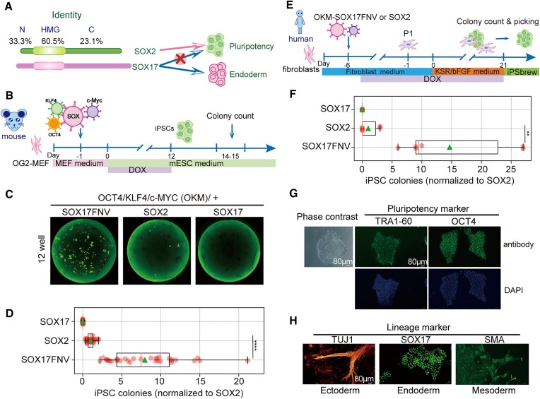
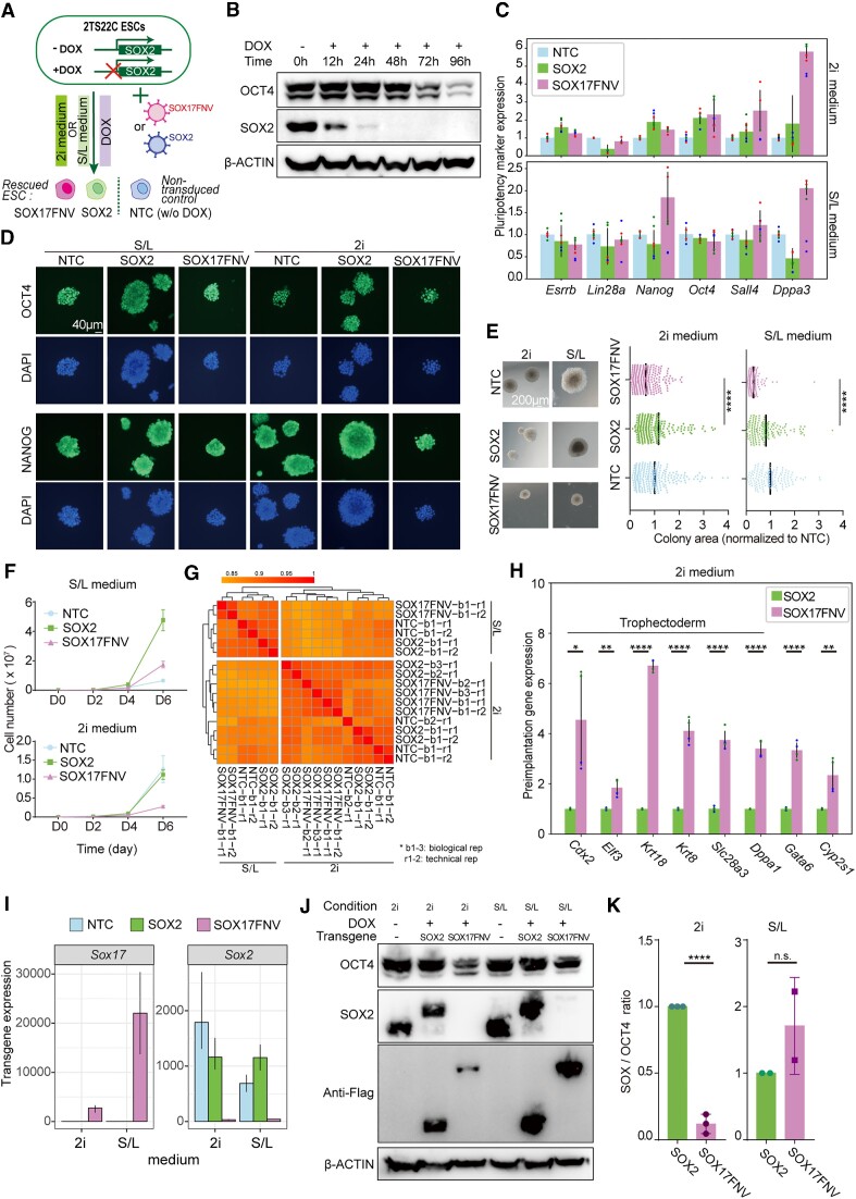
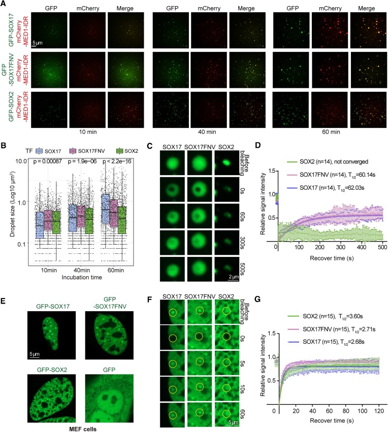
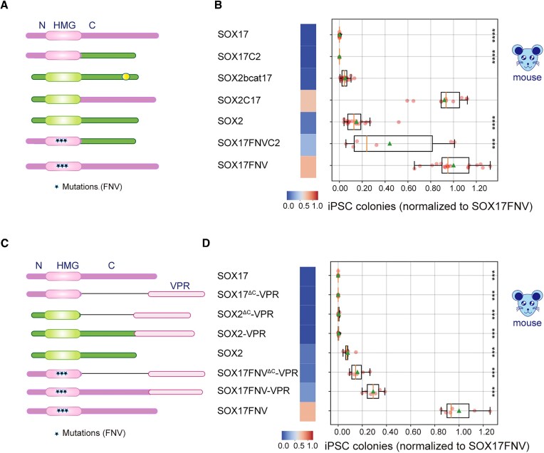
An engineered SOX17 variant with point mutations within its DNA binding domain termed SOX17FNV is a more potent pluripotency inducer than SOX2, yet the underlying mechanism remains unclear. Although wild-type SOX17 was incapable of inducing pluripotency, SOX17FNV outperformed SOX2 in mouse and human pluripotency reprogramming. In embryonic stem cells, SOX17FNV could replace SOX2 to maintain pluripotency despite considerable sequence differences and upregulated genes expressed in cleavage-stage embryos. Mechanistically, SOX17FNV co-bound OCT4 more cooperatively than SOX2 in the context of the canonical SoxOct DNA element. SOX2, SOX17, and SOX17FNV were all able to bind nucleosome core particles in vitro, which is a prerequisite for pioneer transcription factors. Experiments using purified proteins and in cellular contexts showed that SOX17 variants phase-separated more efficiently than SOX2, suggesting an enhanced ability to self-organise. Systematic deletion analyses showed that the N-terminus of SOX17FNV was dispensable for its reprogramming activity. However, the C-terminus encodes essential domains indicating multivalent interactions that drive transactivation and reprogramming. We defined a minimal SOX17FNV (miniSOX) that can support reprogramming with high activity, reducing the payload of reprogramming cassettes. This study uncovers the mechanisms behind SOX17FNV-induced pluripotency and establishes engineered SOX factors as powerful cell engineering tools.


Figures

Graphical Abstract
Graphical Abstract
Figure 1.
Figure 1.
The SOX17 triple-mutant SOX17FNV is a high-performance pluripotency inducer in mice and humans. (A) Sequence identity and functional differences between mouse SOX2 and SOX17. Identity percentages were calculated with the EMBOSS Water tool of EMBL-EBI (https://wwwdev.ebi.ac.uk/Tools/jdispatcher/psa/emboss_water). (B) Schematic of inducible mouse iPSC reprogramming. (C) Representative whole well scanning from a 12-well plate at reprogramming day 14 using Alexa Fluor 488 channel and (D) quantification of mouse reprogramming by counting the GFP positive colony numbers. (E) Scheme of human iPSC reprogramming from adult human dermal fibroblasts (HDFa). (F) Reprogramming efficiency of human reprogramming at day 21. Colony counts were done according to colony morphology. For (D, F), a two-tailed unpaired t-test was used. P > 0.05 as N.A, P < 0.05 as *, P < 0.01 as **, P < 0.001 as ***, P < 0.0001 as ****. (D, F) Efficiency is calculated by normalizing colony numbers to that of the SOX2 condition in given experiments (n ≥ 3 independent experiments each with technical replicates, the orange line represents the median, and the green triangle represents the means). (G) ICC for pluripotency marker expressed by human iPSC colonies. Scale bar, 80 μm. (H) Lineage markers labeling the three germ layers of the spontaneously differentiated human iPSC colonies. Scale bar, 80 μm.
Figure 2.
Figure 2.
SOX17FNV can functionally replace SOX2 in embryonic stem cells. (A) Pluripotency maintenance assay scheme using the 2TS22C ES cell line and constitutive lentiviruses. (B) Western blot for endogenous SOX2, OCT4, and β-ACTIN using non-transduced 2TS22C ES cells (NTC) treated with DOX at indicated time periods. (C) RT-qPCR of pluripotency markers of 2TS22C cells at passage 4 (n = 3 independent experiments, mean ± SD). (D) ICC for pluripotency markers of rescued 2TS22C ES cells at passage 19. Scale bar, 40 μm. (E) Left: Colony morphology of 2TS22C ESCs. Scale bar, 200 μm. Right: colony area after 6 days (2i) or 7 days (S/L) of growth after seeding (n = 246 colonies for 2i, n = 142 for S/L, mean ± SEM). (F) Proliferation rate curves of indicated 2TS22C lines (n = 4 independent cells lines for 2i, n = 3 for S/L, mean ± SEM). (G) Correlation heat map of indicated 2TS22C cell lines with global genes expression matrix as input (‘b’ for biological replicate, ‘r’ for technical replicate). (H) RT-qPCR for preimplantation genes (n = 2 independent experiments, mean ± SD). (I) Expression of transgenes from RNA-seq data. (J, K) Western blot and quantification of SOX transgenes and OCT4 (n = 3 independent cell lines for 2i, n = 2 for S/L, mean ± SD). Blots were from passage 14 cell lysates. For (E, H, K), a two-tailed unpaired t-test was performed using GraphPad. P > 0.05 as n.s., P < 0.05 as *, P < 0.01 as **, P < 0.001 as ***, P < 0.0001 as ****.
Figure 3.
Figure 3.
SOX17FNV induces a comparable chromatin opening status as SOX2 but interacts more efficiently with OCT4. (A) Heatmaps for ATAC-seq signals around common or unique ATAC-seq peaks defined by MAnorm. (B) Fraction of peaks in the three categories with motifs (Sox monomer, canonical SoxOct, compressed SoxOct). (C) Nucleosome occupancy signals derived from NucleoATAC around the specified motifs (Sox monomer and canonical SoxOct) and (D) filtered for binding by SOX2 or SOX17EK as determined in a previous ChIP-seq analysis (23). (E) Quantification of the binding affinity of SOX2, SOX17 and SOX17FNV to 1 nM of a SOX consensus motif using bacterially purified HMG domains (n = 3 independent experiments). (F) Nucleosome binding EMSA using the same batch of purified HMG proteins and a sequence derived from the LIN28B locus. Probe concentration range: 0, 10, 20, 40, 60, 120 nM for SOX2 and SOX17; 0, 60, 120, 240, 480 nM for SOX17FNV. Hollow triangles represent supershifted free DNA and solid triangles for supershifted nucleosomes. (G) Whole-cell extract EMSA scheme. (H) Whole-cell extract EMSA of SOX17FNV, SOX17 and SOX2 binding in the absence or presence of OCT4 to 280nM Cy5 labeled SoxOct canonical or compressed motifs using HEK293T cell lysates. ‘*’ marks a SOX17FNV/OCT4 heterodimers, ‘#’ a SOX2/OCT4 heterodimers, and ‘>’ an OCT4 monomer. (I) Table summarising the interactions between SOX variants and OCT4 on composite DNA elements.
Figure 4.
Figure 4.
SOX17FNV shows enhanced capacities to dynamically self-organise. (A) Representative images of droplet formation of GFP-SOX2, GFP-SOX17, and GFP-SOX17FNV at 900 nM mixed with 2.5 μM mCherry-MED1-IDR in binding buffer with 125 mM NaCl and 10% PEG8000. (B) Quantification of droplet sizes. One-way ANOVA method was used to determine the statistical significance. (C) Representative images and (D) quantification of FRAP recovery of phase-separated condensates. 14 different bleaching areas were included for the analysis. (E) Representative images of nuclei expressing GFP-tagged SOX variants in MEF cells. (F) Representative enlarged images before and after photobleaching and (G) quantification of FRAP recovery of GFP-SOX2, GFP-SOX17 and GFP-SOX17FNV. 15 different bleaching areas were included for the analysis.
Figure 5.
Figure 5.
The outperformance of SOX17FNV relies on its potent C-terminus. (A, B) iPSC reprogramming of C-terminal swap constructs. (C, D) iPSC reprogramming of chimeric constructs with VPR domains. The designs of constructs were shown schematically (A, C), and reprogramming efficiency is summarised using a heat map and boxplots (B, D). Efficiency is calculated by normalizing colony numbers of other conditions to that of the (B, D) SOX17FNV condition (n ≥ 3 independent experiments, the orange line represents the median, and the green triangle represents the means. See also Supplementary Figure S5 (B, D) for the adjusted P value of each variant. ‘*’ represents the significance level for each variant to SOX17FNV condition.
Figure 6.
Figure 6.
A miniaturised SOX17FNV (miniSOX) potently drives reprogramming. (A) Disorder analysis of SOX2 and SOX17FNV sequences using AlphaFold2 pLDDT scores (orange) and Metapredict scores (blue) (https://github.com/idptools/metapredict). The HMG box regions were marked, and the predicted disordered regions were shaded with red. (B) AlphaFold prediction of the SOX17 protein structure. Model confidence is color coded. (C, D) Mouse iPSC reprogramming using indicated domain deletions of SOX17FNV. Constructs are schematised in (C) and reprogramming efficiency is shown as a heat map and boxplot in (D). Reprogramming efficiencies are normalized to the SOX17FNV condition for each replicate (n ≥ 3 independent experiments). See also Supplementary Figure S5(c) for the adjusted P value of each variant. The miniSOX is highlighted with a green box. (E) RT-qPCR of pluripotency marker (top) and preimplantation gene expressions of 2TS22C cells in 2i medium (n = 2 independent experiments, mean ± SD). (F) Quantification of FRAP recovery of phase-separated condensates. 14 different bleaching areas were included for analysis. The SOX17FNV and SOX17 data were adopted from Figure 4C for comparison. (G) Human iPSC reprogramming efficiency calculated by normalizing colony numbers of other conditions to that of the SOX17FNV condition (n = 3 independent experiments each including technical replicates). Colony counts were based on colony morphology. (H) ICC for pluripotency markers of human iPSC stable cell line generated with miniSOX (left) and lineage markers labeling the three germ layers of the spontaneously differentiated human iPSC colonies generated with miniSOX (right).

Similar articles

Cited by

References

    1. Takahashi K., Yamanaka S.. Induction of pluripotent stem cells from mouse embryonic and adult fibroblast cultures by defined factors. Cell. 2006; 126:663–676. - PubMed
    1. Thier M., Wörsdörfer P., Lakes Y.B., Gorris R., Herms S., Opitz T., Seiferling D., Quandel T., Hoffmann P., Nöthen M.M.et al. .. Direct conversion of fibroblasts into stably expandable neural stem cells. Cell Stem Cell. 2012; 10:473–479. - PubMed
    1. Thier M.C., Hommerding O., Panten J., Pinna R., García-González D., Berger T., Wörsdörfer P., Assenov Y., Scognamiglio R., Przybylla A.et al. .. Identification of embryonic neural plate border stem cells and their generation by direct reprogramming from adult human blood cells. Cell Stem Cell. 2019; 24:166–182. - PubMed
    1. Nakagawa M., Koyanagi M., Tanabe K., Takahashi K., Ichisaka T., Aoi T., Okita K., Mochiduki Y., Takizawa N., Yamanaka S.. Generation of induced pluripotent stem cells without Myc from mouse and human fibroblasts. Nat. Biotechnol. 2008; 26:101–106. - PubMed
    1. An Z., Liu P., Zheng J., Si C., Li T., Chen Y., Ma T., Zhang M.Q., Zhou Q., Ding S.. Sox2 and Klf4 as the functional core in pluripotency induction without exogenous Oct4. Cell Rep. 2019; 29:1986–2000. - PubMed

Publication types

MeSH terms