. 2023 Aug 22;14(1):5083.
doi: 10.1038/s41467-023-40794-w.

A ferroptosis-targeting ceria anchored halloysite as orally drug delivery system for radiation colitis therapy

Affiliations

A ferroptosis-targeting ceria anchored halloysite as orally drug delivery system for radiation colitis therapy

Yue Feng et al. Nat Commun. .

Abstract

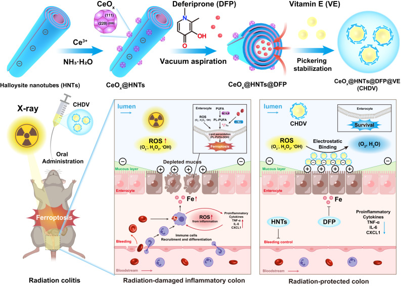
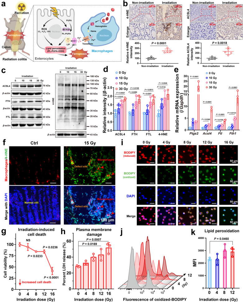
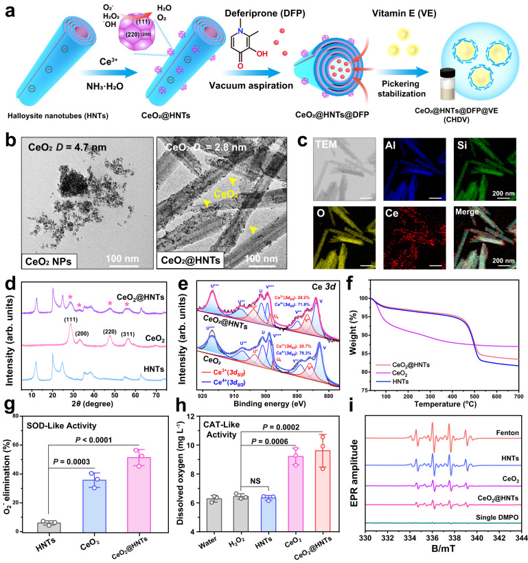
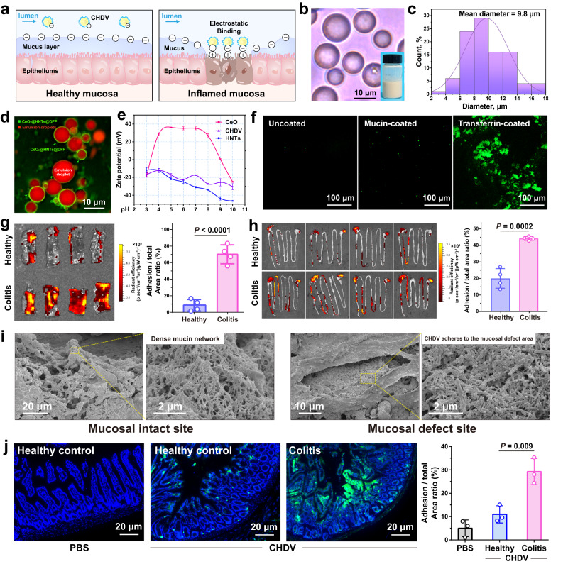
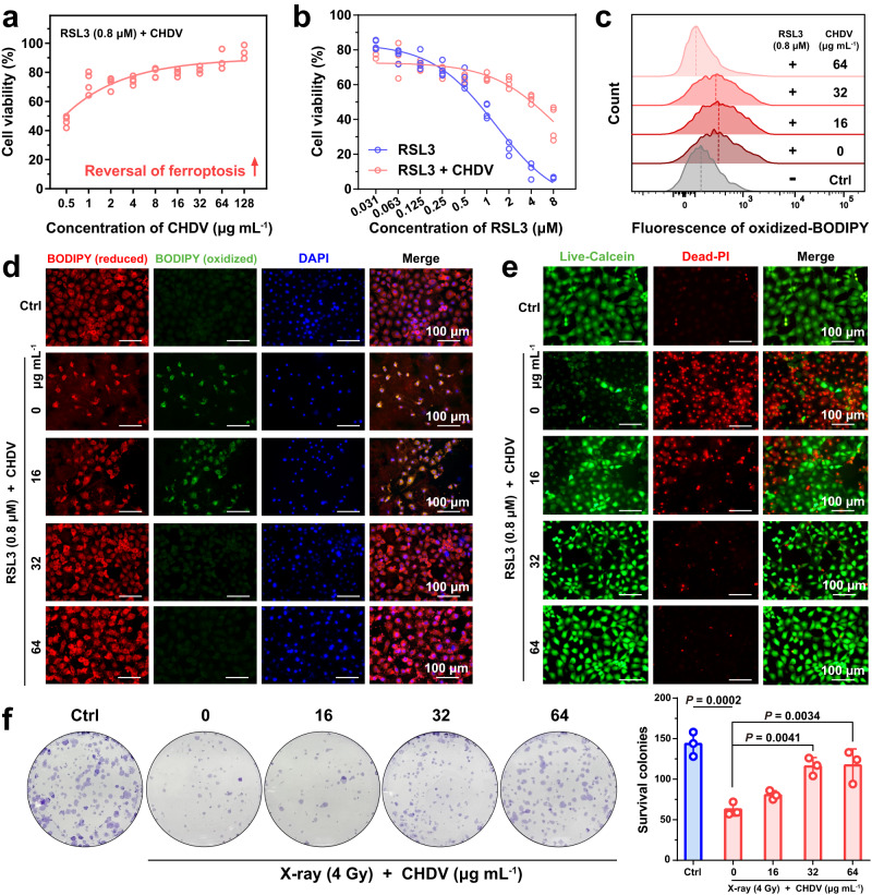
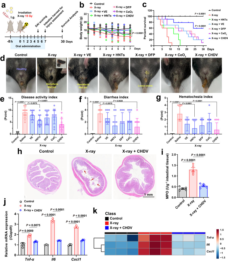
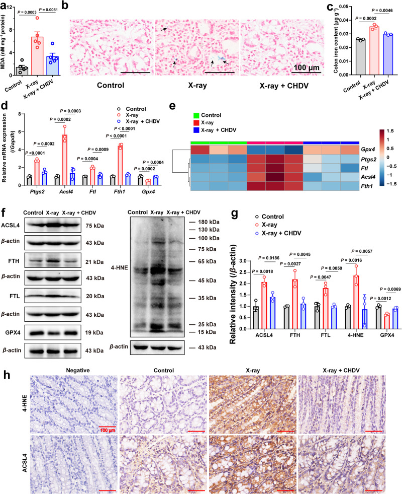
Radiation colitis is the leading cause of diarrhea and hematochezia in pelvic radiotherapy patients. This work advances the pathogenesis of radiation colitis from the perspective of ferroptosis. An oral Pickering emulsion is stabilized with halloysite clay nanotubes to alleviate radiation colitis by inhibiting ferroptosis. Ceria nanozyme grown in situ on nanotubes can scavenge reactive oxygen species, and deferiprone was loaded into the lumen of nanotubes to relieve iron stress. These two strategies effectively inhibit lipid peroxidation and rescue ferroptosis in the intestinal microenvironment. The clay nanotubes play a critical role as either a medicine to alleviate colitis, a nanocarrier that targets the inflamed colon by electrostatic adsorption, or an interfacial stabilizer for emulsions. This ferroptosis-based strategy was effective in vitro and in vivo, providing a prospective candidate for radiotherapy protection via rational regulation of specific oxidative stress.


Conflict of interest statement

The authors declare no competing interests.

Figures

Fig. 1
Fig. 1. Schematic illustration of the synthetic method and therapeutic mechanisms of CHDV Pickering emulsion.
As an oral nanoplatform, CHDV Pickering emulsion can treat radiation colitis via scavenging ROS and relieving ferroptosis.
Fig. 2
Fig. 2. Ferroptosis occurs in radiation colitis.
a Schematic showing the ferroptosis-related pathways under radiation colitis. b Representative immunohistochemical staining of 4-HNE and ACSL4 of colonic tissue samples from patients with radiation colitis, and their normalized expression intensity (relative to nonirradiation), n = 5. Scale = 200 μm (top panels) and 50 μm (bottom panels). P values were calculated using a two-tailed unpaired Students t test. c Western blotting analysis of ferroptosis-related protein expression in colon tissue of mice. d Normalized protein expression (relative to β-actin) levels, n = 3. e The ferroptosis-related gene expressions of Ptgs2, Acsl4, Ftl, and Fth1 in colon tissue, n = 3. f Immunofluorescence images (F4/80, 4-HNE, DAPI) of colon tissue. Scale bars, 100 μm. g Cell viability (n = 3), and h lactate dehydrogenase (LDH) release after incremental dose irradiation exposure (n = 3). i Fluorescence images of reduced (red) and oxidized (green) C11-BODIPY (scale bars, 50 μm), j representative flow cytometry histogram of oxidized C11-BODIPY (10,000 cells per tube were collected), and k their mean fluorescence intensity quantification (n = 3). Error bars are presented as mean ± standard deviation (SD). The data were analyzed by one-way ANOVA with Tukey’s post hoc test. The experiments for (b, c, f, i, and j) were repeated three times independently with similar results. Source data are provided as a Source Data file.
Fig. 3
Fig. 3. Synthesis and characterization of CeO2@HNTs.
a Schematic illustration of CHDV Pickering emulsion. b TEM images of CeO2 and CeO2@HNTs, scale bars, 100 nm. c TEM image and elemental mapping of CeO2@HNTs, scale bars, 200 nm. d XRD patterns, e Ce 3d XPS spectra, and f TGA cruces of HNTs, CeO2, and CeO2@HNTs. gi Enzyme-like activity tests of HNTs, CeO2, and CeO2@HNTs, the amounts of materials were normalized by Ce content (100 µg mL−1). g O2- elimination rate by SOD-like activity, h oxygen generation from H2O2 catalyzed by CAT-like activity, and i EPR spectra of the •OH scavenging ability of materials using DMPO as a spin trap agent. Error bars are presented as means ± SD, n = 3 independent repeats. The data were analyzed by one-way ANOVA with Tukey’s post hoc test. NS means no significant difference. The experiments for (b, c) were repeated three times independently with similar results. Source data are provided as a Source Data file.
Fig. 4
Fig. 4. CHDV Pickering emulsion preferentially adheres to inflamed mucosa.
a Schematic diagram of healthy and inflamed mucosa. The latter is characterized by mucosal defects and the accumulation of positively charged proteins. b The microscope images (scale bars, 10 μm), c corresponding particle size distribution statistics, and d fluorescence micrographs of CHDV, scale bars, 10 μm. e Zeta potentials of HNTs, CeO2, and CHDV at different solution pH (n = 3). f CHDV was incubated with uncoated, mucin-coated (simulating healthy epithelium) or transferrin-coated (simulating inflamed epithelium) surfaces. Scale bars, 100 μm. g The distal colon of mice with radiation colitis and healthy control was incubated ex vivo with CHDV, and fluorescence was quantified by an IVIS imaging system (n = 4). h The mice were sacrificed 8 h after administration, and the intestines were dissected and imaged using an IVIS imaging system. The fluorescence images above were quantified by ImageJ software, and the percentage of CHDV retained in the intestine versus the whole intestine was quantified (n = 4). i SEM images of colitis mice mucosa after treatment with CHDV. j The fluorescent images of healthy and colitis mice colon sections after treatment with orally administered CHDV (scale bars, 20 μm), and the percentage of retained material versus the colon was quantified (n = 3). Error bars are presented as mean ± SD. P values were calculated using a two-tailed unpaired Student t test. The experiments for (b, d, f, i, j) were repeated three times independently with similar results. The experiments for (g, h) were repeated four times independently with similar results. Source data are provided as a Source Data file.
Fig. 5
Fig. 5. In vitro ferroptosis reversal and radiation protection.
a CDHV reverses RSL3 (0.8 μM)-induced ferroptosis in intestinal epithelial cells (n = 4). b CHDV (64 mg mL−1) reversed various degrees of ferroptosis induced by gradient concentrations of RSL3 (n = 4). c The fluorescence intensity of ferroptosis-dependent C11-BODIPY oxidation decreased with increasing CHDV (10,000 cells per tube were collected). d The fluorescence images and e Calcein-AM/PI staining of cells after rescue by CHDV, scale bars, 100 μm. f Crystal violet staining and quantification of the surviving colonies irradiated with 4 Gy X-ray and different doses of CHDV treatment (n = 3). Error bars are presented as mean ± SD. The data were analyzed by one-way ANOVA with Tukey’s post hoc test. The experiments for (df) were repeated three times independently with similar results. Source data are provided as a Source Data file.
Fig. 6
Fig. 6. Radioprotection of CHDV alleviates radiation colitis.
a Schematic illustration for in vivo radioprotection evaluation. b Body weight of mice (n = 6). c Survival curves at day 30 (n = 10), the data were analyzed by Log-rank test. P < 0.0001 vs the X-ray group. d Representative photographs of the wet tail and hematochezia on day 7 in various groups. e DAI of radiation colitis, f diarrhea index, and g hematochezia index in each group (n = 10). h Representative photographs of H&E staining of colon tissue after 7 days of treatment. Scale bars, 1 mm. The experiment was repeated six times independently with similar results. i The activity of MPO in colon tissue (n = 5). j The gene expressions of Cxcl1, Tnf-α, and Il6 in colon tissue were detected by the q-PCR assay (n = 3), and k they were quantified and visualized by heatmap. The data are represented as mean ± SD. The data were analyzed by one-way ANOVA with Tukey’s post hoc test. Source data are provided as a Source Data file.
Fig. 7
Fig. 7. CHDV protects against irradiation-induced colon injury by inhibiting ferroptosis.
a MDA levels in colon tissue (n = 5). b Representative images of colon sections detected by Perls Prussian blue staining. The blue spots (the black arrows) indicate the iron deposits in colon tissue. Scale bars, 50 μm. The experiment was repeated three times independently with similar results. c Histogram of colonic iron quantification analysis (n = 3). d The gene expressions of ferroptosis pivotal genes such as Ptgs2, Acsl4, Ftl, Fth1, and Gpx4 in colon tissue (n = 3), and e they were quantified and visualized by heatmap. f, g The protein expressions of ACSL4, FTL, FTH, 4-HNE, and GPX4 in colon tissue were determined by western blotting analysis (n = 3). h Immunohistochemistry of colon sections labeled with antibodies of 4-HNE (top row) and ACSL4 (bottom row) (n = 3). Scale bars, 100 μm. The data are represented as mean ± SD. The data were analyzed by one-way ANOVA with Tukey’s post hoc test. The experiments for (b, f, h) were repeated three times independently with similar results. Source data are provided as a Source Data file.

References

    1. Hauer-Jensen M, Denham JW, Andreyev HJN. Radiation enteropathy-pathogenesis, treatment and prevention. Nat. Rev. Gastroenterol. Hepatol. 2014;11:470–479. - PMC - PubMed
    1. Kumagai T, Rahman F, Smith AM. The microbiome and radiation induced-bowel injury: evidence for potential mechanistic role in disease pathogenesis. Nutrients. 2018;10:1405. - PMC - PubMed
    1. Lei G, et al. Ferroptosis, radiotherapy, and combination therapeutic strategies. Protein Cell. 2021;12:836–857. - PMC - PubMed
    1. Stockwell BR. Ferroptosis turns 10: Emerging mechanisms, physiological functions, and therapeutic applications. Cell. 2022;185:2401–2421. - PMC - PubMed
    1. Ji Q, et al. ACSL4 is essential for radiation-induced intestinal injury by initiating ferroptosis. Cell Death Discov. 2022;8:1–9. - PMC - PubMed

Publication types