. 2023 Aug 22;14(1):5092.
doi: 10.1038/s41467-023-40896-5.

Convergent somatic evolution commences in utero in a germline ribosomopathy

Affiliations

Convergent somatic evolution commences in utero in a germline ribosomopathy

Heather E Machado et al. Nat Commun. .

Abstract

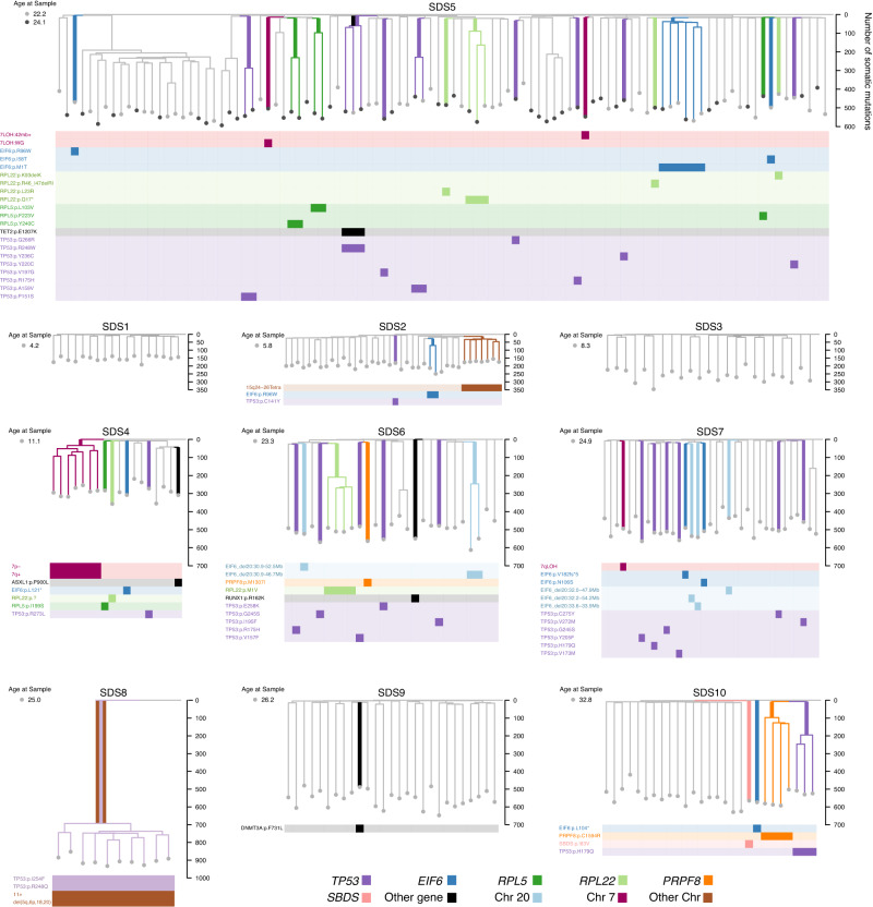
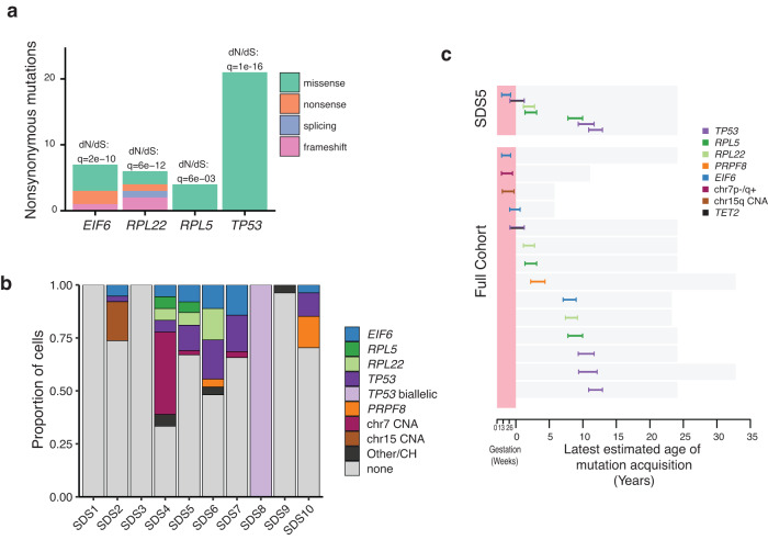
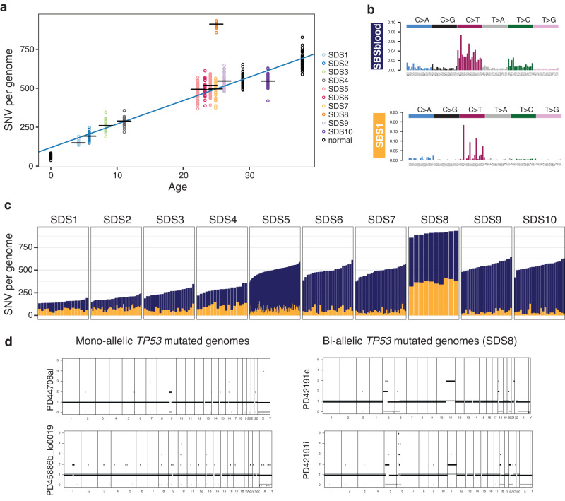
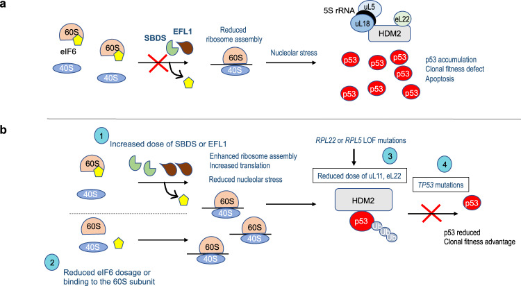
Clonal tracking of cells using somatic mutations permits exploration of clonal dynamics in human disease. Here, we perform whole genome sequencing of 323 haematopoietic colonies from 10 individuals with the inherited ribosomopathy Shwachman-Diamond syndrome to reconstruct haematopoietic phylogenies. In ~30% of colonies, we identify mutually exclusive mutations in TP53, EIF6, RPL5, RPL22, PRPF8, plus chromosome 7 and 15 aberrations that increase SBDS and EFL1 gene dosage, respectively. Target gene mutations commence in utero, resulting in a profusion of clonal expansions, with only a few haematopoietic stem cell lineages (mean 8, range 1-24) contributing ~50% of haematopoietic colonies across 8 individuals (range 4-100% clonality) by young adulthood. Rapid clonal expansion during disease transformation is associated with biallelic TP53 mutations and increased mutation burden. Our study highlights how convergent somatic mutation of the p53-dependent nucleolar surveillance pathway offsets the deleterious effects of germline ribosomopathy but increases opportunity for TP53-mutated cancer evolution.


Conflict of interest statement

A.J.W. and S.T. are consultants for SDS Therapeutics. P.J.C. is a cofounder and shareholder of FL86 Inc. The remaining authors declare no competing interests.

Figures

Fig. 1
Fig. 1. Study design and cohort.
a Schematic of experimental design. Single haematopoietic stem and progenitor cells (HSPC) and mononuclear cells (MNCs) from peripheral blood or bone marrow from individuals with SDS were expanded into colonies in vitro and each colony underwent whole-genome sequencing (WGS). Somatic mutations were used to reconstruct haematopoietic phylogenies. The timing of acquisition, clonal dynamics and functional consequences were investigated for driver mutations associated with SDS. Inkscape. b Age at sampling and SBDS genotype for each individual with SDS. The two bi-coloured columns represent the two parental alleles, with all individuals having biallelic germline mutations in SBDS. Highlighted samples (SDS2, SDS7, SDS9 and SDS10) were measured for frequency of CD34+ HSPCs, shown in (c). N the number of haematopoietic colonies analysed per individual. c Frequency of CD34+ HSPCs in bone marrow samples (expressed as a % of total viable MNCs) was analysed by flow cytometry in four of the individuals with SDS and three healthy individuals (black). Source data are provided as a Source Data file.
Fig. 2
Fig. 2. Recurrent mutations across SDS haematopoietic phylogenies.
Phylogenetic trees of haematopoietic colonies for ten individuals with SDS. Each individual had between 10–100 colonies sequenced and included for phylogenetic analysis. Branches with somatic mutations in driver genes previously reported and/or under positive selection in this study are coloured. Branches with known driver mutations of clonal haematopoiesis are shown in black, and those associated with SDS in other colours. The branch harbouring the driver mutation is shown with a thicker coloured line. The Y-axis shows the total number of somatic mutations including driver mutations. Rows beneath phylogenetic trees show the specific driver mutations, with colonies harbouring that mutation more densely coloured. Of note, no known driver mutations were detected amongst the somatic mutations found in SDS1 and SDS3. *SDS8 was diagnosed with transformation to myelodysplasia with trilineage dysplasia 1 month before sampling. All SDS8 colonies had a complex karyotype with many chromosomal (Chr) copy number aberrations (CNA). CNAs confidently shared across SDS8 colonies are shown on the tree.
Fig. 3
Fig. 3. Detecting positive selection and timing of mutation acquisition.
a Number of nonsynonymous mutations in the four genes under significant positive selection (ratio of normalised nonsynonymous mutations (dN) to normalised synonymous mutations (dS) dN:dS > 1, q < 0.01). b Proportion of cells per individual carrying driver mutations classified by gene and chromosomal abnormality. CH genes associated with clonal haematopoiesis, CNA copy number alteration. c Timing of division of the most recent common ancestor (MRCA) of clonal expansions (clade comprising 2 or more colonies) harbouring driver mutations. This gives the latest time point (together with 95% credibility interval) by which the driver mutation was acquired as represented by the inferred timing of the end of the shared branch, but it is possible that the driver mutation occurred at any time along the branch that harbours the driver mutation. The top panel illustrates the diversity of timings within a single individual (SDS5) and the bottom panel shows the timing of all the identified driver based expansions in the full cohort (including SDS5) with the exception of SDS8. A total of 14 branches harbouring driver mutations from the cohort are timed for their latest age of acquisition. The bars span the 95% credibility interval of the MRCAs. Source data are provided as a Source Data file.
Fig. 4
Fig. 4. Mutation burden, mutational processes and biallelic TP53 variants.
a Mutation burden (number of SNVs) as a function of age for ten individuals with SDS. Each circle represents one colony genome, with the black horizontal bars representing the median burden per individual. Two timepoints from SDS5 are shown at different ages. Circles coloured black (normal) representing mutation burdens from three haematopoietically healthy (non-SDS) individuals (published data) are shown for comparison. The blue line represents the regression line through the colonies from individuals with SDS. b Trinucleotide context of somatic mutations. The two mutational signatures were identified across all genomes. SBS1, is characterised by spontaneous deamination of cytosines, and the second mutational signature, termed SBSBlood, , represents mutations typical of endogenous mutations in HSCs. c Number of SNVs attributable to the mutational signatures SBS1 (green) and SBSblood (blue) across each colony from each individual with SDS. Each bar represents the genome from one colony. Note SDS8 has a higher total mutation burden due to increased SBS1 mutations. d Copy number variation for two representative colony genomes with heterozygous/mono-allelic TP53 mutation (from different individuals) and two clonally related colonies from SDS8 with biallelic TP53 mutations. Ploidy is shown on the y-axis and genomic location on the x-axis, for the two parental alleles (green and red). CNA copy number aberration. Source data are provided as a Source Data file.
Fig. 5
Fig. 5. Functional consequences of EIF6 variants.
a Atomic model of human eIF6 bound to the 60S ribosomal subunit (PDBID: 7OW7). CP central protuberance. Stabilising interactions formed by eIF6 residues N106 (b), I58 (c) and R96 (d) are predicted to be lost with somatic mutation. Figures were generated using Pymol v1.2. eL24 is coloured salmon; uL14, gold; eIF6, green. e Cell extracts from HEK293T cells expressing empty vector, human eIF6-WT-FLAG or eIF6-M1T-FLAG mutant for 24 h were immunoblotted to detect eIF6, FLAG and actin as loading control. f, g Overexpression of eIF6 variants in WT flies. Genotypes of fly samples are indicated in Supplementary Table 2. f Extracts from larvae with the stated genotypes were immunoblotted to detect the indicated proteins (minimum 3 replicates). Control, da-GAL4 line. g Proportion of indicated fly genotypes that eclosed (minimum 5 replicates, minimum n = 216; error bars represent mean ± SD). h Genetic complementation of Sbds-deficient Drosophila (Sbds P/P) with SDS-related eIF6 variants. Proportion of indicated genotypes developing to the pupal stage is shown (minimum 5 replicates, minimum n = 256; error bars represent mean ± SD; ***two tailed t-test, p(1) = 0.00060711; p(2) = 2.56426E−07; p(3) = 2.3141E−13. Source data are provided as a Source Data file.
Fig. 6
Fig. 6. TP53 and the nucleolar surveillance pathway as targets for convergent somatic mutation.
a Defective germline ribosome assembly in SDS promotes nucleolar stress through inhibitory binding of the 5S RNP complex (consisting of the 5S rRNA, uL5, encoded by RPL11 and uL18, encoded by RPL5) to the nuclear E3 ligase HDM2 (enhanced by eL22, encoded by RPL22), promoting p53 accumulation and apoptosis. b Convergent evolution of somatic mutations restores ribosome homoeostasis, favouring HDM2-dependent p53 ubiquitination and degradation, through multiple independent somatic genetic rescue events including: (1) increased dose of SBDS or EFL1 proteins (2) reduced eIF6 dosage or eIF6 binding to the 60S subunit; (3) disrupted inhibitory binding of HDM2 to p53 through mutations in RPL5 and RPL22 ; (4) TP53 mutations. Ub ubiquitin.

References

    1. Lee-Six H, et al. Population dynamics of normal human blood inferred from somatic mutations. Nature. 2018;561:473–478. - PMC - PubMed
    1. Osorio FG, et al. Somatic mutations reveal lineage relationships and age-related mutagenesis in human hematopoiesis. Cell Rep. 2018;25:2308–2316.e4. - PMC - PubMed
    1. Mitchell E, et al. Clonal dynamics of haematopoiesis across the human lifespan. Nature. 2022;606:343–350. - PMC - PubMed
    1. Williams N, et al. Life histories of myeloproliferative neoplasms inferred from phylogenies. Nature. 2022;602:162–168. - PubMed
    1. Fabre MA, et al. The longitudinal dynamics and natural history of clonal haematopoiesis. Nature. 2022;606:335–342. - PMC - PubMed

Publication types