Review
. 2023 Aug 22;27(1):80.
doi: 10.1186/s40824-023-00415-5.

3D biofabrication of diseased human skin models in vitro

Affiliations
Review

3D biofabrication of diseased human skin models in vitro

Minjun Ahn et al. Biomater Res. .

Abstract

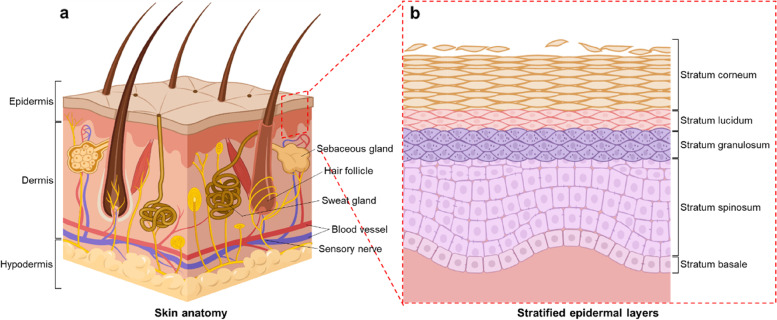
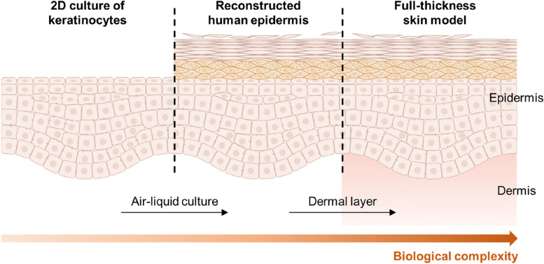
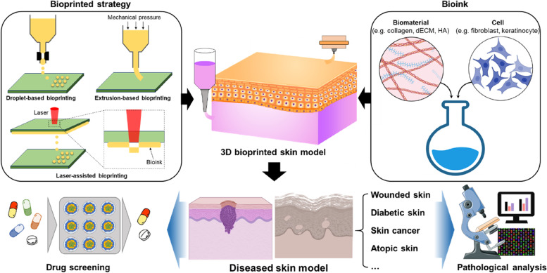
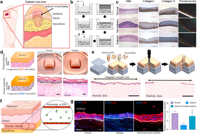
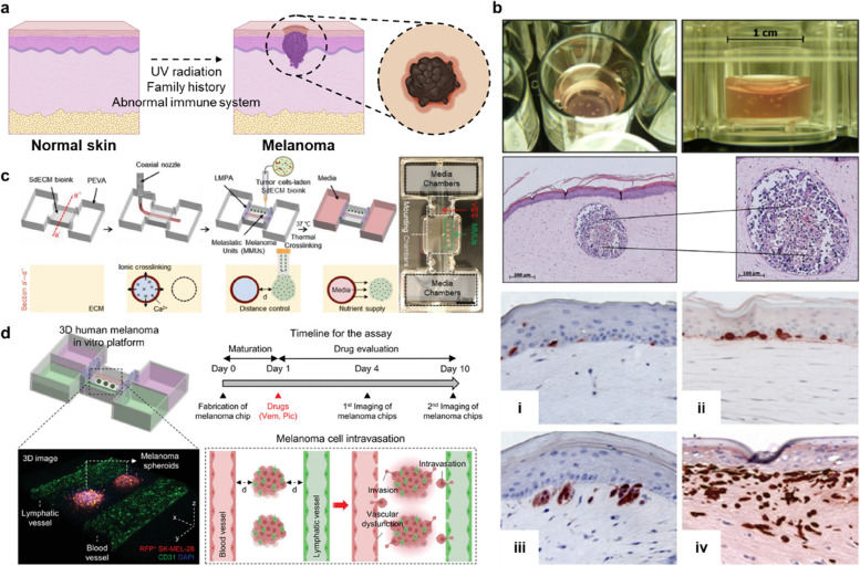
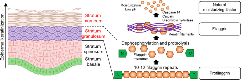
Human skin is an organ located in the outermost part of the body; thus, it frequently exhibits visible signs of physiological health. Ethical concerns and genetic differences in conventional animal studies have increased the need for alternative in vitro platforms that mimic the structural and functional hallmarks of natural skin. Despite significant advances in in vitro skin modeling over the past few decades, different reproducible biofabrication strategies are required to reproduce the pathological features of diseased human skin compared to those used for healthy-skin models. To explain human skin modeling with pathological hallmarks, we first summarize the structural and functional characteristics of healthy human skin. We then provide an extensive overview of how to recreate diseased human skin models in vitro, including models for wounded, diabetic, skin-cancer, atopic, and other pathological skin types. We conclude with an outlook on diseased-skin modeling and its technical perspective for the further development of skin engineering.

Keywords: Diseased-skin model; In vitro modeling; Skin engineering; Tissue engineering.


Conflict of interest statement

The authors declare that they have no competing interests.

Figures

Fig. 1
Fig. 1
Schematic representing the skin structure. a Anatomy of human skin. b Stratified epidermis consisting of five distinct layers
Fig. 2
Fig. 2
Advancement of an in vitro human skin model based on the tissue-engineering approach
Fig. 3
Fig. 3
Schematic of the fundamental elements used for creating a 3D bioprinted skin model. These approaches can also be used for the biofabrication of diseased skin in vitro, which may be applied to drug screening and pathological analysis
Fig. 4
Fig. 4
Representative studies on wounded-skin models. a 2D-based in vitro wound assays and wound-fabrication method using mechanical, optical, electrical, and thermal tools. b Workflow of wound-healing analysis using laser ablation [38]. First, cells were cultured on the plate until full confluency. A circular wound was then created using laser ablation with LEAP™, followed by rinsing to remove cell debris. Wound closure owing to cellular proliferation and migration was observed by LEAP™ in the bright-field mode daily. The wound area was calculated and quantified using the texture-segmentation algorithm developed by Smith et al. c Wound-healing analysis was conducted using a laser-irradiated skin model to demonstrate the effect of calcium pantothenate on re-epithelialization [42]. Reprinted with permission from Ref. [38, 42]
Fig. 5
Fig. 5
Representative studies for diabetic-skin models. a Schematic showing that even a small wound can become an ulcer owing to the poor wound-healing ability in patients with diabetes [47]. b Biofabrication process for the diabetic-skin model [48]. First, diabetic foot-ulcer fibroblasts contained in type I collagen were seeded onto the hanging cell-culture insert, which was followed by maturation for the dermal equivalent for three weeks of culture. Keratinocytes were then seeded on the dermal equivalent and allowed to proliferate for 5 d. Finally, an air–liquid interface culture was used to differentiate the keratinocytes, followed by the formation of stratified epidermal layers. c Comparison of skin models comprising human-foreskin, nondiabetic adult-foot, and diabetic foot-ulcer fibroblasts [48]. d Biofabrication strategy to create the diabetic-skin model via dermal–epidermal crosstalk [47]. e Wound fabrication using a 3D bioprinting device and wound-healing analysis via hematoxylin and eosin staining [47]. f Perfusion of metformin via a bioprinted vascular channel in the diabetic-skin model [47]. g Application of the diabetic-skin model as a drug-testing platform [47]. Reprinted with permission from Ref. [47, 48]
Fig. 6
Fig. 6
Examples of melanoma-skin models. a The main risk factors for melanoma. b Tissue-engineering approaches for the fabrication of in vitro melanoma models. Three-dimensional skin substitutes representing various stages of melanoma progression: (i) melanocyte located between epidermis and dermis; (ii) cell cluster formation by melanoma cells at the basement membrane; (iii) melanoma cell invasion into the dermis; (iv) aggressive invasion of melanoma cells into the dermis [53, 54]. c) In-bath bioprinting of melanoma cell aggregates with perfusable vascular channel [55]. d) In-bath bioprinting of melanoma stroma with blood and lymphatic vessel pair [56]. Reprinted with permission from Ref. [–56]
Fig. 7
Fig. 7
Pathology of AD. Filaggrin is located in the stratum granulosum of the epidermal layers. Profilaggrin is dephosphorylated and degrades into filaggrin monomers under the keratinocyte differentiation. Keratin filaments aggregate the cleaved filaggrin molecules, forming a dense matrix. The monomers are then degraded into natural moisturizing factors in the middle of the stratum corneum, which is regulated by proteases such as caspase 14, calpain, and bleomycin hydrolase

References

    1. Mansbridge J. Skin tissue engineering. J Biomater Sci Polym Ed. 2008;19:955–968. doi: 10.1163/156856208784909417. - DOI - PubMed
    1. Kolarsick PA, Kolarsick MA, Goodwin C. Anatomy and physiology of the skin. J Dermatol Nurs Assoc. 2011;3:203–213.
    1. McLafferty E, Hendry C, Farley A. The integumentary system: anatomy, physiology and function of skin. Nurs Stand (through 2013) 2012;27:35. doi: 10.7748/ns2012.09.27.3.35.c9299. - DOI - PubMed
    1. Lee MC, Lee WJ, Lee BI, Chung KY, Kim JW, Kang EH, Kim YO. Adipose tissue formation utilizing fat flap distraction technique. Sci Rep. 2017;7:1–10. - PMC - PubMed
    1. Ahn M, Cho WW, Kim BS, Cho DW. Engineering densely packed adipose tissue via environmentally controlled in-bath 3D sioprinting. Adv Funct Mater. 2022;32(28):2200203. doi: 10.1002/adfm.202200203. - DOI