

Ipsilateral Foot Drop After Leg Traction on Fracture Table for Mid-Shaft Femur Fracture Nailing: A Rare Case Report
- PMID: 37608904
- PMCID: PMC10440397
- DOI: 10.7759/cureus.43826
Ipsilateral Foot Drop After Leg Traction on Fracture Table for Mid-Shaft Femur Fracture Nailing: A Rare Case Report
Abstract
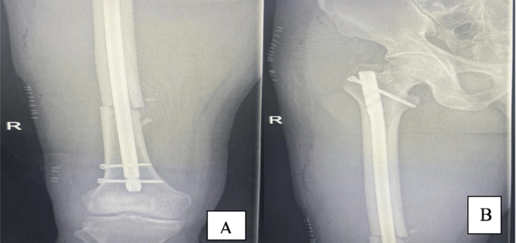
Femoral shaft fracture, one of the most common orthopaedic injuries, is usually treated with intramedullary nailing. During the operative procedure patients are placed on a traction table. Traction tables facilitate the procedure but are associated with some risk. Here we are sharing a case of a 35-year-old male healthy young patient who sustained a foot drop post nailing of femoral shaft fracture on a traction table. This patient has had some recovery in six weeks but is still not fully recovered. We think traction tables are a very helpful tool but carry some risks that should be kept on mind for every surgeon, and for the patients too.
Keywords: drop foot; femur shaft fracture; iotrogenic injury; sciatic nerve injury; traction table complication.
Copyright © 2023, Alzahrani et al.
Conflict of interest statement
The authors have declared that no competing interests exist.
Figures


References
-
- Closed nailing of a femoral fracture followed by sciatic nerve palsy. Britton JM, Dunkerley DR. J Bone Joint Surg Br. 1990;72:318. - PubMed
-
- Sciatic nerve palsy after operative treatment of subtrochanteric femur fracture resulting from postoperative hematoma: a case report. Pedri TG, Treme GP. https://digitalrepository.unm.edu/unm_jor/vol6/iss1/38 UNM Orthopaed Res J. 2017;6
-
- Delayed sciatic nerve palsy due to hematoma related with anticoagulants prophylaxis in the femur intramedullary nailing - a case report. Kim YM, Joo YB, Song SH. J Korean Fracture Soc. 2017;30:198–202.
-
- Sensory neurapraxia of the foot after leg traction on fracture table. Madhu T, Dunsmuir R. http://10.1016/j.injury.2005.04.002 Injury Extra. 2005;36:306–307.
Publication types
LinkOut - more resources
Full Text Sources