. 2023 Aug 21:9:104.
doi: 10.1038/s41378-023-00569-1. eCollection 2023.

A microfluidic biosensor for the diagnosis of chronic wasting disease

Affiliations

A microfluidic biosensor for the diagnosis of chronic wasting disease

Sura A Muhsin et al. Microsyst Nanoeng. .

Abstract

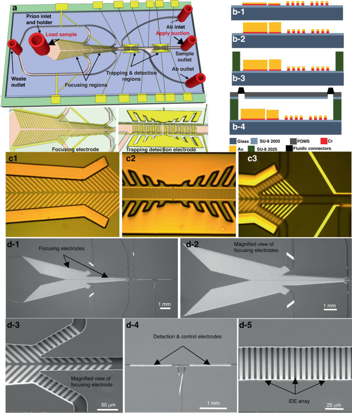
Cervids are affected by a neurologic disease that is always fatal to individuals and has population effects. This disease is called chronic wasting disease (CWD) and is caused by a misfolded prion protein. The disease is transmitted via contact with contaminated body fluids and tissue or exposure to the environment, such as drinking water or food. Current CWD diagnosis depends on ELISA screening of cervid lymph nodes and subsequent immunohistochemistry (IHC) confirmation of ELISA-positive results. The disease has proven to be difficult to control in part because of sensitivity and specificity issues with the current test regimen. We have investigated an accurate, rapid, and low-cost microfluidic microelectromechanical system (MEMS) biosensing device for the detection of CWD pathologic prions in retropharyngeal lymph nodes (RLNs), which is the current standard type of CWD diagnostic sample. The device consists of three novel regions for concentrating, trapping, and detecting the prion. The detection region includes an array of electrodes coated with a monoclonal antibody against pathologic prions. The experimental conditions were optimized using an engineered prion control antigen. Testing could be completed in less than 1 hour with high sensitivity and selectivity. The biosensor detected the engineered prion antigen at a 1:24 dilution, while ELISA detected the same antigen at a 1:8 dilution. The relative limit of detection (rLOD) of the biosensor was a 1:1000 dilution of a known strong positive RLN sample, whereas ELISA showed a rLOD of 1:100 dilution. Thus, the biosensor was 10 times more sensitive than ELISA, which is the currently approved CWD diagnostic test. The biosensor's specificity and selectivity were confirmed using known negative RPLN samples, a negative control antibody (monoclonal antibody against bovine coronavirus BCV), and two negative control antigens (bluetongue virus and Epizootic hemorrhagic disease virus). The biosensor's ability to detect pathogenic prions was verified by testing proteinase-digested positive RLN samples.

Keywords: Electrical and electronic engineering; Nanoparticles.


Conflict of interest statement

Conflict of interestThe authors declare no competing interests.

Figures

Fig. 1
Fig. 1. Description of the Biosensor.
a 3-D view, b1b4 sideview. c1c3 Optical images of the biosensor after fabrication of the focusing electrode pair, the sensing and trapping electrodes array, and the SU8 microchannel. d1d5 Scanning electron microscope (SEMs) micrographs of the fabricated biosensor. d-1 The two set focusing electrodes, detection electrode, and control electrode embedded in SU-8 microchannel, d-2 a magnified view of the two-set focusing electrode, d-3 magnified view of one focusing electrode, d-4 detection and control electrodes, d-5 a magnified view of the detection IDE array
Fig. 2
Fig. 2. Modeling and Simulation of the Electric field.
An electric field (E-Field) modeling and simulation using COMSOL Multiphysics software of the three regions making the biosensor, i.e., a focusing. b1b5 Detection IDE array. The finger width and spacing between fingers are b-1 5 µm and 2 µm, b-2 4 µm and 4 µm, b-3 10 µm and 2 µm, b-4 10 µm and 4 µm, respectively. c Trapping. d An equivalent electrical circuit of the biosensor, e experimental and simulation results after the CWD prion antibody-antigen binding in the microchannel
Fig. 3
Fig. 3. Device Operation.
a A cartoon showing the experimental setup. Top-view cartoon showing the flow direction during: b-1 antibody coating where it was first placed at the antibody inlet and suction was applied to the antibody outlet while all other inlets were closed, b-2 CWD prion antigen loading at the sample inlet while suction was applied to the waste outlet. The flow continued toward the detection region. The process flow for antibody immobilization, and the antibody/ antigen binding on the interdigitated microelectrode: c-1 the antibody was loaded from the antibody inlet while suction was applied from the antibody outlet, c-2 the microchannel was washed after adhesion of antibody to the IDE array, c-3 the CWD prion protein sample was loaded into the sample inlet while suction was applied to the sample outlet, c-4 the microchannel was washed again after antibody antigen binding was completed, d a package biosensor
Fig. 4
Fig. 4. Demonstration of the focusing and trapping capabilities.
a Fluorescent images before focusing the nanobeads (diameter < 1 μm) into the centerline of the focusing region, b fluorescent images after focusing the nanobeads into the centerline of the focusing region. c Magnified view of image b, d fluorescent images after focusing and before trapping the nanobeads onto the surface of the detection electrode array. e Fluorescent images after focusing and trapping the nanobeads onto the surface of the detection electrode array. f Magnified view of images e. g Fluorescent images after focusing the nanobeads (diameter < 200 nm) into the centerline of the focusing region, h fluorescent images after trapping the nanobeads onto the surface of the detection electrode array, i Magnified view of image h
Fig. 5
Fig. 5. Sensitivity measurement.
This includes detection of a Different antibody concentrations from 0.25 µg/mL to 10 µg/mL against CWD prion antigen (Ag). b Different antibody (Ab) coating time at a fixed Ab concentration of 2 µg/mL (optimum value). c Prion antigen from the ELSIA kit at different dilutions. d Detection of CWD Prion antigen as a function of concentration (1:4, 1:8, 1:16, 1:24) at 100 KHz. The data was fitted with a polynomial. This shows that the impedance change was not linear
Fig. 6
Fig. 6. Study of a known CWD positive retropharyngeal lymph nodes (RLN) sample.
a Relative limit of detection (rLOD) based on a known CWD positive retropharyngeal lymph nodes (RLN) sample (20015350). b Known CWD-negative RLN samples
Fig. 7
Fig. 7. Study of specificity, selectivity, and negative control of the biosensor.
a Specificity testing using anti-prion antibody and Bluetongue (BT) virus. The negative control electrode was not coated with antibody and exposed to BT virus. b Specificity testing using anti-prion antibody and Epizootic hemorrhagic disease virus (EHDV). The negative control electrode was not coated with antibody and exposed to EHDV, c Selectivity testing of a known positive RLN sample against anti-prion mAb (high impedance) and anti-BCV antibody (baseline impedance)
Fig. 8
Fig. 8. Confirmation of the Detection of Pathogenic Prions.
Detection of CWD pathogenic prion in RLNs: includes 2 strong positives (OD = 4), 2 moderate strong positive (OD = 2.678, and 2.459), and 2 weak strong positives (OD = 0.614 and 0.237). a Untreated RLN samples and b RLN samples treated with proteinase K to remove non-prion proteins in the samples

References

    1. Haley NJ, et al. Detection of chronic wasting disease in the lymph nodes of free-ranging cervids by real-time quaking-induced conversion. J. Clin. Microbiol. 2014;52:3237–3243. doi: 10.1128/JCM.01258-14. - DOI - PMC - PubMed
    1. Kramm C, et al. Detection of Prions in Blood of Cervids at the Asymptomatic Stage of Chronic Wasting Disease. Sci. Rep. 2017;7:1–8. doi: 10.1038/s41598-017-17090-x. - DOI - PMC - PubMed
    1. Schmitz M, et al. The real-time quaking-induced conversion assay for detection of human prion disease and study of other protein misfolding diseases. Nat. Protoc. 2016;11:2233–2242. doi: 10.1038/nprot.2016.120. - DOI - PubMed
    1. Miller MW, Williams ES. Chronic wasting disease of cervids. Curr. Top. Microbiol Immunol. 2004;284:193–214. - PubMed
    1. Rivera, N. A., Brandt, A. L., Novakofski, J. E., Mateus-Pinilla, N. E. Chronic wasting disease in cervids: prevalence, impact and management strategies, Veter. Med. Res. Rep. 123–139. 10.2147/VMRR.S197404 (2019). - PMC - PubMed