. 2023 Aug 7:10:1242146.
doi: 10.3389/fvets.2023.1242146. eCollection 2023.

Therapeutic efficacy of tylvalosin combined with Poria cocos polysaccharides against porcine reproductive and respiratory syndrome

Affiliations

Therapeutic efficacy of tylvalosin combined with Poria cocos polysaccharides against porcine reproductive and respiratory syndrome

Hong Shi et al. Front Vet Sci. .

Abstract

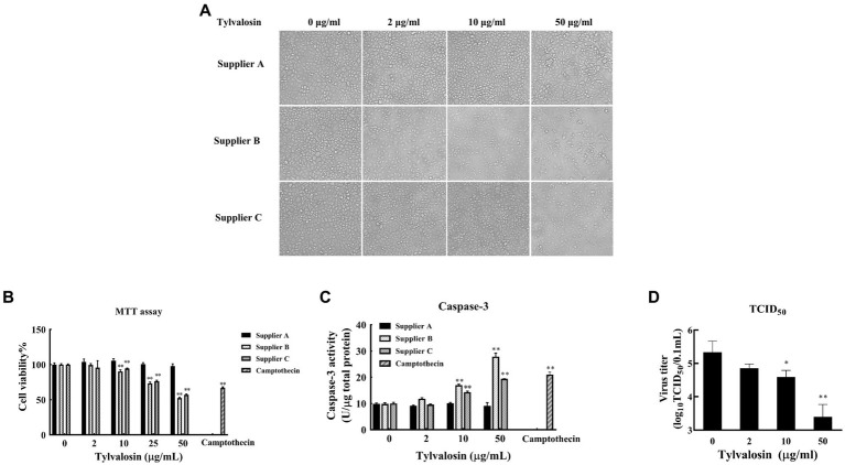
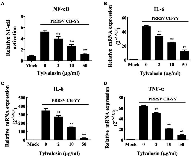
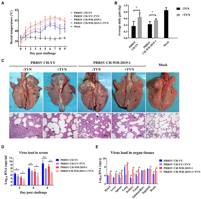
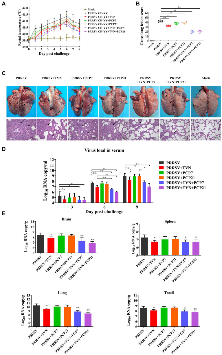
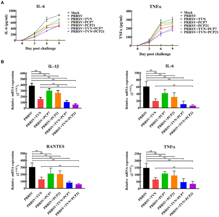
Porcine reproductive and respiratory syndrome (PRRS) is one of the most economically important infectious diseases of pigs worldwide. Vaccination and various management measures have been implemented to control PRRS. However, due to high genetic diversity and insufficient understanding of the pathogenesis and immunological mechanisms, PRRS is still a challenge to the pig industry. Therefore, it is important to develop novel strategies to combat PRRS virus (PRRSV) infection. In this study, our data show that tylvalosin, a third-generation animal-specific macrolide, could inhibit PRRSV replication in MARC-145 cells, and suppress the PRRSV-induced NF-κB activation and cytokines expression. The pig infection experiment further demonstrated that tylvalosin could significantly reduce the virus loads in serum and tissues, and alleviate lung lesions of pigs infected with highly pathogenic PRRSV strains. The fever and loss of daily gain (LoDG) of the pigs were decreased as well. Considering the feature of immune suppression of PRRSV, a combination of tylvalosin with the immunopotentiator Poria cocos polysaccharides (PCP) was developed. Pig experiment showed this combination had a better therapeutic efficacy against PRRSV infection than tylvalosin and PCP alone in attenuating lung lesions, alleviating fever, and suppressing cytokines production. This study suggests that tylvalosin has significant antiviral and anti-inflammatory effects against PRRSV infection, and the combination of tylvalosin and PCP provides a promising strategy for PRRS treatment.

Keywords: NF-κB; Poria cocos polysaccharides (PCP); antiviral; cytokines; pig; porcine reproductive and respiratory syndrome (PRRS); tylvalosin.


Conflict of interest statement

The authors declare that the research was conducted in the absence of any commercial or financial relationships that could be construed as a potential conflict of interest.

Figures

Figure 1
Figure 1
Cytotoxicity and effect on PRRSV replication of tylvalosin. (A) Influence of tylvalosin on growth and morphology of MARC-145 cells. Tylvalosin from suppliers A, B, or C was incubated with MARC-145 cells at each indicated concentration. The cells were cultured in normal conditions and imaged after culture for 24 h. (B) Cytotoxicity by MTT assay. Tylvalosin from suppliers A, B, or C was incubated with MARC-145 cells at each indicated concentration for 3 h. Camptothecin was used as a positive control. The cytotoxicity was determined using an MTT kit according to the manufacturer’s instructions. The assay was performed in triplicate and the data was shown as the mean ± standard deviation (SD). The statistical significance was calculated using the ANOVA by comparing the data obtained at each indicated time point with that obtained at time point 0 h. (C) Caspase-3 activity assay. Tylvalosin from suppliers A, B, or C was incubated with MARC-145 cells at each indicated concentration for 3 h. Caspase-3 activity was measured by using Caspase 3 Activity Assay Kit. The assay was performed in triplicate and the data was shown as the mean ± SD. The statistical significance was calculated using the ANOVA by comparing the data obtained at each indicated time point with that obtained at time point 0 h. (D) TCID50 assay. MARC-145 cells were infected with PRRSV strain CH-YY and then treated with tylvalosin at each indicated concentration for 24 h. The cell culture was then subjected to TCID50 determination. The assay was performed in triplicate and the data was shown as the mean ± SD. The statistical significance was calculated using the ANOVA by comparing the data obtained at each indicated time point with that obtained at time point 0 h. ** indicates that the p value <0.01.
Figure 2
Figure 2
Inhibitory effect of tylvalosin on NF-κB activation and cytokines production induced by PRRSV infection. (A) MARC-145 cells seeded in 48-well plate were co-transfected with 50 ng/well of reporter plasmid NF-κB-Luc along with 50 ng/well of pRL-TK. At 24 h post-transfection, the cells were treated with tylvalosin at different concentrations (0, 2, 10, 25, 50 μg/mL) and infected with PRRSV strain CH-YY. The NF-κB activity was determined using a dual-luciferase reporter assay system. (B–D) MARC-145 cells were infected by PRRSV strain CH-YY and then treated with TVN. The expression of IL-6 (B), IL-8 (C), and TNF-α (D) was determined by qRT-PCR. Mock indicates the samples without PRRSV infection or tylvalosin treatment. The assays were performed in triplicate and the data was shown as the mean ± SD. The statistical significance was calculated using the ANOVA by comparing the data obtained by treatment with each indicated concentration of tylvalosin with that obtained without tylvalosin treatment. ** indicates that the p value <0.01.
Figure 3
Figure 3
Tylvalosin shows therapeutic efficacy against PRRSV infection in pigs. (A) Rectal temperature. The JXA1-like strain CH-YY or NADC30-like strain CH-WH-2019-1 was used to infect pigs treated with or without TVN. The rectal temperature was recorded at each indicated time point. The data was shown as the mean ± SD. (B) Average daily gain. The body weight of each pig was monitored and the average daily gain was calculated. (C) Gross and histopathological analysis. At the end of the experiment, the pigs were sacrificed and the lungs were collected and imaged (Upper panel). The lung tissues were then fixed and subjected to histopathological analysis (lower panel). The lung lesions are marked with black arrows. The scale bar is 100 μm. (D) Virus load in serum. The blood was collected from the pigs at the indicated time point. The serum was isolated and virus load was determined by qRT-PCR. The data were shown as the mean ± SD. The statistical significance was calculated using the ANOVA. (E) Virus load in organ tissues. After the piglets were sacrificed, each indicated organ was collected. The total RNA was extracted and the virus load was determined by qRT-PCR. The statistical significance was calculated using the ANOVA. * indicates that the p value <0.05. ** indicates that the p value <0.01.
Figure 4
Figure 4
Efficacy of the combination of TVN and PCP on PRRSV infection in pigs. (A) Rectal temperature. The rectal temperature was recorded at each indicated time point. The data was shown as the mean ± SD. (B) Gross lung lesion score. The pigs were sacrificed at the end of the experiment and the lung lesion was scored according to Halbur et al. (53). (C) Gross and histopathological analysis. At the end of the experiment, the pigs were sacrificed and the lung was collected and imaged (Upper panel). The lung tissues were then fixed and subjected to histopathological analysis (lower panel). The lung lesions are marked with black arrows. The scale bar is 100 μm. (D) Virus load in serum. The blood was collected from the pigs in each group at the indicated time point. The serum was isolated and virus load was determined by qRT-PCR. The data was shown as the mean ± SD. The statistical significance was calculated using the ANOVA. * indicates that the p < 0.05. ** indicates that the p < 0.01. (E) Virus load in organ tissues. The piglets wereFIGURE 4 (Continued)sacrificed at the end of the experiment and each indicated organ was collected. The total RNA was extracted and the virus load was determined by qRT-PCR. The statistical significance was calculated using the ANOVA. * indicates that the p value <0.05. ** indicates that the p value <0.01. Mock indicates the group without infection or TVN treatment. PRRSV indicates the group with PRRSV CH-YY infection but without TVN treatment. PRRSV+TVN indicates the group with PRRSV CH-YY infection as well as TVN treatment. PRRSV+PCP7 indicates the group with PRRSV CH-YY infection as well as PCP treatment at 7 mg/kg·bw. PRRSV+PCP21 indicates the group with PRRSV CH-YY infection as well as PCP treatment at 21 mg/kg·bw. PRRSV+TVN + PCP7 indicates the group with PRRSV CH-YY infection as well as TVN treatment and PCP treatment at 7 mg/kg·bw. PRRSV+TVN + PCP21 indicates the group with PRRSV CH-YY infection as well as TVN treatment and PCP treatment at 21 mg/kg·bw.
Figure 5
Figure 5
Cytokines quantification. The pigs in each group were sacrificed at the end of the experiment. The lungs were collected. The cytokines in the serum were determined by commercial pig IL-6 and TNF-α ELISA kits (A) and those in the lung tissues were determined by qRT-PCR (B). The statistical significance was calculated using the ANOVA. * indicates that the p value <0.05. ** indicates that the p value <0.01. Mock indicates the group without infection or TVN treatment. PRRSV indicates the group with PRRSV CH-YY infection but without TVN treatment. PRRSV+TVN indicates the group with PRRSV CH-YY infection as well as TVN treatment. PRRSV+PCP7 indicates the group with PRRSV CH-YY infection as well as PCP treatment at 7 mg/kg·bw. PRRSV+PCP21 indicates the group with PRRSV CH-YY infection as well as PCP treatment at 21 mg/kg·bw. PRRSV+TVN + PCP7 indicates the group with PRRSV CH-YY infection as well as TVN treatment and PCP treatment at 7 mg/kg·bw. PRRSV+TVN + PCP21 indicates the group with PRRSV CH-YY infection as well as TVN treatment and PCP treatment at 21 mg/kg·bw.

Similar articles

Cited by

References

    1. Keffaber KK. Reproductive failure of unknown etiology. Am Assoc Swine Pract News. (1989) 1:1–9.
    1. Wensvoort G, Terpstra C, Pol JM, ter Laak EA, Bloemraad M, de Kluyver EP, et al. . Mystery swine disease in the Netherlands: the isolation of Lelystad virus. Vet Q. (1991) 13:121–30. doi: 10.1080/01652176.1991.9694296 - DOI - PubMed
    1. Lunney JK, Fang Y, Ladinig A, Chen N, Li Y, Rowland B, et al. . Porcine reproductive and respiratory syndrome virus (PRRSV): pathogenesis and interaction with the immune system. Annu Rev Anim Biosci. (2016) 4:129–54. doi: 10.1146/annurev-animal-022114-111025 - DOI - PubMed
    1. Guo Z, Chen XX, Li R, Qiao S, Zhang G. The prevalent status and genetic diversity of porcine reproductive and respiratory syndrome virus in China: a molecular epidemiological perspective. Virol J. (2018) 15:2. doi: 10.1186/s12985-017-0910-6, PMID: - DOI - PMC - PubMed
    1. Holtkamp DJ, Kliebenstein JB, Neumann EJ, Zimmerman JJ, Rotto HF, Yoder TK, et al. . Assessment of the economic impact of porcine reproductive and respiratory syndrome virus on United States pork producers. J Swine Health Prod. (2013) 21:72–84. doi: 10.2460/javma.2005.227.385 - DOI

LinkOut - more resources