

This is a preprint.
MRIO: The Magnetic Resonance Imaging Acquisition and Analysis Ontology
- PMID: 37609265
- PMCID: PMC10441376
- DOI: 10.1101/2023.08.04.552020
MRIO: The Magnetic Resonance Imaging Acquisition and Analysis Ontology
Update in
-
MRIO: the Magnetic Resonance Imaging Acquisition and Analysis Ontology.Neuroinformatics. 2024 Jul;22(3):269-283. doi: 10.1007/s12021-024-09664-8. Epub 2024 May 20. Neuroinformatics. 2024. PMID: 38763990 Free PMC article.
Abstract
Objective: Magnetic resonance imaging of the brain is a useful tool in both the clinic and research settings, aiding in the diagnosis and treatments of neurological disease and expanding our knowledge of the brain. However, there are many challenges inherent in managing and analyzing MRI data, due in large part to the heterogeneity of data acquisition.
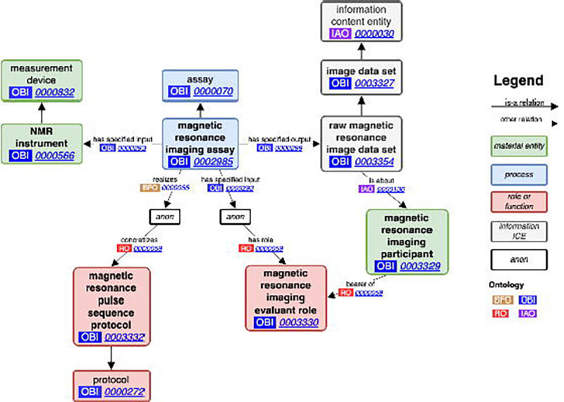
Materials and methods: To address this, we have developed MRIO, the Magnetic Resonance Imaging Acquisition and Analysis Ontology.
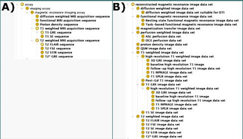
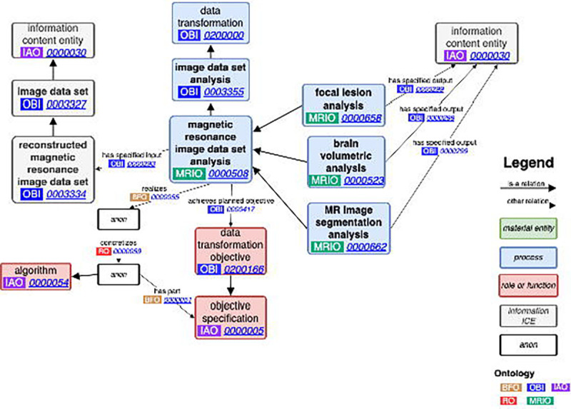
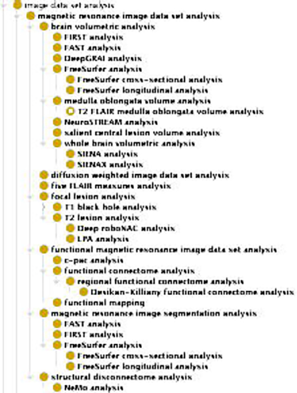
Results: MRIO provides well-reasoned classes and logical axioms for the acquisition of several MRI acquisition types and well-known, peer-reviewed analysis software, facilitating the use of MRI data. These classes provide a common language for the neuroimaging research process and help standardize the organization and analysis of MRI data for reproducible datasets. We also provide queries for automated assignment of analyses for given MRI types.
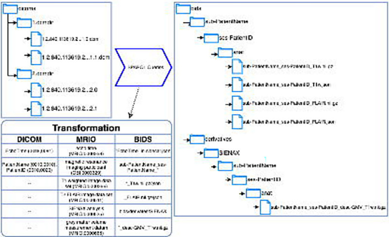
Discussion: MRIO aids researchers in managing neuroimaging studies by helping organize and annotate MRI data and integrating with existing standards such as Digital Imaging and Communications in Medicine and the Brain Imaging Data Structure, enhancing reproducibility and interoperability. MRIO was constructed according to Open Biomedical Ontologies Foundry principals and has contributed several terms to the Ontology for Biomedical Investigations to help bridge neuroimaging data to other domains.
Conclusion: MRIO addresses the need for a "common language" for MRI that can help manage the neuroimaging research, by enabling researchers to identify appropriate analyses for sets of scans and facilitating data organization and reporting.
Keywords: Biomedical; Translational Research; biological ontologies; magnetic resonance imaging; neuroimaging; neuroinformatics.
Conflict of interest statement
Competing interests The authors have no competing interests to report.
Figures
References
-
- Smits M. MRI biomarkers in neuro-oncology. Nature Reviews Neurology. 2021;17(8):486–500. - PubMed
-
- Kehoe EG, McNulty JP, Mullins PG, Bokde ALW. Advances in MRI biomarkers for the diagnosis of Alzheimer’s disease. Biomarkers in medicine. 2014;8(9):1151–1169. - PubMed
-
- Tavazzi E, Zivadinov R, Dwyer MG, et al. MRI biomarkers of disease progression and conversion to secondary-progressive multiple sclerosis. Expert Review of Neurotherapeutics. 2020;20(8):821–834. - PubMed
Publication types
Grants and funding
LinkOut - more resources
Full Text Sources