. 2023 Sep 26;42(9):113024.
doi: 10.1016/j.celrep.2023.113024. Epub 2023 Aug 22.

Staphylococcus epidermidis activates keratinocyte cytokine expression and promotes skin inflammation through the production of phenol-soluble modulins

Affiliations

Staphylococcus epidermidis activates keratinocyte cytokine expression and promotes skin inflammation through the production of phenol-soluble modulins

Michael R Williams et al. Cell Rep. .

Abstract

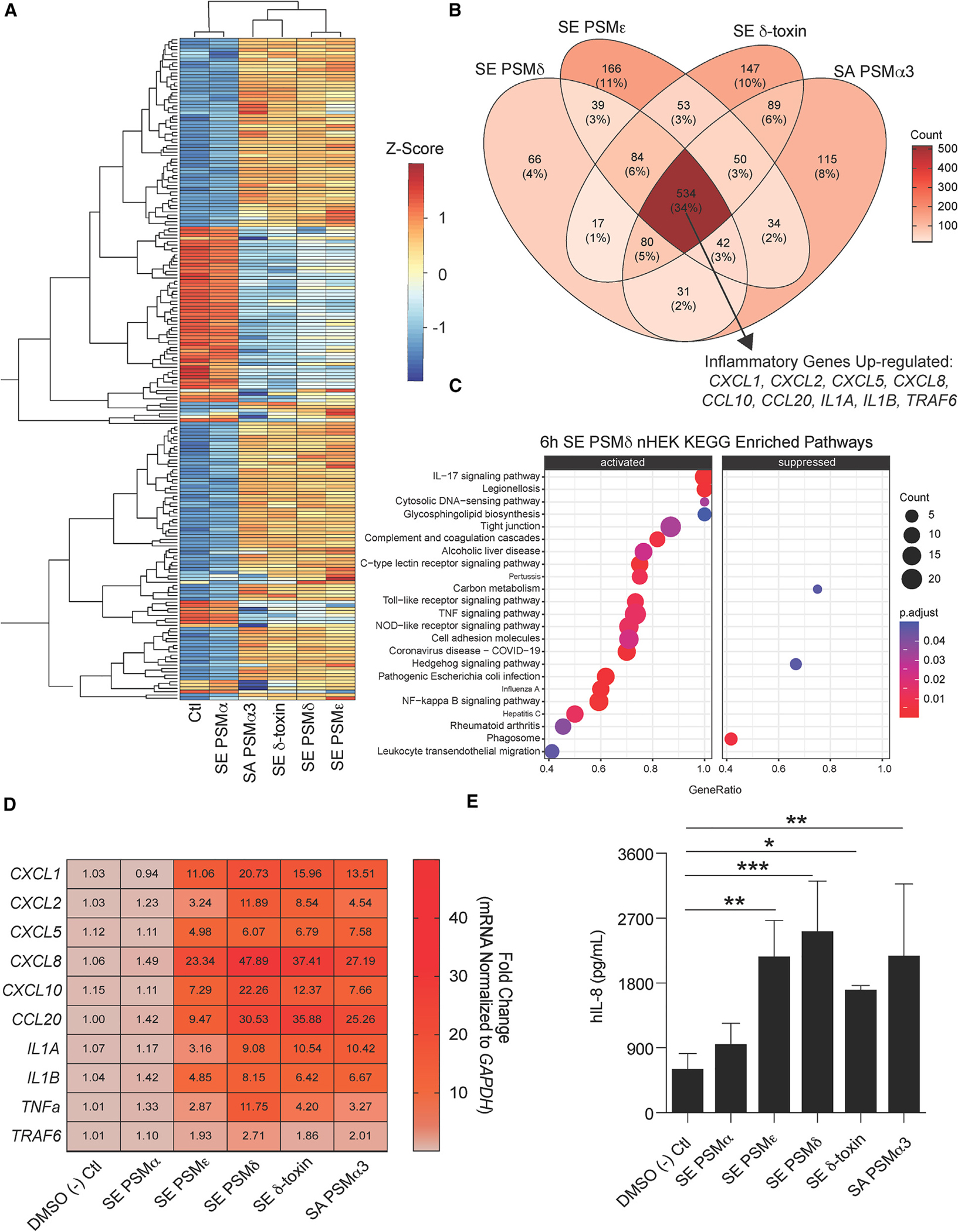
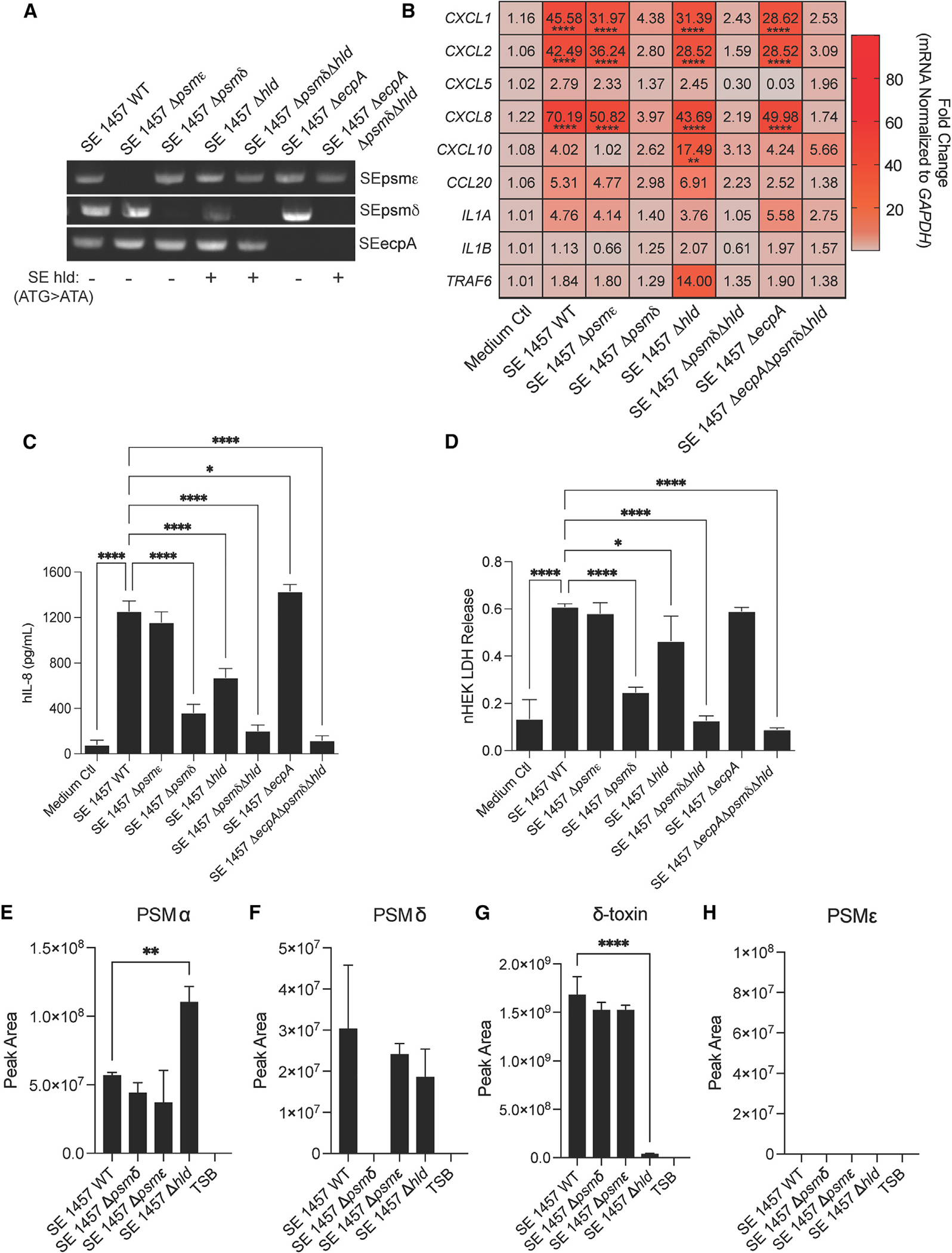
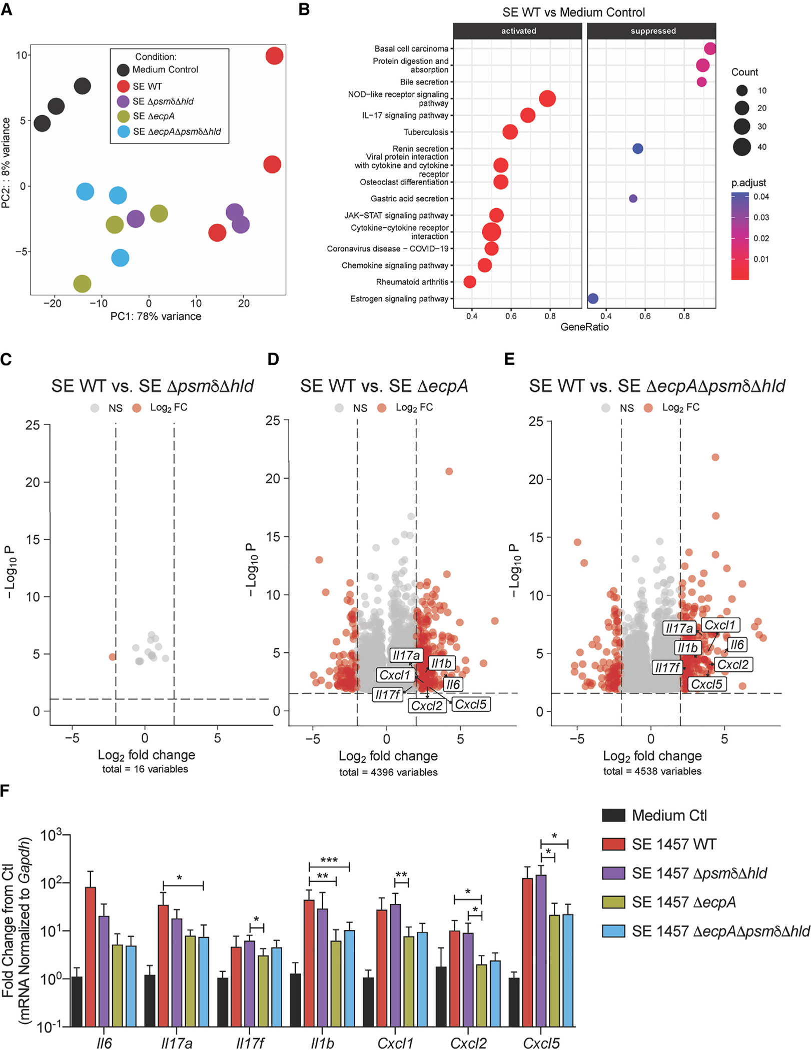
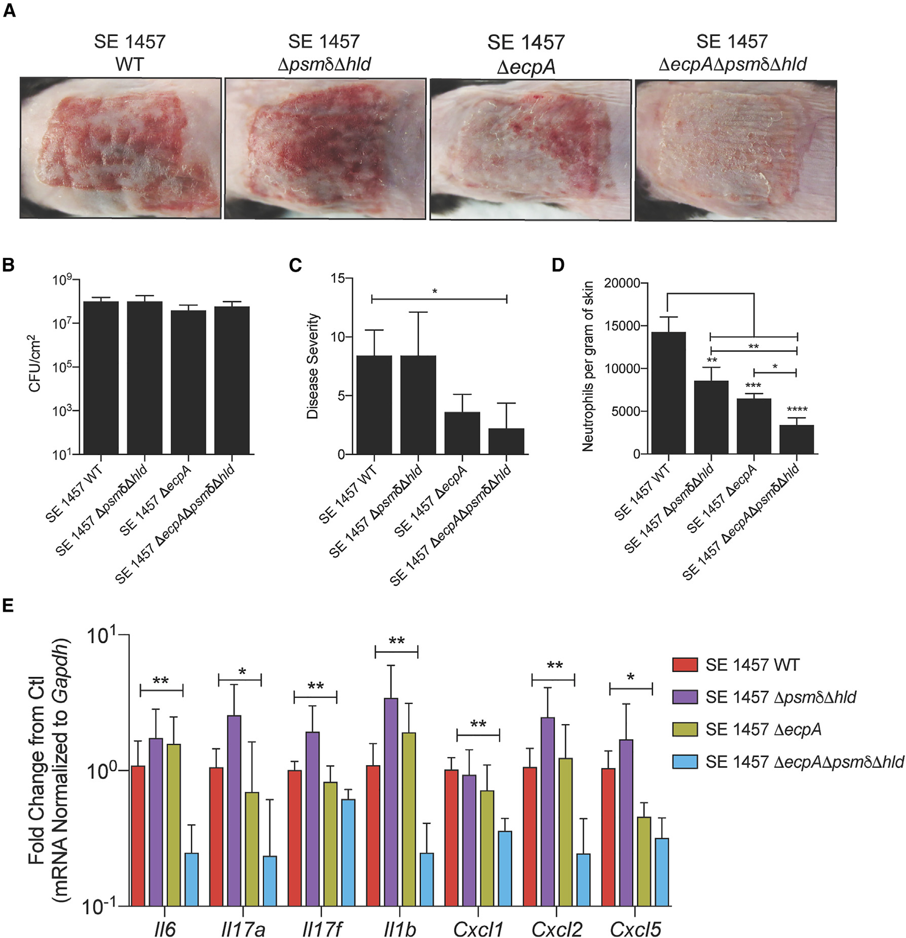
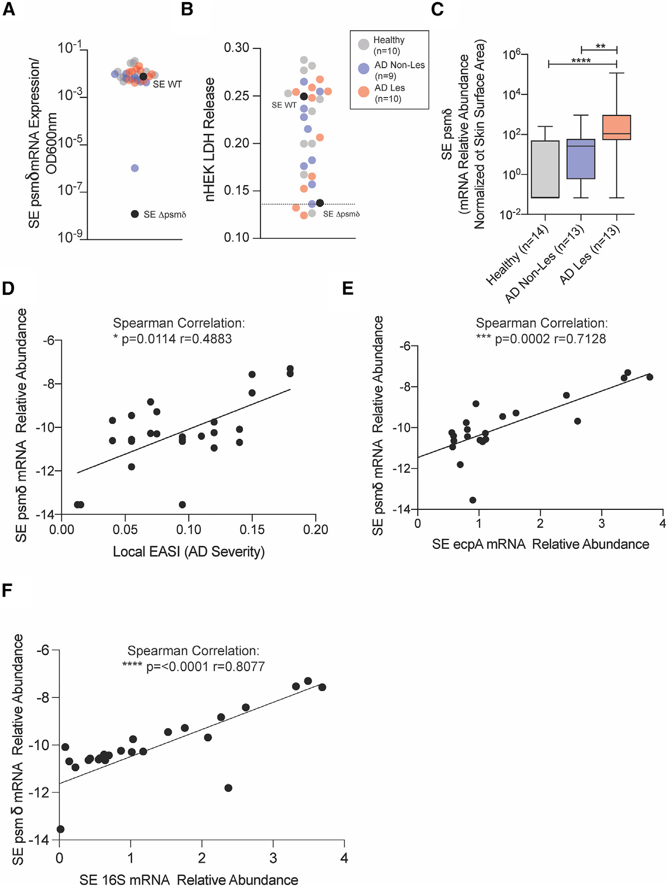
Staphylococcus epidermidis is a common microbe on human skin and has beneficial functions in the skin microbiome. However, under conditions of allergic inflammation, the abundance of S. epidermidis increases, establishing potential danger to the epidermis. To understand how this commensal may injure the host, we investigate phenol-soluble modulin (PSM) peptides produced by S. epidermidis that are similar to peptides produced by Staphylococcus aureus. Synthetic S. epidermidis PSMs induce expression of host defense genes and are cytotoxic to human keratinocytes. Deletion mutants of S. epidermidis lacking these gene products support these observations and further show that PSMs require the action of the EcpA bacterial protease to induce inflammation when applied on mouse skin with an intact stratum corneum. The expression of PSMδ from S. epidermidis is also found to correlate with disease severity in patients with atopic dermatitis. These observations show how S. epidermidis PSMs can promote skin inflammation.

Keywords: CP: Immunology; CP: Microbiology; Staphylococcus; atopic dermatitis; inflammation; microbiome; phenol-soluble modulins; skin.


Conflict of interest statement

Declaration of interests R.L.G. is a co-founder, scientific advisor, and consultant and has equity in MatriSys Biosciences.

Figures

Figure 1.
Figure 1.. SE synthetic phenol-soluble modulins drive keratinocyte inflammation
(A–C) RNA-seq data from primary human keratinocytes treated for 6 h at 10 μM with different S. epidermidis (SE) and S. aureus (SA) phenol-soluble modulins (PSMs). (A) Heatmap of top 250 variable genes. (B) Venn diagram of 2-fold up-regulated genes among PSMs differing from the control treatment with specific emphasis on up-regulated inflammatory genes shared among all PSMs. (C) Kyoto Encyclopedia of Genes and Genomes enriched pathway analysis of SE PSMδ (10 μM) treated keratinocytes. (D) qPCR results of keratinocytes treated for 6 h with SE and SA synthetic PSMs (10 μM) for gene significantly up-regulated in RNA-seq data above (n = 4). (E) ELISA analysis of human IL-8 cytokine (hIL-8) levels in conditioned medium from keratinocytes treated for 24 h with SE and SA synthetic PSMs (10 μM) (n = 4). Results are representative of at least two independent experiments. Mean ± SEM and a parametric unpaired one-way ANOVA analysis was used to determine statistical significance: *p < 0.05, **p < 0.01, ***p < 0.001, ****p < 0.0001. See also Figures S1A and S1B.
Figure 2.
Figure 2.. SE isogenic mutant strains reveal specific PSMs drive keratinocyte inflammation
(A) PCR confirmation gel for specific SE knockout strains generated for this study. SE hld point mutation in start codon for gene confirmed through DNA sequencing and indicated by a +/−. For knockout strain growth curves, see also Figures S1C and S1D. (B) qPCR analysis of up-regulated inflammatory markers in keratinocytes treated for 3 h with 10% sterile-filtered conditioned medium of overnight growths of SE wild-type (WT) and specific knockout strains (n = 4). (C and D) (C) hIL-8 ELISA and (D) LDH release analysis from conditioned medium of keratinocytes treated for 24 h with 10% sterile-filtered conditioned medium of overnight growths of SE wild-type (WT) and specific knockout strains (n = 4). (E–H) Mass spectrometric validation of the presence or absence of PSMs in spent media for S. epidermidis 1457 WT and knockout strains. Results are averages of selected ion chromatogram peak areas for injections of biological triplicates of S. epidermidis spent media (TSB). The ions were detected in the quadropoly protonated form, [M+4H]4+. Results are representative of at least two independent experiments. Mean ± SEM and a parametric unpaired one-way ANOVA analysis was used to determine statistical significance: *p < 0.05, **p < 0.01, ***p < 0.001, ****p < 0.0001. See also Figure S2.
Figure 3.
Figure 3.. SE protease EcpA primarily drives early inflammation in epicutaneous mouse skin model
(A) PCA analysis of bulk RNA-seq data from murine dorsal skin treated with epicutaneous application of 1 × 10⁷ CFU/cm2 of SE wild-type or mutant strains SE ΔpsmδΔhld, SEDecpA, or SE ΔpsmδΔhlecpA for 24 h (n = 3 per group). (B) Kyoto Encyclopedia of Genes and Genomes enriched pathway analysis of SE WT versus control treated mouse skin at 24 h (C–E) Volcano plots of log2fold changed genes (red) between SE WT and various knockout (KO) strains. (F) qPCR analysis of up-regulated inflammatory genes in murine skin treated for 24 h with epicutaneous application of 1 × 10⁷ CFU/cm2 of SE wild-type or mutant strains SE ΔpsmδΔhld, SE ΔecpA, or SE ΔpsmδΔhldΔecpA for 24 h (n = 6 per group). Results are representative of at least two independent experiments. Mean ± SEM and a non-parametric unpaired Kruskal-Wallis analysis was used to determine statistical significance: *p < 0.05, **p < 0.01, ***p < 0.001, ****p < 0.0001.
Figure 4.
Figure 4.. Both SE PSMs and EcpA promote skin inflammation in late inflammatory epicutaneous mouse model
(A) Representative pictures of murine back skin after epicutaneous application of 1 × 10⁷ CFU/cm2 of SE wild-type (WT), SE ΔpsmδΔhld, SE ΔecpA, or SE ΔpsmδΔhldΔecpA for 72 h (n = 5 per group). (B and C) CFU/cm2 of live bacteria and single-blinded assessment of skin disease severity following the 72-h application of epicutaneous bacteria. See also Figure S3. (D) Flow cytometric analysis of neutrophils (CD45+,CD11b+,Ly6G+) per gram of skin following the 72-h application of epicutaneous bacteria. Results represent mean ± SEM and a parametric unpaired one-way ANOVA analysis was used to determine statistical significance: *p < 0.05, **p < 0.01, ***p < 0.001, ****p < 0.0001. (E) qPCR analysis of up-regulated inflammatory genes in murine skin treated for 72 h with epicutaneous application of 1 × 10⁷ CFU/cm2 of SE wild-type or mutant strains SE ΔpsmδΔhld, SE ΔecpA, or SE ΔpsmδΔhldΔecpA (n = 5 per group). Results are representative of at least two independent experiments. Mean ± SEM and a non-parametric unpaired Kruskal-Wallis analysis was used to determine statistical significance: *p < 0.05, **p < 0.01, ***p < 0.001, ****p < 0.0001.
Figure 5.
Figure 5.. Staphylococcus epidermidis PSMs are produced in clinical isolates and elevated on diseased skin
(A) SE psmδ transcript level expression compared with SE 1457 WT and SE 1457 Δpsmδ control strains across healthy (n = 10), atopic dermatitis non-lesional (AD Non-Les, n = 9), and atopic dermatitis lesional (AD Les, n = 10) clinical isolates. (B) Assessment of LDH release from keratinocytes treated for 24 h with the sterile-filtered conditioned medium from individual SE clinical isolates. (C) qPCR analysis of SE psmδ mRNA expression levels from skin swabs of healthy (n = 14), AD Non-Les (n = 13), and AD Les (n = 13) skin. (D–F) Spearman correlations between SE psmδ transcript levels and (D) local EASI disease severity scores, (E) SE ecpA transcript levels, and (F) SE 16s transcript levels on AD lesional skin swabs. Mean ± SEM and a non-parametric unpaired Kruskal-Wallis analysis was used to determine statistical significance: *p < 0.05, **p < 0.01, ***p < 0.001, ****p < 0.0001. See also Figure S4.

References

    1. Byrd AL, Belkaid Y, and Segre JA (2018). The human skin microbiome. Nat. Rev. Microbiol. 16, 143–155. 10.1038/nrmicro.2017.157. - DOI - PubMed
    1. Cogen AL, Yamasaki K, Sanchez KM, Dorschner RA, Lai Y, MacLeod DT, Torpey JW, Otto M, Nizet V, Kim JE, and Gallo RL (2010). Selective antimicrobial action is provided by phenol-soluble modulins derived from Staphylococcus epidermidis, a normal resident of the skin. J. Invest. Dermatol. 130, 192–200. 10.1038/jid.2009.243. - DOI - PMC - PubMed
    1. Nakatsuji T, Chen TH, Butcher AM, Trzoss LL, Nam SJ, Shirakawa KT, Zhou W, Oh J, Otto M, Fenical W, and Gallo RL (2018). A commensal strain of Staphylococcus epidermidis protects against skin neoplasia. Sci. Adv. 4, eaao4502. 10.1126/sciadv.aao4502. - DOI - PMC - PubMed
    1. Williams MR, Costa SK, Zaramela LS, Khalil S, Todd DA, Winter HL, Sanford JA, O’Neill AM, Liggins MC, Nakatsuji T, et al. (2019). Quorum sensing between bacterial species on the skin protects against epidermal injury in atopic dermatitis. Sci. Transl. Med. 11, eaat8329. 10.1126/scitranslmed.aat8329. - DOI - PMC - PubMed
    1. Naik S, Bouladoux N, Linehan JL, Han SJ, Harrison OJ, Wilhelm C, Conlan S, Himmelfarb S, Byrd AL, Deming C, et al. (2015). Commensal-dendritic-cell interaction specifies a unique protective skin immune signature. Nature 520, 104–108. 10.1038/nature14052. - DOI - PMC - PubMed

Publication types