. 2023 Nov 14;44(43):4592-4604.
doi: 10.1093/eurheartj/ehad456.

Severe aortic stenosis detection by deep learning applied to echocardiography

Affiliations

Severe aortic stenosis detection by deep learning applied to echocardiography

Gregory Holste et al. Eur Heart J. .

Abstract

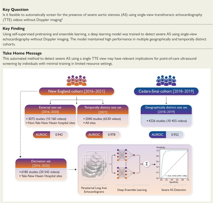
Background and aims: Early diagnosis of aortic stenosis (AS) is critical to prevent morbidity and mortality but requires skilled examination with Doppler imaging. This study reports the development and validation of a novel deep learning model that relies on two-dimensional (2D) parasternal long axis videos from transthoracic echocardiography without Doppler imaging to identify severe AS, suitable for point-of-care ultrasonography.

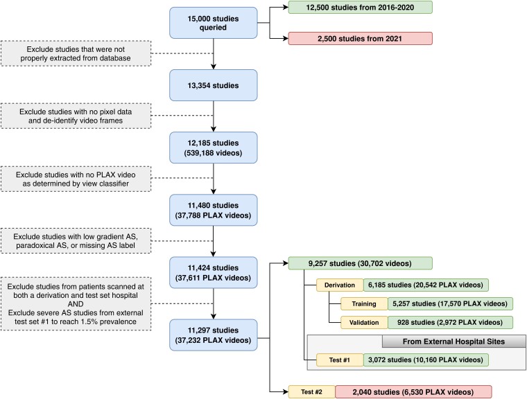
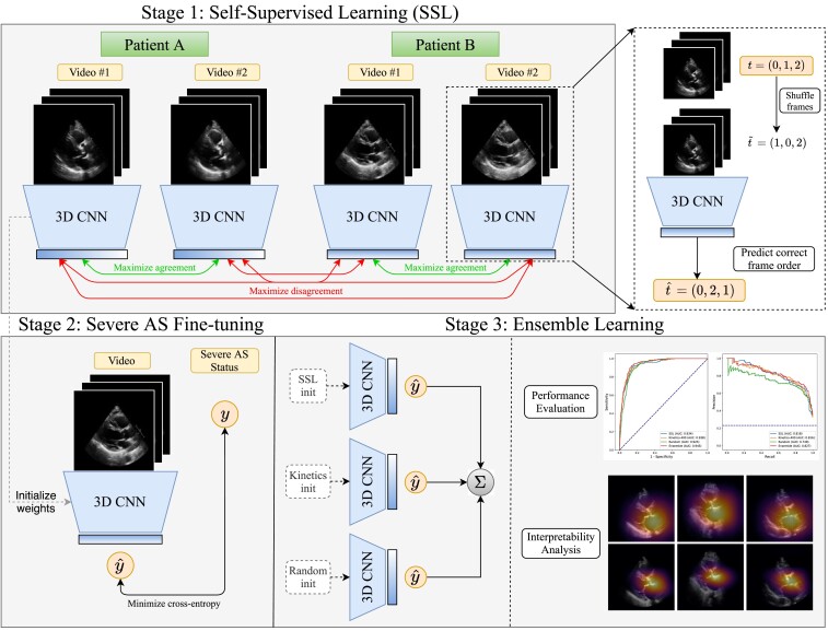
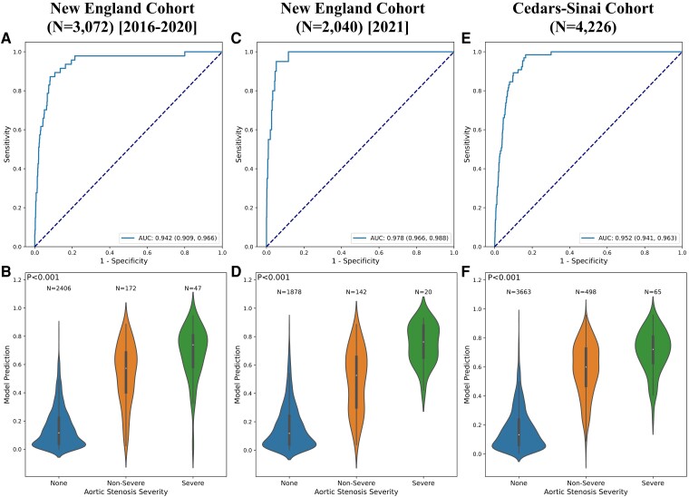
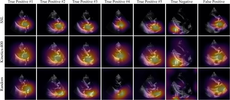
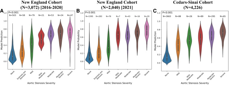
Methods and results: In a training set of 5257 studies (17 570 videos) from 2016 to 2020 [Yale-New Haven Hospital (YNHH), Connecticut], an ensemble of three-dimensional convolutional neural networks was developed to detect severe AS, leveraging self-supervised contrastive pretraining for label-efficient model development. This deep learning model was validated in a temporally distinct set of 2040 consecutive studies from 2021 from YNHH as well as two geographically distinct cohorts of 4226 and 3072 studies, from California and other hospitals in New England, respectively. The deep learning model achieved an area under the receiver operating characteristic curve (AUROC) of 0.978 (95% CI: 0.966, 0.988) for detecting severe AS in the temporally distinct test set, maintaining its diagnostic performance in geographically distinct cohorts [0.952 AUROC (95% CI: 0.941, 0.963) in California and 0.942 AUROC (95% CI: 0.909, 0.966) in New England]. The model was interpretable with saliency maps identifying the aortic valve, mitral annulus, and left atrium as the predictive regions. Among non-severe AS cases, predicted probabilities were associated with worse quantitative metrics of AS suggesting an association with various stages of AS severity.

Conclusion: This study developed and externally validated an automated approach for severe AS detection using single-view 2D echocardiography, with potential utility for point-of-care screening.

Keywords: Aortic stenosis; Deep learning; Digital health; Echocardiography.


Figures

Structured Graphical abstract
An automated deep learning approach for severe AS detection from single-view echocardiography evaluated across geographically and temporally distinct cohorts. AUROC, area under the receiver operating characteristic curve.
Figure 1
Figure 1
Inclusion-exclusion flowchart for the New England study population. Exclusion criteria for transthoracic echocardiography (TTE) studies and videos included in this study from the Yale-New Haven Health network. Studies with valid pixel data were de-identified frame by frame, and the parasternal long axis (PLAX) view was determined by an automated view classifier. A sample of 12 500 studies from 2016 to 2020 were split into a derivation set and external test set, which comprised studies from hospital sites not encountered during model development. An independent random sample of 2500 studies from 2021 was used as an additional test set to evaluate robustness to temporal shift
Figure 2
Figure 2
Overview of the proposed approach. We first perform self-supervised pretraining on parasternal long axis (PLAX) echocardiogram videos, selecting different PLAX videos from the same patient as ‘positive samples’ for contrastive learning. After this representation learning step, we then use these learned weights as the initialization for a model that is fine-tuned to predict severe aortic stenosis (AS) in a supervised fashion
Figure 3
Figure 3
Model performance in the external validation sets. Receiver operating characteristic curves (first row) and violin plots showing relationship of model output with aortic stenosis severity (second row) for the external New England cohort (first column), temporally distinct New England cohort (second column), and external Cedars-Sinai cohort (third column)
Figure 4
Figure 4
Saliency map visualization. Spatial attention maps for the self-supervised learning (SSL)-pretrained model (top row), Kinetics-pretrained model (middle row), and randomly initialized model (bottom row) for five true positives (first five columns), a true negative (sixth column), and a false positive (last column). As determined by the Kinetics-pretrained model, the first five columns represent the five most confident severe AS predictions, the sixth column represents the most confident ‘normal’ (no severe AS) prediction, and the seventh column represents the most confident incorrect severe AS prediction. Saliency maps were computed with the GradCAM method and reduced to a single 2D heatmap by maximum intensity projection along the temporal axis
Figure 5
Figure 5
Comparison between model predictions and echocardiographic left ventricular and aortic valve assessment among patients without severe aortic stenosis. Violin plots demonstrating the distribution of LVEF (left ventricular ejection fraction, (A) peak aortic valve velocity (B), mean aortic valve gradient (C) and mean aortic valve area (D) for patients without severe AS, stratified based on the predicted class based on the final ensemble model. These results are based on the temporally distinct cohort of patients scanned in 2021, without oversampling for severe aortic stenosis cases
Figure 6
Figure 6
Correlation of model probabilities with fine-grained AS severity. Violin plots depicting the distribution of predicted model probabilities stratified by cardiologist-determined AS severity for the New England 2016–2020 (A), New England 2021 (B), and Cedars-Sinai (C) cohorts

References

    1. Marc E, Piotr D, Bernard P, Olaf W, Cécile L, Jean-Luc M, et al. Contemporary management of severe symptomatic aortic stenosis. J Am Coll Cardiol 2021;78:2131–43. 10.1016/j.jacc.2021.09.864 - DOI - PubMed
    1. Otto CM, Prendergast B. Aortic-valve stenosis—from patients at risk to severe valve obstruction. N Engl J Med 2014;371:744–56. 10.1056/NEJMra1313875 - DOI - PubMed
    1. Smith CR, Leon MB, Mack MJ, Miller DC, Moses JW, Svensson LG, et al. Transcatheter versus surgical aortic-valve replacement in high-risk patients. N Engl J Med 2011;364:2187–98. 10.1056/NEJMoa1103510 - DOI - PubMed
    1. Reardon MJ, Van Mieghem NM, Popma JJ, Kleiman NS, Søndergaard L, Mumtaz M, et al. Surgical or transcatheter aortic-valve replacement in intermediate-risk patients. N Engl J Med 2017;376:1321–31. 10.1056/NEJMoa1700456 - DOI - PubMed
    1. Kang D-H, Park S-J, Lee S-A, Lee S, Kim D-H, Kim H-K, et al. Early surgery or conservative care for asymptomatic aortic stenosis. N Engl J Med 2020;382:111–9. 10.1056/NEJMoa1912846 - DOI - PubMed

Publication types