. 2023 Aug 23;19(8):e1011579.
doi: 10.1371/journal.ppat.1011579. eCollection 2023 Aug.

Candida albicans stimulates formation of a multi-receptor complex that mediates epithelial cell invasion during oropharyngeal infection

Affiliations

Candida albicans stimulates formation of a multi-receptor complex that mediates epithelial cell invasion during oropharyngeal infection

Quynh T Phan et al. PLoS Pathog. .

Abstract

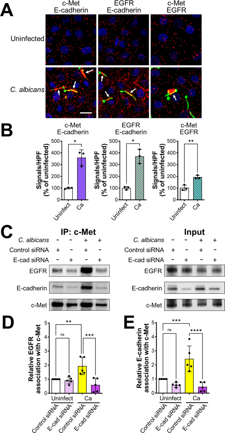
Fungal invasion of the oral epithelium is central to the pathogenesis of oropharyngeal candidiasis (OPC). Candida albicans invades the oral epithelium by receptor-induced endocytosis but this process is incompletely understood. We found that C. albicans infection of oral epithelial cells induces c-Met to form a multi-protein complex with E-cadherin and the epidermal growth factor receptor (EGFR). E-cadherin is necessary for C. albicans to activate both c-Met and EGFR and to induce the endocytosis of C. albicans. Proteomics analysis revealed that c-Met interacts with C. albicans Hyr1, Als3 and Ssa1. Both Hyr1 and Als3 are required for C. albicans to stimulate c-Met and EGFR in oral epithelial cells in vitro and for full virulence during OPC in mice. Treating mice with small molecule inhibitors of c-Met and EGFR ameliorates OPC, demonstrating the potential therapeutic efficacy of blocking these host receptors for C. albicans.


Conflict of interest statement

The authors have declared that no competing interests exist.

Figures

Fig 1
Fig 1. C. albicans activates c-Met in oral epithelial cells.
(A) Confocal microscopic images of OKF6/TERT-2 oral epithelial cells infected with C. albicans SC5314 for 20 min. c-Met, the epidermal growth factor receptor (EGFR), and E-cadherin were detected by indirect immunofluorescence using specific antibodies. Arrows point to the organisms in the magnified insets. Scale bar 10 μm. (B and C) Immunoblot analysis showing the time course of the phosphorylation of c-Met and EGFR in oral epithelial cells induced by C. albicans germ tubes (B). Densitometric analysis of 4 immunoblots (C). Data are mean ± SD. (D and E) Knockdown of c-Met with siRNA (D) or inhibition of c-Met signaling with SG523 (E) in oral epithelial cells inhibits the endocytosis of C. albicans. (F) Stimulation of oral epithelial cells with recombinant hepatocyte growth factor (HGF) enhances the endocytosis of C. albicans. Data in (D-F) are the mean ± SD of three experiments, each performed in triplicate. **p < 0.01, ****p < 0.0001, ns; not significant (two-tailed Student’s t test [D and E] or one-way ANOVA with Sidak’s multiple comparisons test [F]).
Fig 2
Fig 2. Functional interactions among c-Met, EGFR, and E-cadherin during the endocytosis of C. albicans.
(A-C) Immunoblot analysis showing the effects of the EGFR inhibitor gefitinib and the c-Met inhibitor SGX523 on C. albicans-induced phosphorylation of c-Met and EGFR in oral epithelial cells after 20 min of infection. Representative immunoblot (A), densitometric analysis of 4 immunoblots showing the phosphorylation of EGFR (B) and c-Met (C). Data are mean ± SD. (D) Effects of SGX523 and gefitinib on the endocytosis of C. albicans by oral epithelial cells. (E and F) Endocytosis of C. albicans by NIH/3T3 cells that expressed human c-Met (E) or human c-Met, EGFR, and HER2 (F). (G) Confocal micrographs of NIH3T3 cells expressing no human receptors (control), human c-Met, human EGFR and HER2, or human c-Met, EGFR, and HER2. The cells were infected with wild-type C. albicans germ tubes for 20 min, after which phosphorylated c-Met was detected with a phosphospecific anti-c-Met antibody. Scale bar 10 μm. Results in (D-F) are the mean ± SD of three experiments, each performed in triplicate. *p < 05, **p < 0.01, ***p < 0.001, ****p < 0.0001, ns; not significant (one-way ANOVA with Sidak’s multiple comparisons test [B-D] or two-tailed Student’s t test [E and F]).
Fig 3
Fig 3. E-cadherin is required for C. albicans to activate c-Met and EGFR during the endocytosis of C. albicans.
(A-C) Knockdown of E-cadherin by siRNA inhibits the phosphorylation of c-Met and EGFR in oral epithelial cells infected with C. albicans. Representative immunoblot (A). Densitometric analysis of 5 immunoblots showing the phosphorylation of c-Met (B) and EGFR (C). Results are mean ± SD. (D and E) Effects of inhibiting c-Met (D) and EGFR (E) in combination with siRNA knockdown of E-cadherin on the endocytosis of C. albicans by oral epithelial cells. Results in (D and E) are the mean ± SD of three experiments, each performed in triplicate. *p < 05, **p < 0.01, ***p < 0.001, ns; not significant (one-way ANOVA with Sidak’s multiple comparisons test.
Fig 4
Fig 4. C. albicans induces c-Met, EGFR, and E-cadherin to form a multiprotein complex.
(A-C) Proximity ligation assay showing the interaction of c-Met with E-cadherin, EGFR with E-cadherin, and c-Met with EGFR in oral epithelial cells with and without 20-min infection with C. albicans. Confocal microscopic images (A). Scale bar 10 μm. Signal counts (B). Proximity ligation assay showing the interaction of c-Met with E-cadherin, EGFR with E-cadherin, and c-Met with EGFR in oral epithelial cells with and without 20-min infection with C. albicans. (A) Confocal microscopic images. Scale bar 10 μm. (B) Signal counts. (C-E) Co-immunoprecipitation experiments in oral epithelial cells transfected with control or E-cadherin (E-cad) siRNA and then infected with C. albicans for 20 min. (C) Representative immunoblots of proteins obtained by immunoprecipitation with an anti-c-Met antibody. (D and E) Densitometric analysis of 5 immunoblots. Results are mean ± SD. **p < 0.01, ***p < 0.001, ****p < 0.0001, ns; not significant (two-tailed Student’s t test [B] or one-way ANOVA with Sidak’s multiple comparisons test [D and E]).
Fig 5
Fig 5. Hyr1 interacts with c-Met.
(A-C). C. albicans germ tubes stimulate phosphorylation of c-Met and EGFR after 20 min of infection. Representative immunoblots (A). Densitometric analysis of 4 immunoblots showing the phosphorylation of c-Met (B) and EGFR (C) induced by C. albicans yeast and hyphae. (D-F) An als3 Δ/Δ ssa1Δ/Δ mutant does not induce phosphorylation of c-Met and EGFR. Representative immunoblots (D). Densitometric analysis of 4 immunoblots showing the phosphorylation of c-Met (E) and EGFR (F) induced by the indicated strains of C. albicans. (G) Far Western blot showing proteins from the indicated C. albicans morphotypes and strains that were recognized by recombinant c-Met. Arrow indicates the protein band. (H-J) Hyr1 is required for maximal phosphorylation of c-Met and EGFR. Representative immunoblots (H). Densitometric analysis of 4 immunoblots showing the phosphorylation of c-Met (I) and EGFR (J) induced by the indicated strains of C. albicans. (K) Hyr1 is required for maximal endocytosis of C. albicans. Results in (B, C, E, F, I-K) are mean ± SD. WT, wild type; *p < 0.05, **p < 0.01, ***p < 0.001, ****p, 0.0001, ns; not significant (one-way ANOVA with Sidak’s multiple comparisons test).
Fig 6
Fig 6. Model of how C. albicans induces its own endocytosis by oral epithelial cells.
In uninfected epithelial cells, c-Met, E-cadherin, and EGFR/HER2 are not associated with each other in the cell membrane. When a cell is infected with C. albicans, Hyr1 and Als3 on the fungal cell surface cause c-Met, E-cadherin, and EGFR/HER2 to form a multi-protein complex. This complex contains activated (phosphorylated) c-Met and EGFR, which induce epithelial cell cytoskeletal rearrangement and the subsequent endocytosis of the fungus.
Fig 7
Fig 7. Interactions of c-Met with Als3.
(A-C) Both Hyr1 and Als3 are required for phosphorylation of c-Met and EGFR. Representative immunoblots from oral epithelial cells infected with the indicated strains of C. albicans (A). Densitometric analysis of 5 immunoblots showing the phosphorylation of c-Met (B) and EGFR (C). (D) Epithelial cell endocytosis of the indicated strains of C. albicans. (E) Damage to oral epithelial cells caused by the indicated strains of C. albicans. (F) Invasion of live and fixed epithelial cells by the wild-type (WT) hyr1Δ/Δ mutant strains. (G-J) Induction of epithelial cell secretion of IL-1α (G), IL-1β (H), IL-8 (I) and GM-CSF (J) by the indicated strains of C. albicans. Results are mean ± SD. WT, wild type; ns, not significant; *p < 0.05, **p < 0.01, ***p, 0.001, ****p < 0.0001 (one-way ANOVA with Sidak’s multiple comparisons test).
Fig 8
Fig 8. Hyr1 and Als3 are required for C. albicans to resist killing by neutrophils.
(A) Human neutrophils were infected with the indicated C. albicans strains constructed in the SC5314 strain background. Results are mean ± SD of neutrophils from 3 donors, tested in triplicate. (B) The hyr1Δ/Δ mutant is resistant to killing by mouse neutrophils, even in the absence of c-Met. Killing of wild-type and hyr1Δ/Δ mutant strains in the SN250 strain background by neutrophils from Mrp8;Metfl/fl mice, which have a neutrophil-specific deletion in c-Met, and their littermates. Results are mean ± SD of neutrophils from 2 experiments performed in triplicate. ns, not significant; *p < 0.05, **p < 0.01, ***p < 0.001, ****p < 0.0001 (one-way ANOVA with Sidak’s multiple comparisons test).
Fig 9
Fig 9. Als3 and Hyr1 are required for C. albicans to activate c-Met and EGFR during OPC in mice.
Confocal micrographs of the tongues of immunocompetent mice after 1 d of infection with the indicated strains of C. albicans. The samples were stained for c-Met, C. albicans, and DAPI (A) or EGFR, C. albicans, and DAPI (B). Epithelial cells are in the region above the dotted lines. Scale bar 50 μm.
Fig 10
Fig 10. Hyr1 and Als3 mediate virulence during OPC.
(A and B) Oral fungal burden of immunocompetent mice infected with the indicated C. albicans strains for 1 (A) and 2 (B) days. (C and D) Effects of phagocyte depletion on susceptibility to OPC, as determined by oral fungal burden after 1 (C) and 2 (D) days of infection. (E) Oral fungal burden after 5 d of infection with the wild-type strain of mice that had been immunosuppressed with cortisone acetate and treated with gefitinib and/or SGX523. (F) Histopathology of the tongues. Scale bar 200 μm. Data are the mean ± SD combined results from 2 independent experiments, each using 4 mice per C. albicans strain (A-D) or 8 mice per condition in one experiment and 5 mice per condition in the other (E). ns, not significant; *p < 0.05, **p < 0.01, ***p < 0.001, ****p < 0.0001 (one-way ANOVA with Sidak’s multiple comparisons test).

Update of

References

    1. Berberi A, Noujeim Z, Aoun G. Epidemiology of oropharyngeal candidiasis in human immunodeficiency virus/acquired immune deficiency syndrome patients and CD4+ counts. J Int Oral Health. 2015;7(3):20–3. Epub 2015/04/17. ; PubMed Central PMCID: PMC4385720. - PMC - PubMed
    1. Serrano J, Lopez-Pintor RM, Ramirez L, Fernandez-Castro M, Sanz M, Melchor S, et al. Risk factors related to oral candidiasis in patients with primary Sjogren’s syndrome. Med Oral Patol Oral Cir Bucal. 2020;25(5):e700–e5. Epub 2020/07/20. doi: 10.4317/medoral.23719 ; PubMed Central PMCID: PMC7473438. - DOI - PMC - PubMed
    1. Saito H, Shodo R, Yamazaki K, Katsura K, Ueki Y, Nakano T, et al. The association between oral candidiasis and severity of chemoradiotherapy-induced dysphagia in head and neck cancer patients: A retrospective cohort study. Clin Transl Radiat Oncol. 2020;20:13–8. Epub 2019/11/19. doi: 10.1016/j.ctro.2019.10.006 ; PubMed Central PMCID: PMC6849117. - DOI - PMC - PubMed
    1. Guilbert TW, Colice G, Grigg J, van Aalderen W, Martin RJ, Israel E, et al. Real-life outcomes for patients with asthma prescribed spacers for use with either extrafine- or fine-particle inhaled corticosteroids. J Allergy Clin Immunol Pract. 2017;5(4):1040–9. doi: 10.1016/j.jaip.2016.11.026 - DOI - PubMed
    1. Pappas PG, Kauffman CA, Andes DR, Clancy CJ, Marr KA, Ostrosky-Zeichner L, et al. Clinical practice guideline for the management of candidiasis: 2016 update by the Infectious Diseases Society of America. Clin Infect Dis. 2016;62(4):16. - PMC - PubMed

Publication types

Supplementary concepts