

Thinner inner retinal layers are associated with lower cognitive performance, lower brain volume, and altered white matter network structure-The Maastricht Study
- PMID: 37611119
- PMCID: PMC10917009
- DOI: 10.1002/alz.13442
Thinner inner retinal layers are associated with lower cognitive performance, lower brain volume, and altered white matter network structure-The Maastricht Study
Abstract
Introduction: The retina may provide non-invasive, scalable biomarkers for monitoring cerebral neurodegeneration.
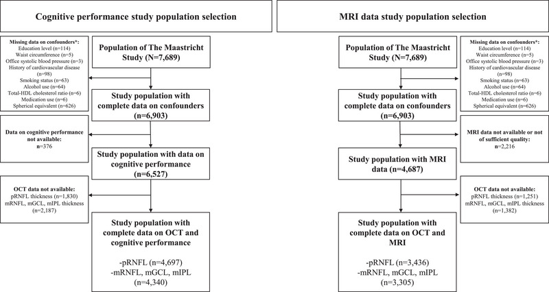
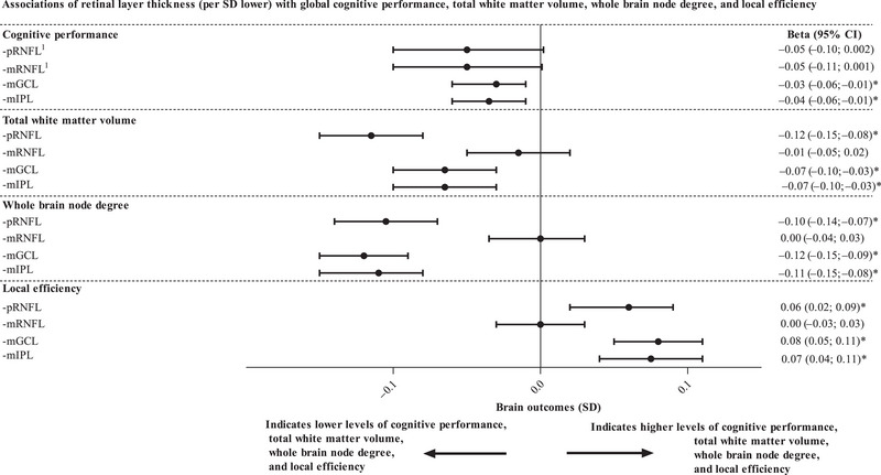
Methods: We used cross-sectional data from The Maastricht study (n = 3436; mean age 59.3 years; 48% men; and 21% with type 2 diabetes [the latter oversampled by design]). We evaluated associations of retinal nerve fiber layer, ganglion cell layer, and inner plexiform layer thicknesses with cognitive performance and magnetic resonance imaging indices (global grey and white matter volume, hippocampal volume, whole brain node degree, global efficiency, clustering coefficient, and local efficiency).
Results: After adjustment, lower thicknesses of most inner retinal layers were significantly associated with worse cognitive performance, lower grey and white matter volume, lower hippocampal volume, and worse brain white matter network structure assessed from lower whole brain node degree, lower global efficiency, higher clustering coefficient, and higher local efficiency.
Discussion: The retina may provide biomarkers that are informative of cerebral neurodegenerative changes in the pathobiology of dementia.
Keywords: brain structural connectivity; brain volume; clustering coefficient; cognitive function; cognitive performance; global efficiency; graph theory; grey matter; local efficiency; magnetic resonance imaging (MRI); optical coherence tomography (OCT); retinal imaging; retinal neurodegeneration; white matter; whole brain node degree.
© 2023 The Authors. Alzheimer's & Dementia published by Wiley Periodicals LLC on behalf of Alzheimer's Association.
Conflict of interest statement
The authors declare no conflicts of interest. Author disclosures are available in the Supporting information.
Figures
References
Publication types
MeSH terms
Substances
Grants and funding
- 31O.041/OP-Zuid, the Province of Limburg, the Dutch Ministry of Economic Affairs
- Stichting De Weijerhorst (Maastricht, the Netherlands), the Pearl String Initiative Diabetes (Amsterdam, the Netherlands), the Cardiovascular Center (CVC, Maastricht, the Netherlands), CARIM School for Cardiovascular Diseases (Maastricht, the Netherlands), CAPHRI School for Public Health and Primary Care (Maastricht, the Netherlands), NUTRIM School for Nutrition and Translational Research in Metabolism (Maastricht, the Netherlands), Stichting Annadal (Maastricht, the Netherlands), Health Foundation Limburg (Maastricht, the Netherlands), Perimed (Järfälla, Sweden), and by unrestricted grants from Janssen-Cilag B.V. (Tilburg, the Netherlands), Novo Nordisk Farma B.V. (Alphen aan den Rijn, the Netherlands), and Sanofi-Aventis Netherlands B.V. (Gouda, the Netherlands)
- 916.19.074/VENI research
- 2018T025/Netherlands Organization for Scientific Research and the Netherlands Organization for Health Research and Development, and a Dutch Heart Foundation research
- 2021.81.004/Diabetes Fonds Fellowship
LinkOut - more resources
Full Text Sources
Medical
