Review
. 2023 Nov;299(11):105182.
doi: 10.1016/j.jbc.2023.105182. Epub 2023 Aug 22.

Structural insights of the p97/VCP AAA+ ATPase: How adapter interactions coordinate diverse cellular functionality

Affiliations
Review

Structural insights of the p97/VCP AAA+ ATPase: How adapter interactions coordinate diverse cellular functionality

Julian R Braxton et al. J Biol Chem. 2023 Nov.

Abstract

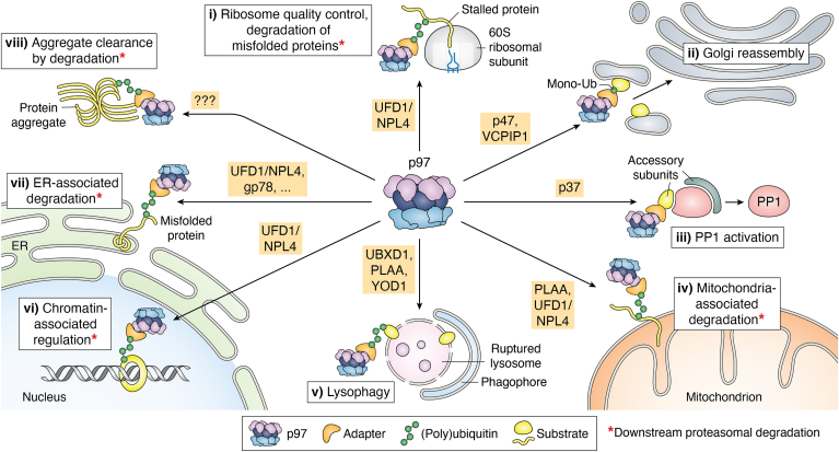
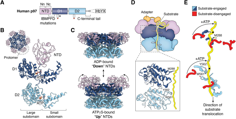
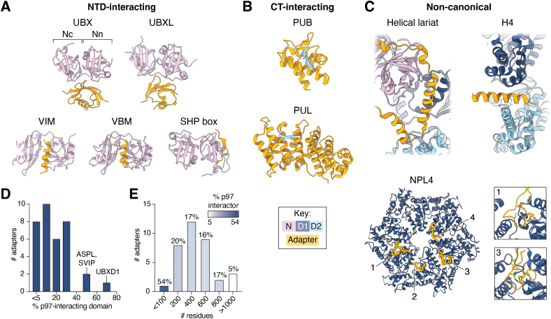
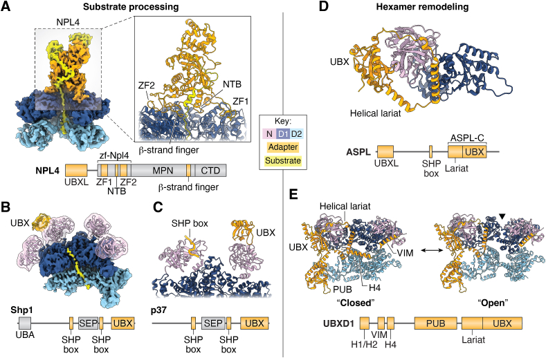
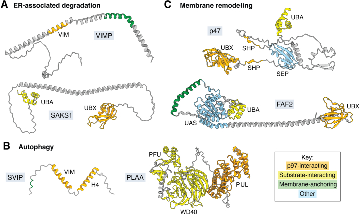
p97/valosin-containing protein is an essential eukaryotic AAA+ ATPase with diverse functions including protein homeostasis, membrane remodeling, and chromatin regulation. Dysregulation of p97 function causes severe neurodegenerative disease and is associated with cancer, making this protein a significant therapeutic target. p97 extracts polypeptide substrates from macromolecular assemblies by hydrolysis-driven translocation through its central pore. Growing evidence indicates that this activity is highly coordinated by "adapter" partner proteins, of which more than 30 have been identified and are commonly described to facilitate translocation through substrate recruitment or modification. In so doing, these adapters enable critical p97-dependent functions such as extraction of misfolded proteins from the endoplasmic reticulum or mitochondria, and are likely the reason for the extreme functional diversity of p97 relative to other AAA+ translocases. Here, we review the known functions of adapter proteins and highlight recent structural and biochemical advances that have begun to reveal the diverse molecular bases for adapter-mediated regulation of p97 function. These studies suggest that the range of mechanisms by which p97 activity is controlled is vastly underexplored with significant advances possible for understanding p97 regulation by the most known adapters.

Keywords: AAA+ ATPase; AlphaFold; ERAD; VCP; adapter; adaptor; autophagy; cofactor; cryo-EM; molecular chaperone; p97; protein structure prediction; proteostasis; unfoldase.


Conflict of interest statement

Conflict of interest The authors declare that they have no conflicts of interest with the contents of this article.

Figures

Figure 1
Figure 1
Cellular functions of p97. Overview of p97-dependent pathways, with associated adapters listed. p97 is colored by domain (NTD: purple, D1: dark blue, D2: light blue), and adapters are in orange. Substrates (yellow) are shown as spherical shapes to indicate folded, native states or as strands to indicate misfolded or unfolded states. Pathways where p97 activity promotes downstream proteasomal degradation are indicated (∗). p97-dependent processes are as follows (going clockwise from ribosome quality control): (i) as a component of ribosome quality control p97 with the adapter UFD1/NPL4 (UN) dislodges stalled substrates from 60S ribosomal subunits (28); (ii) during Golgi reassembly p97 with adapters p47 and VCPIP1 promotes fusion of postmitotic Golgi fragments through targeting of monoubiquitylated syntaxin 5 on the Golgi membrane (38, 39); (iii) in PP1 activation p97 with the p37 adapter extracts an inhibitory subunit (inhibitor 3) from an inactive PP1 precursor complex enabling formation of active holoenzymes (41, 42, 43); (iv) in mitochondria-associated degradation p97 with the adapters Doa1 (PLAA in humans) and UN extracts misfolded proteins from the outer mitochondrial membrane (101); (v) in lysophagy, extraction of (currently unknown) surface proteins on ruptured lysosomes by p97 with adapters UBXD1, PLAA, and YOD1 is required for phagophore formation (blue) and subsequent autophagic clearance (34); (vi) as an example of a chromatin-related function, after DNA double-strand break repair p97 with UN removes trapped Ku70/80 rings from intact DNA (33); (vii) in ER-associated degradation, p97 with UN, gp78, and many other adapters extracts misfolded proteins from the ER lumen (26, 102); (viii) in the clearance of tau fibrillar aggregates p97 may target ubiquitylated tau, though the relevant adapter(s) are unknown (45). ER, endoplasmic reticulum; NTD, N-terminal domain; PP1, protein phosphatase 1.
Figure 2
Figure 2
Structure and substrate threading mechanism of p97.A, domain schematic of human p97 colored by domain (NTD: purple, D1: dark blue, and D2: light blue). Location of pore loops in D1 and D2 are shown; degenerate residues in D1 are indicated (∗). The two lobes of the NTD (N- and C-terminal lobes, Nn and Nc) are indicated, as is the location of most IBMPFD mutations, namely the NTD-D1 interface. The HbYX motif at the end of the C-terminal tail is also shown. B, protomer of ATPγS-bound p97 from an intact hexamer (PDB 5FTN) (52), colored as in (A). A downscaled top view of the full hexamer is also shown, with one protomer circled to delineate protomer boundaries. C, side views of ADP-bound (top, PDB 5FTK) and ATPγS-bound (bottom, PDB 5FTN) p97 hexamers (52), showing rotation and elevation of the NTDs above the D1 ring in the ATPγS-bound state. D, illustration of adapter-mediated substrate threading through the p97 central pore, with D1 and D2 pore loops shown. The adapter is colored in orange, and the substrate in yellow. Below, an enlarged view of pore loop contacts is shown (PDB 6OA9) (23). E, view of substrate in the Cdc48 channel (PDB 6OA9) (23), showing a spiral arrangement of pore loops in D1 and D2. Pore loops engaged with substrate are shown in blue; those not engaged are in red. The highest substrate contacts in D1 and D2 are marked by black dots; hydrolysis and subsequent ATP binding by the disengaged protomers is proposed to drive translocation by two amino acid steps whereby conformational changes enable pore loop engagement with the next site along the substrate (gray dots). IBMPFD, inclusion body myopathy associated with Paget disease of bone and frontotemporal dementia; NTD, N-terminal domain; PDB, Protein Data Bank.
Figure 3
Figure 3
Architecture and interaction modes of p97 adapters. Structures of individual p97-interacting domains bound to the p97 NTD, CT tail, or other sites are shown, highlighting the diversity of adapter interaction modes. A, structures of the p97 NTD bound to NTD-interacting domains. The p97 NTD lobes are indicated in the UBX-bound structure. PDB IDs: UBX: 5X4L (63), UBXL: 4KDI (66), VIM: 3TIW (67), VBM: 5EPP (68), and SHP box: 5GLF (69). B, structures of the HbYX motif at the end of the p97 CT tail bound to CT-interacting domains. PDB IDs: PUB: 2HPL (73) and PUL: 3EBB (71). C, structures of noncanonical p97-interacting domains bound to p97 or a homolog thereof. PDB IDs: helical lariat: 8FCL (24), H4: 8FCR (24), and NPL4: 6OA9 (23). The four interacting motifs in NPL4 are numbered counterclockwise: zinc finger 1 (ZF1, 1), N-terminal bundle (NTB, 2), zinc finger 2 (ZF2, 3), and β-strand finger (4). Enlarged views of ZF1 and ZF2 are shown at right; Zn2+ is colored in teal. D, histogram of the proportion of adapters corresponding to p97-interacting domains (determined by % of total sequence length). E, histogram of adapter length. Bars are colored by average % p97 interactor of adapters in that bin (determined by % of total sequence length). The color key indicates p97 domains, as in Figure 2, and adapters. CT, C-terminal; NTD, N-terminal domain; PDB, Protein Data Bank; PUB, peptide:N-glycanase and UBA or UBX-containing proteins; PUL, PLAP, Ufd3p, and Lub1p; UBX, ubiquitin regulatory X; UBXL, UBX-like; VBM, VCP-binding motif; VIM, VCP-interacting motif.
Figure 4
Figure 4
Structures of intact p97-adapter complexes. Structures of p97-adapter complexes involved in substrate processing (left) or hexamer remodeling (right). Domain colors are indicated by the key. Domain schematics (not to scale) of each adapter are shown below the corresponding structure, with p97-interacting domains colored in orange and other domains in gray. A, (left) cryo-EM structure of yeast Cdc48 bound to the UFD1/NPL4 adapter (PDB 6OA9) (23), unfolding an ubiquitylated substrate and threading it through the Cdc48 central channel. (Right) Enlarged view of contacts made between NPL4 and Cdc48. B, cryo-EM structure of Cdc48 bound to substrate and the Shp1 adapter (PDB 6OPC) (56). A filtered transparent map and model of the NTDs and UBX is shown over the sharpened map of the D1, D2, and substrate. C, cryo-EM structure of a SHP box and UBX domain of p37 bound to adjacent NTDs of an actively processing p97 complex (PDB 8B5R) (43). D, crystal structure of an ASPL truncation construct containing the UBX and helical lariat domains (ASPL-C) bound to a human p97 construct (PDB 5IFS) (74). E, cryo-EM structures of human p97 bound to UBXD1 (closed: PDB 8FCR, open: PDB 8FCM) (24), showing separation of adjacent p97 protomers coordinated by multiple UBXD1 contacts. The hexamer seam is indicated in the open state by a black triangle. NTB, N-terminal bundle; NTD, N-terminal domain; PDB, Protein Data Bank; UBX, ubiquitin regulatory X; ZF, zinc finger.
Figure 5
Figure 5
AlphaFold2 predictions of selected p97 adapters. AlphaFold2 models of p97 adapters implicated in (A) ER-associated degradation, (B) membrane remodeling, and (C) autophagy. Domain colors are indicated by the key. ER, endoplasmic reticulum.

References

    1. Peters J.M., Walsh M.J., Franke W.W. An abundant and ubiquitous homo-oligomeric ring-shaped ATPase particle related to the putative vesicle fusion proteins Sec18p and NSF. EMBO J. 1990;9:1757–1767. - PMC - PubMed
    1. Stach L., Freemont P.S. The AAA+ ATPase p97, a cellular multitool. Biochem. J. 2017;474:2953–2976. - PMC - PubMed
    1. Prakash S., Tian L., Ratliff K.S., Lehotzky R.E., Matouschek A. An unstructured initiation site is required for efficient proteasome-mediated degradation. Nat. Struct. Mol. Biol. 2004;11:830–837. - PubMed
    1. Olszewski M.M., Williams C., Dong K.C., Martin A. The Cdc48 unfoldase prepares well-folded protein substrates for degradation by the 26S proteasome. Commun. Biol. 2019;2:29. - PMC - PubMed
    1. Meyer H.H., Wang Y., Warren G. Direct binding of ubiquitin conjugates by the mammalian p97 adaptor complexes, p47 and Ufd1–Npl4. EMBO J. 2002;21:5645–5652. - PMC - PubMed

Publication types

MeSH terms

Substances