Case Reports
. 2024 May 1;63(9):1277-1280.
doi: 10.2169/internalmedicine.2514-23. Epub 2023 Aug 23.

Cerebral Venous Thrombosis Mimicking Limbic Encephalitis

Affiliations
Case Reports

Cerebral Venous Thrombosis Mimicking Limbic Encephalitis

Toshiki Tezuka et al. Intern Med. .

Abstract

Cerebral venous thrombosis (CVT) is challenging to diagnose, as it presents with variable symptoms. We encountered a complicated case of CVT that mimicked limbic encephalitis due to sensory aphasia. Based on the characteristic magnetic resonance imaging findings, this 72-year-old Japanese man was later confirmed to have CVT, the cause of which was periodontitis due to Eikenella corrodens, a Gram-negative facultative anaerobic that is part of the mouth's normal flora. The symptoms improved without sequelae following anticoagulation treatment and antibiotics. Clinicians should consider CVT as a differential diagnosis when unexplainable neurological symptoms suggesting limbic encephalitis are observed.

Keywords: cerebral venous thrombosis; infectious disease; limbic encephalitis; periodontal infection.


Conflict of interest statement

The authors state that they have no Conflict of Interest (COI).

Figures

Figure.
Figure.
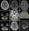
The characteristic radiological findings of CVT. A: Head CT showed a high-density area in the straight sinus, suggesting cerebral venous sinus thrombosis (yellow arrowhead). B, C: Brain MRI identified a high signal intensity in the superior sagittal sinus on DWI (yellow arrowhead), and the corresponding area on a T2-weight image was missing the signal void (yellow arrowhead). D: On DWI-MRI, a low signal intensity in the bilateral cerebellar hemisphere was observed, reflecting decreased perfusion in the affected area (yellow arrowheads). E: The ADC values were high in the bilateral cerebellar region (yellow arrowheads). F: On an intracranial 4D-CT venogram, thrombotic occlusion was detected within the left sigmoid, transverse, superior sagittal, and straight sinuses (yellow arrowheads). G: After treatment for a week, the low-DWI-intensity signal in the bilateral cerebellar hemisphere disappeared. H: After treatment for one month, the thrombi in the transverse sinus disappeared on DWI-MRI. I: On MR venography, the thrombi improved, recanalizing the cerebral venous sinus, and collateral circulation emerged in the cerebral venous system.

References

    1. Bousser MG, Ferro JM. Cerebral venous thrombosis: an update. Lancet Neurol 6: 162-170, 2007. - PubMed
    1. Ferro JM, Aguiar de Sousa D. Cerebral venous thrombosis: an update. Curr Neurol Neurosci Rep 19: 74, 2019. - PubMed
    1. Silvis SM, de Sousa DA, Ferro JM, Coutinho JM. Cerebral venous thrombosis. Nat Rev Neurol 13: 555-565, 2017. - PubMed
    1. Das S, Dubey S, Pandit A, Ray BK. Bilateral thalamic lesion presenting as Broca's type subcortical aphasia in cerebral venous thrombosis: index case report. BMJ Case Rep 14: e240196, 2021. - PMC - PubMed
    1. Bradshaw MJ, Venkatesan A. Herpes simplex virus-1 encephalitis in adults: pathophysiology, diagnosis, and management. Neurotherapeutics 13: 493-508, 2016. - PMC - PubMed

Publication types