. 2023 Aug 23;9(1):88.
doi: 10.1038/s41421-023-00577-5.

A novel recombination protein C12ORF40/REDIC1 is required for meiotic crossover formation

Affiliations

A novel recombination protein C12ORF40/REDIC1 is required for meiotic crossover formation

Suixing Fan et al. Cell Discov. .

Abstract

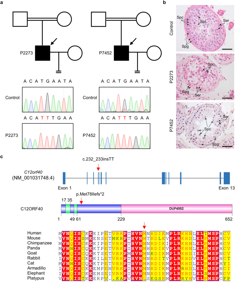
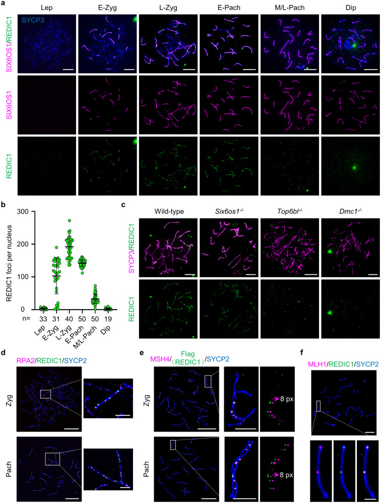
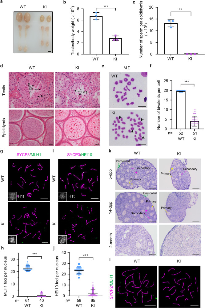
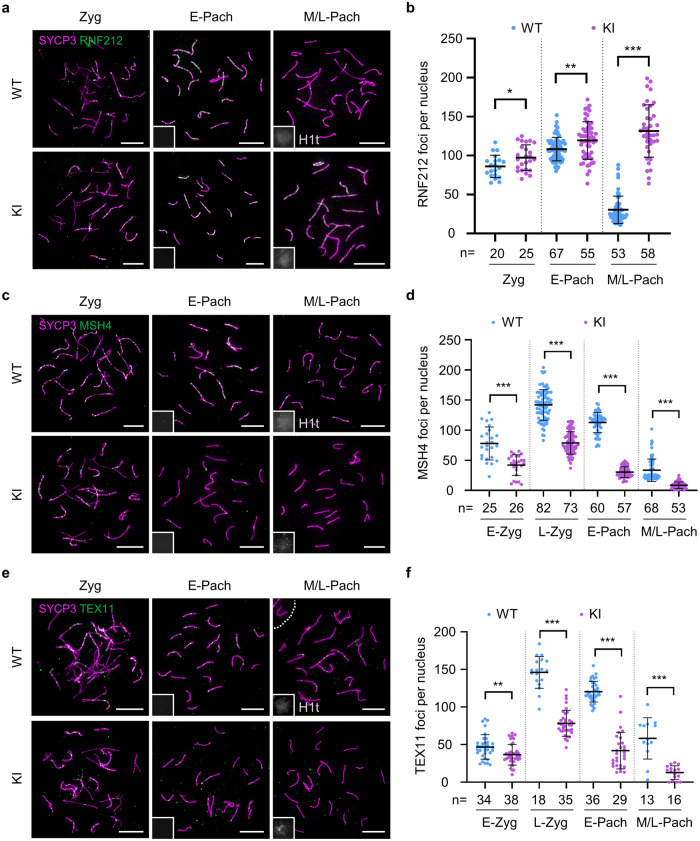
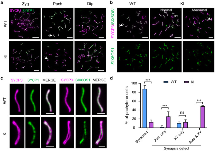
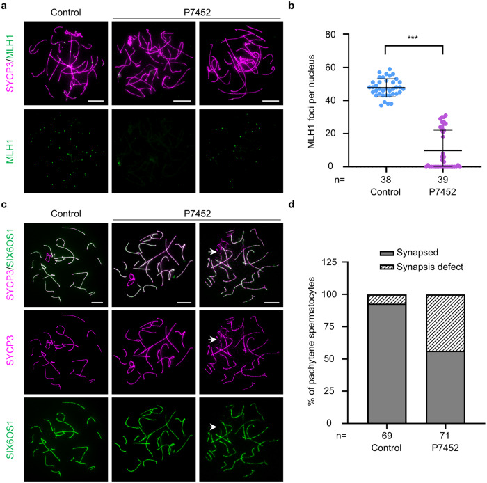
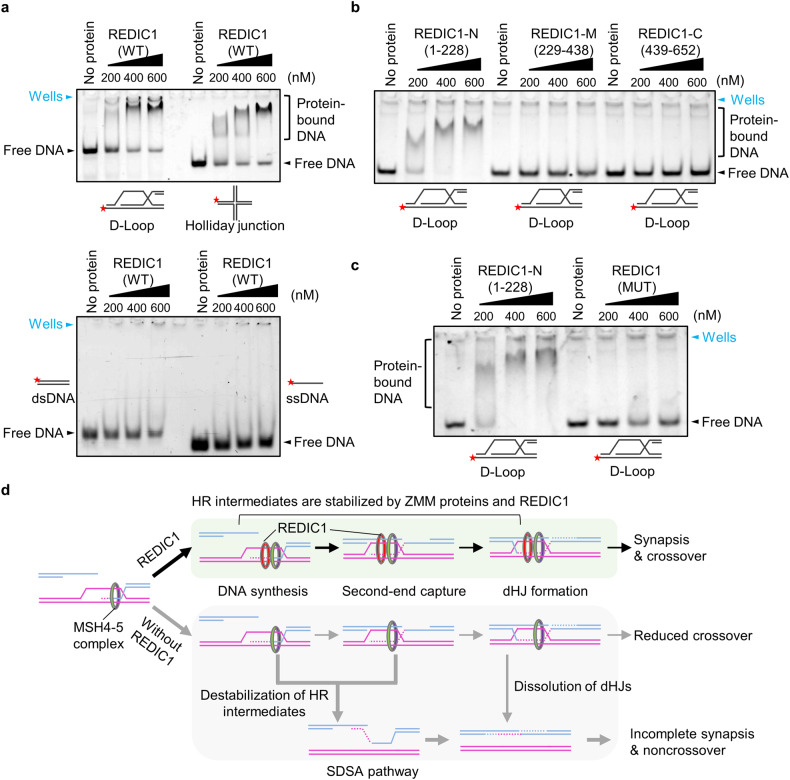
During meiosis, at least one crossover must occur per homologous chromosome pair to ensure normal progression of meiotic division and accurate chromosome segregation. However, the mechanism of crossover formation is not fully understood. Here, we report a novel recombination protein, C12ORF40/REDIC1, essential for meiotic crossover formation in mammals. A homozygous frameshift mutation in C12orf40 (c.232_233insTT, p.Met78Ilefs*2) was identified in two infertile men with meiotic arrest. Spread mouse spermatocyte fluorescence immunostaining showed that REDIC1 forms discrete foci between the paired regions of homologous chromosomes depending on strand invasion and colocalizes with MSH4 and later with MLH1 at the crossover sites. Redic1 knock-in (KI) mice homozygous for mutation c.232_233insTT are infertile in both sexes due to insufficient crossovers and consequent meiotic arrest, which is also observed in our patients. The foci of MSH4 and TEX11, markers of recombination intermediates, are significantly reduced numerically in the spermatocytes of Redic1 KI mice. More importantly, our biochemical results show that the N-terminus of REDIC1 binds branched DNAs present in recombination intermediates, while the identified mutation impairs this interaction. Thus, our findings reveal a crucial role for C12ORF40/REDIC1 in meiotic crossover formation by stabilizing the recombination intermediates, providing prospective molecular targets for the clinical diagnosis and therapy of infertility.


Conflict of interest statement

The authors declare no competing interests.

Figures

Fig. 1
Fig. 1. Identification of a homozygous frameshift mutation in C12orf40 from two NOA-affected men born to consanguineous parents.
a Pedigrees of two Chinese families with a C12orf40 mutation. The double horizontal lines indicate the consanguineous marriage. Squares and circles denote male and female members, respectively. Solid symbols indicate the members with nonobstructive azoospermia. Members indicated by arrows were selected for WES. Sanger sequencing chromatograms of C12orf40 are shown at the bottom. The 2-bp insertion in C12orf40 is marked in red. b Histological analysis of human testicular sections by hematoxylin and eosin staining. A man who was diagnosed with obstructive azoospermia served as the control. Spg, spermatogonium; Spc, spermatocyte; Spt, spermatid; Ser, Sertoli cell. Scale bars: 50 μm. c Location and conservation of the C12ORF40 mutation. The gene composition is based on the Ensembl database (GRCh38, transcript ID: ENST00000324616.9; NCBI RefSeq ID: NM_001031748.4). The blue solid squares represent exons. The domains were predicted by the SMART web server. The conservation of the mutated amino acid is evaluated by the sequence alignment of orthologs from the indicated species. The red arrow shows the identified mutation. The results of the full-length protein multiple sequence alignment are shown in Supplementary Fig. S2.
Fig. 2
Fig. 2. REDIC1 is localized to recombination intermediates in meiotic prophase cells.
a Representative spread spermatocytes of 30 dpp WT mice stained with antibodies against SYCP3 (blue), REDIC1 (green), and SIX6OS1 (magenta). Lep, leptotene; E-Zyg, early zygotene; L-Zyg, late zygotene; E-Pach, early pachytene; M/L-Pach, mid- or late pachytene; Dip, diplotene. Scale bars: 10 μm. b Quantification of the number of REDIC1 foci on chromosome axes at the indicated substages from WT mice. n, the total number of cells analyzed from two animals. The bars represent mean ± SD. c Representative images of zygotene (like) spermatocyte spreads from adult WT, Six6os1–/–, Top6bl–/–, and Dmc1–/– mice stained for SYCP3 (magenta) and REDIC1 (green). The experiments were repeated two times with similar results. Scale bars: 10 μm. d Representative images of surface-spread chromosomes from zygotene and pachytene mouse spermatocytes stained for RPA2 (magenta), REDIC1 (green), and the chromosomal axis marker SYCP2 (blue). The areas in the white rectangles are enlarged. e Structured illumination microscopy of spread zygotene and pachytene spermatocytes of Redic1-Flag/Myc mouse stained for MSH4 (magenta), Flag (green), and chromosomal axis marker SYCP2 (blue). The areas in the white rectangles are enlarged. The panel on the far right shows the magenta and green signals shifted by 8 pixels. Images are representatives from experiments with two adult animals. f Representative images of surface-spread chromosomes from a mid-pachytene spermatocyte stained for MLH1 (magenta), REDIC1 (green), and SYCP2 (blue) in WT mice. The magnified panels show a pair of synapsed chromosomes on which two MLH1 foci colocalized with two REDIC1 signals. The experiments were performed at least twice with similar results. Scale bars in d, e, and f are 10 μm for the original images and 2 μm for the enlarged images, respectively.
Fig. 3
Fig. 3. Mouse models mimicking the patients’ mutation exhibited defects in crossover formation.
a Representative images of testis and epididymis morphology from 2-month-old WT and Redic1 KI mice. Scale bar: 2 mm. b, c Ratios of the testis to body weight (b) and sperm counts per epididymis (c) in 2-month-old WT and Redic1 KI mice. The data are from three adult mice for each genotype and represent the mean ± SD. **P < 0.01; ***P < 0.001; two-tailed Student’s t-test. d Representative images of hematoxylin and eosin-stained testicular and epididymal sections from adult Redic1 KI and WT mice. The metaphase I spermatocytes (MI) and spermatids (spt) are indicated by the arrows. Scale bars: 50 μm. e Representative metaphase I (MI) spermatocytes from adult WT and Redic1 KI mice stained with Giemsa. The bivalent chromosomes in Redic1 KI mice are indicated by the arrows. Scale bars: 10 μm. f Quantification of bivalents per metaphase I spermatocyte from 2-month-old WT and KI mice. n, the total number of MI cells scored from two animals for each genotype. Data are presented as the mean ± SD. ***P < 0.001; Welch’s t-test. g Representative spreads of mid-pachytene spermatocytes from WT and KI mice stained for SYCP3 (magenta), MLH1 (green), and H1t (white). Scale bars: 10 μm. h Quantification of MLH1 foci in mid-pachytene spermatocytes. n, the total number of cells analyzed from two animals per genotype. The bars represent mean ± SD. ***P < 0.001; Mann–Whitney test. i Representative spreads of mid-pachytene spermatocytes from WT and KI mice stained for SYCP3 (magenta), HEI10 (green), and H1t (white). Scale bars: 10 μm. j Quantification of HEI10 foci in mid-pachytene spermatocytes. n, the total number of cells analyzed from three animals per genotype. The bars represent mean ± SD. ***P < 0.001; Mann–Whitney test. k Representative images of the histology of ovaries from WT and Redic1 KI female mice at the indicated ages. The primordial (green arrows), primary (yellow arrows), and secondary (magenta arrows) follicles are indicated in the ovarian sections of 5 dpp and 14 dpp mice. Scale bars, 50 μm. l Representative images of surface-spread oocytes from 18.5 dpc WT and Redic1 KI mice stained for MLH1 (green) and SYCP2 (magenta). Scale bars: 10 μm.
Fig. 4
Fig. 4. The abnormal dynamics of ZMM proteins in Redic1 KI mice.
a Representative images of spread spermatocytes from adult WT and Redic1 KI mice immunostained for SYCP3 (magenta), RNF212 (green), and H1t (white, in the insets). Zygotene, early pachytene (H1t-negative), and mid- or late pachytene (H1t-positive) spermatocytes are shown. b Quantification of RNF212 foci in spermatocytes at the indicated substages. n, the total number of nuclei analyzed from two animals for each genotype. c Representative spread spermatocytes of WT and Redic1 KI mice immunostained with antibodies against SYCP3 (magenta), MSH4 (green), and H1t (white, in the insets). Zygotene, early pachytene (H1t-negative), and mid/late pachytene (H1t-positive) spermatocytes are shown. d Quantification of MSH4 foci per cell at the indicated substages. n, the total number of nuclei analyzed from three animals for each genotype. e Representative images of spread spermatocytes from adult WT and Redic1 KI mice immunostained for SYCP3 (magenta), TEX11 (green), and H1t (white, in the insets). Zygotene, early pachytene (H1t-negative), and mid/late pachytene (H1t-positive) spermatocytes are shown. f Quantification of TEX11 foci in spermatocytes at the indicated substages. n, the total number of nuclei analyzed from two animals for each genotype. Scale bars in a, c, and e indicate 10 μm. H1t (white) was used to differentiate the early and mid/late pachytene spermatocytes. The bars in b, d, and f indicate mean ± SD. P values were calculated by the Mann–Whitney test. ns, not significant; *P < 0.05; **P < 0.01; ***P < 0.001. E-Zyg, early zygotene; L-Zyg, late zygotene; E-Pach, early pachytene; M/L-Pach, mid- or late pachytene.
Fig. 5
Fig. 5. Redic1-deficient spermatocytes show synaptic defects.
a Representative spread zygotene (Zyg), pachytene (Pach), and diplotene (Dip) spermatocytes of 2-month-old WT and Redic1 KI mice immunostained for SYCP3 (magenta) and SYCP1 (green). The SCs indicated by white arrows are enlarged in panel (c). Scale bars, 10 μm. b Immunofluorescence staining of spread pachytene spermatocytes from adult WT and KI mice with antibodies against SYCP3 (magenta) and SIX6OS1 (green). For Redic1 KI mice, both normal (left) and abnormal (right) spermatocytes are shown. The SCs indicated by white arrows are enlarged in panel (c). Scale bars: 10 μm. c Enlarged view of the chromosomes indicated by white arrows in panels (a) and (b). Scale bars: 2 μm. d The percentage of pachytene spermatocytes with or without synaptic defects in WT and Redic1 KI mice. The cells were divided into four groups: with fully synapsed chromosomes; with synapsis defects only on autosomes; with synapsis defects only on XY chromosomes; and with synapsis defects on both autosomes and XY chromosomes. The experiments were performed twice on two animals for each genotype. For WT mice, 169 pachytene spermatocytes were analyzed totally; for KI mice, 196 pachytene cells were analyzed totally. The bars indicate mean ± SD. ns, not significant (P > 0.05); ***P < 0.001; two-way ANOVA. Redic1 KI mice represent mice carrying the homozygous mutation c.232_233insTT in Redic1.
Fig. 6
Fig. 6. The man carrying the C12orf40 homozygous mutation also presented reduced meiotic crossovers and synaptic defects.
a Representative images of surface-spread spermatocytes from the control and individual P7452 immunostained for SYCP3 (magenta) and MLH1 (green). A mid-pachytene spermatocyte with no MLH1 foci and a mid-pachytene spermatocyte with a reduced number of MLH1 foci in individual P7452 are shown. The spermatocytes from a man who was diagnosed with obstructive azoospermia were used as the control. Scale bars: 10 μm. b Quantification of MLH1 foci per spermatocyte in the control and individual P7452. n, the number of cells scored. The bars indicate mean ± SD. ***P < 0.001; Mann–Whitney test. c Immunofluorescence staining of pachytene spermatocytes from individual P7452 and the control (the same man used in a and b) with antibodies against SYCP3 (magenta) and SIX6OS1 (green). A pachytene spermatocyte with normal synapsis and a pachytene cell with synapsis defect on one pair of autosomes (indicated by the white arrow) are shown. Scale bars: 10 μm. d The proportion of normal (with all chromosomes synapsed) and abnormal (containing at least one chromosome with synapsis defect) cells in panel (c). n, the total number of pachytene cells scored.
Fig. 7
Fig. 7. The frameshift mutation in C12ORF40/REDIC1 largely impairs its ability to bind branched recombination intermediates.
a EMSA with purified REDIC1 protein (WT) and the indicated DNA substrates. The experiments were repeated three times with similar results. For each group, 100 nmol of 5-FAM-labeled DNA substrates were used. b EMSA with purified REDIC1 N-fragment (1–228 aa), M-fragment (229–438 aa), or C-fragment (439–652 aa) and D-loop. The experiments were repeated twice with similar results. For each group, 100 nmol of 5-FAM-labeled DNA substrates were used. c EMSA with purified REDIC1 N-fragment (1–228 aa) or mutant (p.Met78Ilefs*2) and D-loop. d Schematic diagram showing the proposed function of REDIC1 in meiotic recombination. Following the strand invasion, the nascent D-loop is first bound and stabilized by the MSH4–5 complex. The binding of REDIC1 not only promotes chromosome synapsis but also further stabilizes HR intermediates, allowing some DSBs to be processed into dHJs, ultimately leading to the formation of crossover. In the absence of REDIC1, some recombination intermediates are destabilized, thereby forming a reduced number of crossovers via the DSBR pathway for a few DSBs; other DSBs may form non-crossovers via the SDSA pathway or the dissolution of dHJs. In addition, the destabilization of recombination intermediates can lead to synaptic defects.

References

    1. Page SL, Hawley RS. Chromosome choreography: the meiotic ballet. Science. 2003;301:785–789. - PubMed
    1. Zelazowski MJ, et al. Age-dependent alterations in meiotic recombination cause chromosome segregation errors in spermatocytes. Cell. 2017;171:601–614.e13. - PMC - PubMed
    1. Yatsenko AN, et al. X-linked TEX11 mutations, meiotic arrest, and azoospermia in infertile men. N. Engl. J. Med. 2015;372:2097–2107. - PMC - PubMed
    1. Yang F, et al. TEX11 is mutated in infertile men with azoospermia and regulates genome-wide recombination rates in mouse. EMBO Mol. Med. 2015;7:1198–1210. - PMC - PubMed
    1. Yang F, et al. Meiotic failure in male mice lacking an X-linked factor. Genes Dev. 2008;22:682–691. - PMC - PubMed

LinkOut - more resources