Review
. 2023 Aug;55(8):1644-1651.
doi: 10.1038/s12276-023-01070-5. Epub 2023 Aug 23.

After cell death: the molecular machinery of efferocytosis

Affiliations
Review

After cell death: the molecular machinery of efferocytosis

Byeongjin Moon et al. Exp Mol Med. 2023 Aug.

Abstract

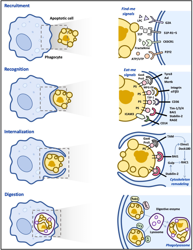
Cells constituting a multicellular organism die in a variety of ways throughout life, and most of them die via apoptosis under normal conditions. The occurrence of apoptosis is especially prevalent during development and in tissues with a high cellular turnover rate, such as the thymus and bone marrow. Interestingly, although the number of apoptotic cells produced daily is known to be innumerable in a healthy adult human body, apoptotic cells are rarely observed. This absence is due to the existence of a cellular process called efferocytosis that efficiently clears apoptotic cells. Studies over the past decades have focused on how phagocytes are able to remove apoptotic cells specifically, swiftly, and continuously, resulting in defined molecular and cellular events. In this review, we will discuss the current understanding of the clearance of apoptotic cells at the molecular level.


Conflict of interest statement

The authors declare no competing interests.

Figures

Fig. 1
Fig. 1. Molecular basis of efferocytosis.
In the first step of efferocytosis, chemoattractants such as nucleotides and LPC released by apoptotic cells recruit macrophages. In the second step, the recruited macrophages directly or indirectly recognize apoptotic cells through ligand‒receptor interactions. This interaction leads to the activation of downstream signaling pathways, which ultimately results in the rearrangement of the cytoskeleton and internalization of apoptotic cells in the third step. In the final step of efferocytosis, internalized apoptotic cells form phagosomes and are degraded by lysosomal enzymes. LC3-associated phagocytosis (LAP) is more efficient in degrading apoptotic cells than non-LAP.
Fig. 2
Fig. 2. Calcium signaling in efferocytosis.
Store-operated calcium entry (SOCE) is induced upon apoptotic cell stimulation. Mertk binding to PS on apoptotic cells activates the downstream signaling pathway, inducing the release of calcium from the ER and thus the interaction between Stim1 and Orai1, which ultimately causes calcium entry into phagocytes during efferocytosis. In addition, Drp-1 upregulation during efferocytosis induces mitochondrial fission, which impedes MCU-mediated mitochondrial calcium sequestration. This SOCE and impairment of mitochondrial calcium sequestration elevate the intracellular calcium level in phagocytes during efferocytosis.

References

    1. Elmore S. Apoptosis: a review of programmed cell death. Toxicol. Pathol. 2007;35:495–516. doi: 10.1080/01926230701320337. - DOI - PMC - PubMed
    1. Singh R, Letai A, Sarosiek K. Regulation of apoptosis in health and disease: the balancing act of BCL-2 family proteins. Nat. Rev. Mol. Cell Biol. 2019;20:175–193. doi: 10.1038/s41580-018-0089-8. - DOI - PMC - PubMed
    1. deCathelineau AM, Henson PM. The final step in programmed cell death: phagocytes carry apoptotic cells to the grave. Essays Biochem. 2003;39:105–117. doi: 10.1042/bse0390105. - DOI - PubMed
    1. Ravichandran KS. Beginnings of a good apoptotic meal: the find-me and eat-me signaling pathways. Immunity. 2011;35:445–455. doi: 10.1016/j.immuni.2011.09.004. - DOI - PMC - PubMed
    1. Hogquist KA, Baldwin TA, Jameson SC. Central tolerance: learning self-control in the thymus. Nat. Rev. Immunol. 2005;5:772–782. doi: 10.1038/nri1707. - DOI - PubMed

Publication types