Review
. 2023 Aug;55(8):1595-1619.
doi: 10.1038/s12276-023-01046-5. Epub 2023 Aug 23.

Mitochondria-associated programmed cell death as a therapeutic target for age-related disease

Affiliations
Review

Mitochondria-associated programmed cell death as a therapeutic target for age-related disease

Thanh T Nguyen et al. Exp Mol Med. 2023 Aug.

Abstract

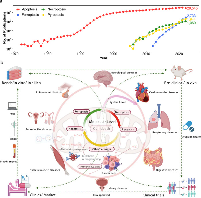
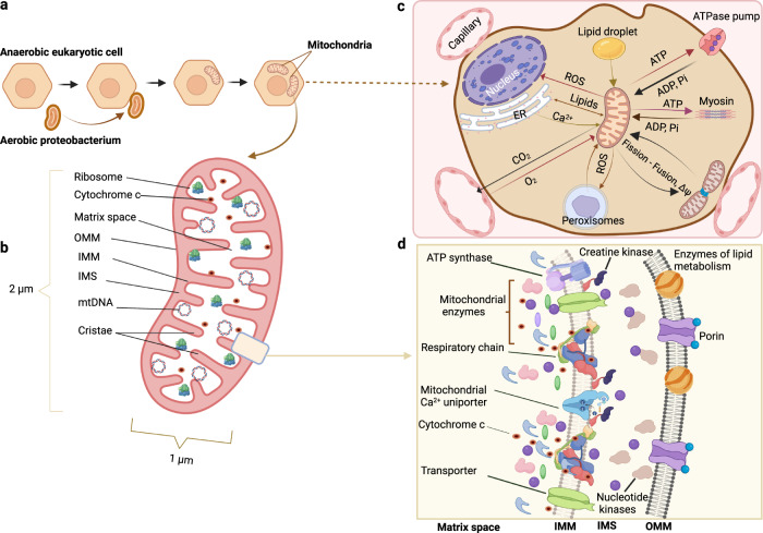
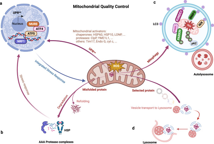
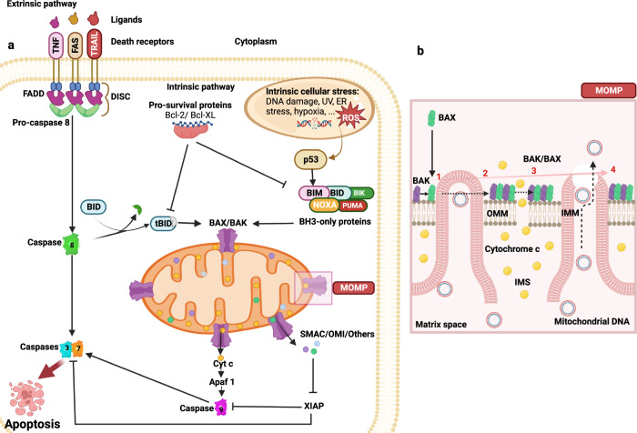
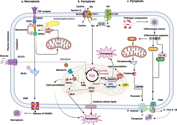
Mitochondria, ubiquitous double-membrane-bound organelles, regulate energy production, support cellular activities, harbor metabolic pathways, and, paradoxically, mediate cell fate. Evidence has shown mitochondria as points of convergence for diverse cell death-inducing pathways that trigger the various mechanisms underlying apoptotic and nonapoptotic programmed cell death. Thus, dysfunctional cellular pathways eventually lead or contribute to various age-related diseases, such as neurodegenerative, cardiovascular and metabolic diseases. Thus, mitochondrion-associated programmed cell death-based treatments show great therapeutic potential, providing novel insights in clinical trials. This review discusses mitochondrial quality control networks with activity triggered by stimuli and that maintain cellular homeostasis via mitohormesis, the mitochondrial unfolded protein response, and mitophagy. The review also presents details on various forms of mitochondria-associated programmed cell death, including apoptosis, necroptosis, ferroptosis, pyroptosis, parthanatos, and paraptosis, and highlights their involvement in age-related disease pathogenesis, collectively suggesting therapeutic directions for further research.


Conflict of interest statement

The authors declare no competing interests.

Figures

Fig. 1
Fig. 1. Overview of mitochondrion-associated programmed cell death and human diseases.
a Publications of typical programmed cell death in the last 50 years. b Mitochondria are vital in regulating cellular metabolism and are involved in the pathogenesis and progression of numerous human diseases, directly or indirectly, through many pathways. Mitochondria are also essential in regulating cell fate via PCD, an evolutionarily conserved process in multicellular organisms that is indispensable for modulating homeostasis, containing diverse patterns, including apoptosis, necroptosis, ferroptosis, and pyroptosis. Mitochondria-associated PCD is extensively involved in the pathological progression of various disorders in different organ systems. In this regard, fresh insights into the interplay between mitochondria and PCD are expected to be inspired in disease treatment development in contexts ranging from in vitro to preclinical and clinical trials. PCD Programmed cell death, EMR Electronic medical record.
Fig. 2
Fig. 2. Mitochondrial structure, function, and communication with other organelles.
a, b Hypothetical depiction of the origin of mitochondria from Aerobic Proteobacterium and mitochondrial simplified structure. c Microstructure and enzyme complexes of mitochondria. d Mitochondrial functions rely on cellular interactions. OMM Outer mitochondrial membrane, IMM Inner mitochondrial membrane, IMS Intermembrane mitochondrial space, mtDNA Mitochondrial DNA, ROS Reactive oxygen species, ER Endoplasmic reticulum, ATP Adenosine triphosphate.
Fig. 3
Fig. 3. Pathways of mitochondrial quality control.
a Activation of the UPRmt pathway is triggered by the accumulation of unfolded proteins in the mitochondrial matrix, expression of mitochondrial protein import components, and upregulation of chaperones and proteases for reestablishing mitochondrial proteostasis. b Molecular chaperones and proteolytic enzymes in the mitochondria perform a major function in refolding or destroying mitochondrial misfolded and unfolded proteins. c Mitochondrial proteins are likely to be decomposed by being transported to lysosomes, where vesicles formed from mitochondrial tubules seize specific mitochondrial cargos for lysis. d Fragmented dysfunctional mitochondria are removed by mitophagy. Bnip3, FUNDC1, and NIX are mitophagy receptors that bind LC3 to tether mitochondria to autophagosomes. OMM proteins and Parkin are ubiquitinated when PINK1 phosphorylates proteins on the surface of depolarized mitochondria. The p62 adaptor can recognize ubiquitinated proteins to initiate mitophagy. UPRmt mitochondrial Unfolded Protein Response, MURE Mitochondrial Unfolded Protein Response Element, ATF Activating Transcription Factor, NRF Nuclear Respiratory Factor, HSP Heat Shock Protein, LC3 microtubule-associated protein Light Chain 3, Bnip3 Bcl-2 interacting protein 3, Ub Ubiquitin, PINK1 PTEN-induced kinases 1.
Fig. 4
Fig. 4. Apoptotic signaling pathways.
a In the extrinsic pathways, death receptor ligands bind to members of the death receptor family to function, for instance, TNF, FAS, and TRAIL receptors. Numerous diverse stressors, such as DNA damage, the removal of growth factors, and mitotic arrest, activate the intrinsic apoptotic pathway, which in turn activates BH3-only members. By activating the effector proapoptotic Bcl-2 proteins Bax and Bak and inhibiting antiapoptotic Bcl-2 proteins, BH3-only proteins cause MOMP. In particular, cyt c is activated by releasing proteins from the mitochondrial intermembrane gap. The heptameric structure known as the apoptosome is created when cyt c interacts with APAF1. As a result, caspase-9 is recruited and activated, which cleaves and activates caspase-3 and -7. The caspase inhibitor XIAP is blocked by proteins such as SMAC, which are released due to MOMP, promoting apoptosis. The extrinsic and intrinsic apoptotic pathways are linked via caspase-8 cleavage and activation of the BH3-only protein Bid. b In normal cases, Bax localizes to the cytoplasm and Bak to the mitochondria. By interacting with BH3-only proteins, Bax and Bak can be directly activated during apoptosis, which leads to their stabilization on the OMM. Their dimer further oligomerizes into a higher-order multimer, contributing to the release of cyt c and other IMS proteins. Over time, Bax and Bak accumulate with macropore formation, which allows the IMM to protrude through the OMM, after which the IMM herniates and ruptures, eventually releasing mtDNA. TNF Tumor Necrosis Factor, FADD Fas-Associated Protein with DD, DISC Death-Inducing Signaling Complex, TRAIL Tumor necrosis factor-Related Apoptosis-Inducing Ligand, SMAC Second Mitochondria-derived Activator of Caspases, tBid truncated Bid, Apaf1 Apoptotic protease-activating Factor 1, XIAP X-linked Inhibitor of Apoptosis Protein, PUMA p53 Upregulated Modulator of Apoptosis, UV Ultraviolet.
Fig. 5
Fig. 5. Mitochondria-associated nonapoptotic cell death.
a Necroptosis is formed from necroptosis-driving factors such as TNF α binding with complementary receptors to recruit RIPK1, where the engagement renders RIPK1 and RIPK3 activated, which fuels necrosome configuration. The necrosome thereafter phosphorylates and activates MLKL, which is translocated to and permeabilizes the plasma membrane and permits DAMP release. b Ferroptosis is driven by iron-dependent lipid peroxidation and ROS overexpression. GPX4 converts excessive peroxides into lipid alcohols to mitigate this process under normal conditions; however, once stimuli occur, overexpressed superoxide from OXPHOS complexes reacts with ferrous ions to yield a slew of unstable radicals, leading to excessive ROS and, afterward, lipid peroxidation reboots to induce ferroptosis. Another route to ferroptosis is the buildup of lipid peroxide by circumstances such as cysteine deprivation, which induces the breakdown of certain iron-binding proteins, including FtMt and heme, to free iron and ROS, therefore triggering nearby MLP and hence ferroptosis. c Pyroptosis is initiated by the pathogen component-activated inflammatory caspase response, which not only converts IL‐1β and IL‐18 to mature forms but also cleaves GSDMD to facilitate pore penetration and enable IL-1β and IL-18 release, thus resulting in cell swelling and pyroptosis. Apart from these, inflammatory stimuli also lead to pyroptosome-mediated caspase-3 activation and GSDME cleavage to unleash membrane permeabilization to favor the release of ions and mtDNA to induce pyroptosis. DAMPs Damage-Associated Molecular Patterns, MLKL Mixed-Lineage Kinase Domain-Like pseudokinase, PDH pyruvate dehydrogenase, GPX4 Glutathione Peroxidase 4, TCA Tricarboxylic acid cycle, ETC Electron Transport Chain, GSH Glutathione, MLP Membrane Lipid Peroxidation, FtMt Mitochondrial Ferritin, α-KG Alpha-ketoglutarate, IDH2 Isocitrate Dehydrogenase 2, NADH Nicotinamide Adenine Dinucleotide Dehydrogenase, OXPHOS mitochondrial Oxidative Phosphorylation System, GSDMD Gasdermin D, GSDME Gasdermin E, IL-18 Interleukin-18, IL-1β Interleukin-1β.

References

    1. Martinez-Reyes I, Chandel NS. Mitochondrial TCA cycle metabolites control physiology and disease. Nat. Commun. 2020;11:102. doi: 10.1038/s41467-019-13668-3. - DOI - PMC - PubMed
    1. Oh CM, Ryu D, Cho S, Jang Y. Mitochondrial Quality Control in the Heart: New Drug Targets for Cardiovascular Disease. Korean Circ. J. 2020;50:395–405. doi: 10.4070/kcj.2019.0416. - DOI - PMC - PubMed
    1. Weinlich R, Oberst A, Beere HM, Green DR. Necroptosis in development, inflammation and disease. Nat. Rev. Mol. Cell Biol. 2017;18:127–136. doi: 10.1038/nrm.2016.149. - DOI - PubMed
    1. Bock FJ, Tait SWG. Mitochondria as multifaceted regulators of cell death. Nat. Rev. Mol. Cell Biol. 2020;21:85–100. doi: 10.1038/s41580-019-0173-8. - DOI - PubMed
    1. Protasoni, M. & Zeviani, M. Mitochondrial Structure and Bioenergetics in Normal and Disease Conditions. Int. J. Mol. Sci.22, 10.3390/ijms22020586 (2021). - PMC - PubMed

Publication types