

MuscleJ2: a rebuilding of MuscleJ with new features for high-content analysis of skeletal muscle immunofluorescence slides
- PMID: 37612778
- PMCID: PMC10463807
- DOI: 10.1186/s13395-023-00323-1
MuscleJ2: a rebuilding of MuscleJ with new features for high-content analysis of skeletal muscle immunofluorescence slides
Abstract
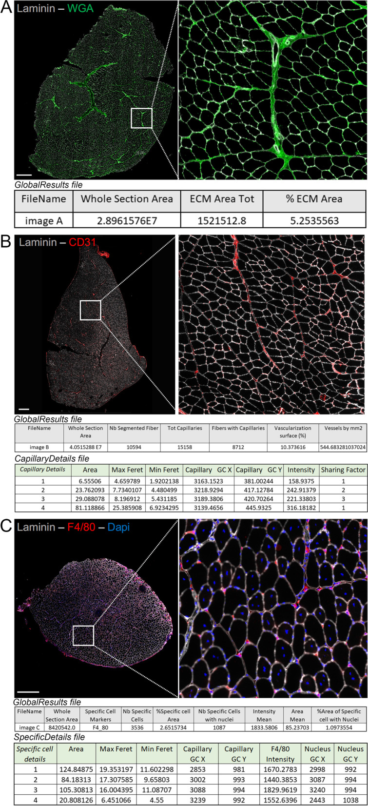
Histological analysis of skeletal muscle is of major interest for understanding its behavior in different pathophysiological conditions, such as the response to different environments or myopathies. In this context, many software programs have been developed to perform automated high-content analysis. We created MuscleJ, a macro that runs in ImageJ/Fiji on batches of images. MuscleJ is a multianalysis tool that initially allows the analysis of muscle fibers, capillaries, and satellite cells. Since its creation, it has been used in many studies, and we have further developed the software and added new features, which are presented in this article. We converted the macro into a Java-language plugin with an improved user interface. MuscleJ2 provides quantitative analysis of fibrosis, vascularization, and cell phenotype in whole muscle sections. It also performs analysis of the peri-myonuclei, the individual capillaries, and any staining in the muscle fibers, providing accurate quantification within regional sublocalizations of the fiber. A multicartography option allows users to visualize multiple results simultaneously. The plugin is freely available to the muscle science community.
Keywords: Centro- and perinuclei; Extracellular matrix; Fiber typing; Histology; Interstitial cells; Muscle fiber morphology; Phenotype cartography; Sarcolemmal staining; Vascularization.
© 2023. BioMed Central Ltd., part of Springer Nature.
Conflict of interest statement
The authors declare no competing interests.
Figures


References
Publication types
MeSH terms
LinkOut - more resources
Full Text Sources
