Primary non leukemic myeloid sarcoma of the ureteral wall: a case report of a rare disease
- PMID: 37614469
- PMCID: PMC10444284
- DOI: 10.1093/jscr/rjad433
Primary non leukemic myeloid sarcoma of the ureteral wall: a case report of a rare disease
Abstract
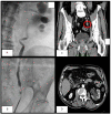
Myeloid sarcoma (MS) is an extramedullary tumor mass causing proliferation of mature or immature blast cells of one or more myeloid lineages. Involvement of the genitourinary tract is rare. We present a case of MS of the ureteral wall. A 74-year-old man was evaluated for left hydronephrosis and ipsilateral low back pain. A computed tomography scan showed a nodular formation in the pelvic ureter. Urinary cytology revealed cellular atypia, so ureteroscopy was performed showing a distal ureteral mass. The histological examination of the biopsy revealed to be malignant neoplasm. The patient underwent left laparoscopic nephroureterectomy with bladder cuff excision. Microscopic histological examination revealed a tumor compatible with MS. A postoperative positron emission tomography revealed residual hypercaptation of the bladder, pelvic muscle and iliac nodes, so the patient started chemotherapy. A multidisciplinary approach was required, taking into account the patient's age, the already poor renal function and the location of the neoplasm.
Keywords: hydronephrosis; myeloid sarcoma; ureter; ureteral neoplasm; ureterectomy.
Published by Oxford University Press and JSCR Publishing Ltd. © The Author(s) 2023.
Conflict of interest statement
None declared.
Figures


References
-
- Shahin OA, Ravandi F. Myeloid sarcoma. Curr Opin Hematol 2020;27:88–94. - PubMed
-
- Almond LM, Charalampakis M, Ford SJ, Gourevitch D, Desai A. Myeloid sarcoma: presentation, diagnosis, and treatment. Clin Lymphoma Myeloma Leuk 2017;17:263–7. - PubMed
-
- Magdy M, Abdel Karim N, Eldessouki I, Gaber O, Rahouma M, Ghareeb M. Myeloid sarcoma. Oncol Res Treat 2019;42:224–9. - PubMed
-
- Bagg MD, Wettlaufer JN, Willadsen DS, Ho V, Lane D, Thrasher JB. Granulocytic sarcoma presenting as a diffuse renal mass before hematological manifestations of acute myelogenous leukemia. J Urol 1994;152:2092–3. - PubMed
Publication types
LinkOut - more resources
Full Text Sources
Research Materials

