Review
. 2023 Jul 25;3(4):556-579.
doi: 10.1016/j.jacasi.2023.05.012. eCollection 2023 Aug.

Echocardiographic Imaging in Transcatheter Structural Intervention: An AAE Review Paper

Affiliations
Review

Echocardiographic Imaging in Transcatheter Structural Intervention: An AAE Review Paper

Raja Ezman Raja Shariff et al. JACC Asia. .

Abstract

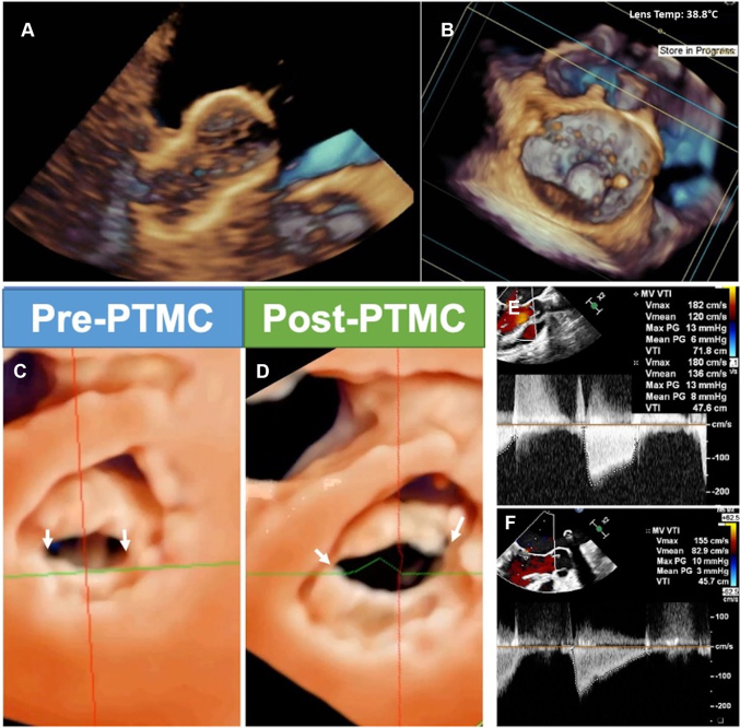
Transcatheter structural heart intervention (TSHI) has gained popularity over the past decade as a means of cardiac intervention in patients with prohibitive surgical risks. Following the exponential rise in cases and devices developed over the period, there has been increased focus on developing the role of "structural imagers" amongst cardiologists. This review, as part of a growing initiative to develop the field of interventional echocardiography, aims to highlight the role of echocardiography in myriad TSHIs available within Asia. We first discuss the various echocardiography-based imaging modalities, including 3-dimensional echocardiography, fusion imaging, and intracardiac echocardiography. We then highlight a selected list of structural interventions available in the region-a combination of established interventions alongside novel approaches-describing key anatomic and pathologic characteristics related to the relevant structural heart diseases, before delving into various aspects of echocardiography imaging for each TSHI.

Keywords: echocardiography; interventional echocardiography; review; transcatheter structural intervention.


Conflict of interest statement

The authors have reported that they have no relationships relevant to the contents of this paper to disclose.

Figures

None
Graphical abstract
Figure 1
Figure 1
Echocardiography-Fluoroscopy Fusion Imaging (A) Biplane transesophageal echocardiography (TEE) showing the mitral valve (MV) in the left ventricular outflow (purple) and bicommissural (green) planes. After co-registration, the TEE data set is aligned in the echocardiographic-fluoroscopy fused images. (B) The purple and green sectors of the corresponding 2-dimensional TEE views. (C) The MV within the boundaries of the 3-dimensional pyramidal data set is aligned in the anterior-posterior fluoroscopic projection. The red arrow denotes the wire in the ventricle. (D) Another example shows the ideal transseptal puncture site being determined by biplane TEE in the bicaval and short-axis views. (E) Placement of fiducial marker (red circle) will be displayed on fluoroscopy. (F) In addition, superimposing the color Doppler TEE data set of the MV during a transcatheter edge-to-edge repair to indicate origin of the regurgitation jet can be performed. LA = left atrium; RA = right atrium; SVC = superior vena cava.
Central Illustration
Developing Interventional Echocardiography in Asia Development in interventional echocardiography requires having a structured curriculum to ensure competency in the field, access to novel echocardiographic modalities, and close networking within the region.
Figure 2
Figure 2
Imaging of the Interatrial Septum (IAS) (A) Three-dimensional TEE shows the IAS seen from the right atrium. The red dashed line corresponds to the bicaval view and the blue dashed line to the short-axis view. (B) Biplane TEE demonstrated tenting of the IAS (white arrows) seen from bicaval view (left) and short-axis (right) planes. Ant = anterior; AoV = aortic valve; IVC = inferior vena cava; Post = posterior; other abbreviations as in Figure 1.
Figure 3
Figure 3
Normal Structure of IAS Three-dimensional (3D) TEE showing (A to C) right and (D) left septal perspectives of the IAS: (A) 3D spatial relation of the IAS to adjacent structures can be assessed in great detail; (B) transillumination imaging with virtual light source placed in the LA behind the IAS allows appreciation of the thinness, location, and limbus of the fossa ovalis (FO); (C) photorealistic transparency (“glass”) rendering of the IAS; and (D) transillumination of the FO from the left septal perspective with virtual light source in the RA. AV = atrioventricular; CS = coronary sinus; TV = tricuspid valve; other abbreviations as in Figures 1 and 2.
Figure 4
Figure 4
Interrogation of the ASD Rims (A) ASD en face from 3D TEE view, looking in from the RA. Colored lines represent the degree of views correlated with the respective TEE 2-dimensional views. (B) corresponds to the blue line in A and shows the posterior rim and the aortic rim (midesophagus, at 0°-45°). (C) corresponds to the green line in A and shows the IVC and aortic rim (midesophagus, at 45°-90°). (D) corresponds to the red line in A and shows the IVC and SVC rim (midesophagus, at 100°-130°). (E) corresponds to the purple line in A and shows the AV valve and the posterior rim (midesophagus, at 150°-180°). Abbreviations as in Figures 1, 2, and 3.
Figure 5
Figure 5
Transseptal Puncture Height A lower puncture is needed when treating lateral lesions, because often the clip will continue to gain height above the valve as it is passes further. Conversely, a higher puncture is used when treating a medial lesion, because the clip will have to “dive down” immediately after transseptal puncture. P1, P2, and P3 = scallops of the posterior mitral leaflets.
Figure 6
Figure 6
Views for Clip Steering and Perpendicularity (A) The bicommissural “index” view allows assessment of medial and lateral device location, as well as precise localization of the area of interest. When the shaft of the delivery system approaches from too much of an anterior position (ie, an “aorta-hugger” position), as seen in (B) the long-axis “grasping” and (C) 3-dimensional en-face views, remedial maneuvers are frequently required (yellow dashed arrow).
Figure 7
Figure 7
Views for Clip Grasping and Closure (A) Biplane imaging of the clip as it is pulled back toward the mitral valve with arms fully extended and the leaflets have been grasped by the grippers. (B) The clip is then closed, and the leaflets are drawn into the mechanism. (C) Color-flow Doppler is used to assess for residual regurgitation. (D) Three-dimensional en-face imaging is used to assess the tissue bridge, the nature of the dual-orifice anatomy, and the position and volume of regurgitant jets.
Figure 8
Figure 8
Transcatheter Mitral Valve-in-Valve (A) Transseptal puncture performed with X-plane imaging. (B) X-Plane imaging using simultaneous midesophageal bicommissural and long-axis views used to guide prosthesis delivery and deployment, and to check leaflet mobility after deployment. (C) Three-dimensional reconstruction with color Doppler is used to assess for paravalvular leaks.
Figure 9
Figure 9
Echocardiographic Guidance of Percutaneous Transluminal Mitral Commissurotomy (PTMC) 3D TEE guides the alignment and orientation of the balloon catheter and further monitors the position of the inflated balloon. These pictures show the inflated Inoue balloon via (A) real-time 3D and (B) en-face views. (C, D) Left ventricular perspective 3D imaging showing splitting of both commissures (arrows) after successful PTMC, with increase in valve area and reduction of transvalvular pressure gradient on Doppler assessment (E, F). Abbreviations as in Figures 1 and 3.
Figure 10
Figure 10
Transcatheter Edge-to-Edge Repair of the Tricuspid Valve (A) Guide catheter and catheter delivery system (CDS) introduction. (B) Steering of the CDS (arrows) toward the tricuspid anteroseptal commissure using an en face view. (C) Multiplane imaging showing positioning of the CDS. (D) Multiplanar reconstruction with the long axes (red and green) aligned to the CDS. (E, F) Clip arm orientation adjusted perpendicular to the line of coaptation between the anterior and septal leaflet. (G, H, I) Leaflet grasping is monitored, and leaflet insertion confirmed with the use of multiple views. 4Ch = four-chamber; AL = anterior leaflet; LAX = long axis; LE = low-esophageal; MPR = multiplanar reconstruction; PL = posterior leaflet; SAX = short axis; SL = septal leaflet; TG = transgastric; TTE = transthoracic echocardiography; other abbreviations as in Figures 1, 2, and 3.
Figure 11
Figure 11
Tricuspid Valve Transcatheter Annuloplasty Procedure (A, B) Preprocedural TTE demonstrating tricuspid regurgitation (TR) on computed tomographic imaging. (C) X-Plane imaging using right ventricular inflow-outflow showing torrential regurgitation. (D) Through the guiding catheter, the K-Clip delivery catheter (KCR) is introduced into the RA and steered toward the tricuspid annulus (TA). (E) Fluoroscopy showing the anchor of the device to the posterior TA. (F) Retraction of the anchor, pulling annular tissue into the device, which then closes to fold the annulus within. (G) Annular and TR reduction immediately after implantation. (H) Coronary angiogram confirming right coronary patency after clipping. (I) Sustained TR reduction at 3-month follow-up. An = anchor; At = annular tissue; GC = guiding catheter; RCA = right coronary artery; other abbreviations as in Figures 1 and 10.
Figure 12
Figure 12
Transcatheter Heterotopic Valve Implantation of the Tricuspid Valve Following fluoroscopic evidence of the desired SVC position (marked using wires and catheters within the right pulmonary artery [RPA] and subclavian and brachiocephalic veins), (A) the guiding wires and catheters (red arrow), and device delivery system (yellow arrow) is advanced into the SVC, where (B) the upper portion of the prosthesis (blue arrow) is positioned above the crossing of the RPA catheter and SVC on simultaneous fluoroscopy. Through TEE, this can be visualized best using the 2-dimensional bicaval window, with 3-dimensional reconstruction if desired. (C) The SVC prosthesis (blue arrows) is fully deployed, and its final positions checked on (D) TEE and (E) fluoroscopy, before being retrieved back along with the RPA catheter. This is followed by (F) IVC prosthesis (white arrows) advancement, positioning, and deployment, before a final check on (G) TEE and (H) fluoroscopy. Abbreviations as in Figures 1, 2, and 10.
Figure 13
Figure 13
TEE in Transcatheter Aortic Valve Implantation (A) Midesophageal plane at 135°, confirming that the wire is located around the left ventricular apex, along the ventricular septum, and not entangled with the MV. This confirmation is useful in preventing complications such as ventricular perforation or MV injury. If the wire entrains the MV, it may affect the balloon’s position and stability, as well as hemodynamics, by causing mitral regurgitation. (B) A case before balloon inflation and (C) after subnominal inflation by 4 mL. During implantation, calcification of the aortic valve can be seen pushing through the sinus of Valsalva and protruding into the LA. Known as the “Mt. Fuji” sign, this is a useful endpoint guide to avoid potential aortic root rupture. Abbreviations as in Figure 1.
Figure 14
Figure 14
MV Prosthesis on TEE (A) The aortic valve (AV) is anteriorly located (12 o’clock), the IAS is medially located (3 o’clock), and the left atrial appendage (LAA) is laterally located (9 o’clock). The MV is then divided into 8 quadrants that serve as a common nomenclature to describe paravalvular leaks (PVLs) among the interventionalists and imagers. Multiplanar 2-dimensional views, including the (B) midesophageal 135° long-axis and (C) 45° bicommissural views, permit localization of the 2 PVLs: a larger PVL is located anterolateral (10 o’clock position), and a smaller PVL is located anteromedial (1 o’clock position). Both leaks are clearly visible on the (D) surgical en-face view, with overlapping color Doppler and “Trueview”. Abbreviations as in Figures 1 and 2.
Figure 15
Figure 15
PVL Closure (A) Under direct 3D TEE guidance, the anterolateral mitral PVL is crossed with a wire (blue arrow). (B) After deployment of an occluder device, color Doppler shows stable placement of the device with trace mitral regurgitation at this site. (C) The mean transmitral gradient is 3 mm Hg, similar to baseline. (D) 3D en-face TEE view confirms the final stable position of the occluder device (asterisk) with no impingement of the prosthetic leaflet motion. (E) A smaller anteromedial PVL is closed with the use of a 12-mm Vascular Plug II (red arrow). (F) This is accompanied by an increase in the mean transmitral gradient to 5 mm Hg. (G) Simultaneous deployment of 2 smaller devices with verification of 2 catheters (blue arrows) crossing the anteromedial PVL on live 3D to ensure correct canalization of the PVL gap. (H) Final position of the 3 deployed occluders (asterisks) with trace residual leak. Abbreviations as in Figures 1, 3, and 14.
Figure 16
Figure 16
TEE of the LAA The LAA segments are illustrated in (A) 2-dimensional TEE view 45° and (B) 3-dimensional TEE 135° photorealistic “glass” view. The ostium is the opening of LA, the neck is located between the ostial region and lobe demarcated by the left circumflex coronary artery (LCx), and the lobe is situated distal to the neck in adjacent to the pulmonary artery (PA). (C) The LAA cauliflower morphology projected by 3-dimensional (3D) TEE photorealistic imaging shares great similarity with (D) contrast angiography at anterior oblique 30°/caudal 30°. LUPV = left upper pulmonary vein; other abbreviations as in Figures 1 and 14.
Figure 17
Figure 17
Echocardiographic Measurements of the Landing Zone (A) On 2-dimensional (2D) TEE, landing zone diameter (red lines) measurements should start from inferior part of the ostium (yellow lines) at the level of LCx to an area 1 cm distal to the ligament of Marshall. The LAA depth (green lines) is measured from the level of landing zone to the distal lobe. For most cases, the maximum landing zone is measured from 2D imaging of 90°-135°, but this is subject to anatomic variation. (B) The state-of-art measurement as shown allows an unbiased and reproducible assessment of the LAA landing zone that is noninferior to cardiac computed tomography. Abbreviations as in Figures 1, 14, and 16.
Figure 18
Figure 18
Echocardiography in the Percutaneous Intramyocardial Septal Radiofrequency Ablation (A) Preoperative assessment is done to confirm left ventricular wall hypertrophy and outflow tract obstruction. (B) Transapical needle insertion is performed under 2-dimensional and color Doppler transthoracic echocardiography guidance to avoid vascular injuries, followed by (C, D) ablation of the anterior and posterior septum. (E) Myocardial contrast echocardiography is performed immediately to evaluate adequacy of ablation, followed by (F) assessment for septal reduction and obstruction relief.

References

    1. Hamid N., Ewe S.H. Interventional echocardiography: current role and progress. Proc Singap Healthc. 2015;24(1):4–15.
    1. Ludman P.F., Moat N., de Belder M.A., et al. Transcatheter aortic valve implantation in the United Kingdom: temporal trends, predictors of outcome, and 6-year follow-up: a report from the UK Transcatheter Aortic Valve Implantation (TAVI) registry, 2007 to 2012. Circulation. 2015;131(13):1181–1190. - PubMed
    1. Durko A.P., Osnabrugge R.L., Van Mieghem N.M., et al. Annual number of candidates for transcatheter aortic valve implantation per country: current estimates and future projections. Eur Heart J. 2018;39(28):2635–2642. - PubMed
    1. Lee C.H., Inohara T., Hayashida K., et al. Transcatheter aortic valve replacement in Asia: present status and future perspectives. J Am Coll Cardiol Asia. 2021;1(3):279–293. - PMC - PubMed
    1. Wong N., Yeo K.K. MitraClip in Asia—current adoption and regional data. Circ Rep. 2019;1(10):397–400. - PMC - PubMed

LinkOut - more resources