

Electrophysiological Study in the Right Upper and Lower Limbs in Infants with Lumbosacral Meningomyelocele and in Normal Infants: A Case-control Study
- PMID: 37614841
- PMCID: PMC10443458
- DOI: 10.4103/ijabmr.ijabmr_484_22
Electrophysiological Study in the Right Upper and Lower Limbs in Infants with Lumbosacral Meningomyelocele and in Normal Infants: A Case-control Study
Abstract
Objective: The study aimed to assess the electrophysiological parameters (Hofmann reflex [H-reflex] and motor nerve conduction velocity [MNCV]) on children's upper and lower limbs with lumbosacral meningomyelocele (MMC) and age-matched control to see the effect of the MMC on the cervical segment of the spinal cord.
Materials and methods: The present study was performed on infants with lumbosacral MMC. Twenty-five infants were examined with a mean age of 50 days of either sex. Out of them, 13 infants were in control and the remaining 12 were diagnosed with MMC. The H-reflex parameter and MNCV were recorded in these children's right upper and lower limbs.
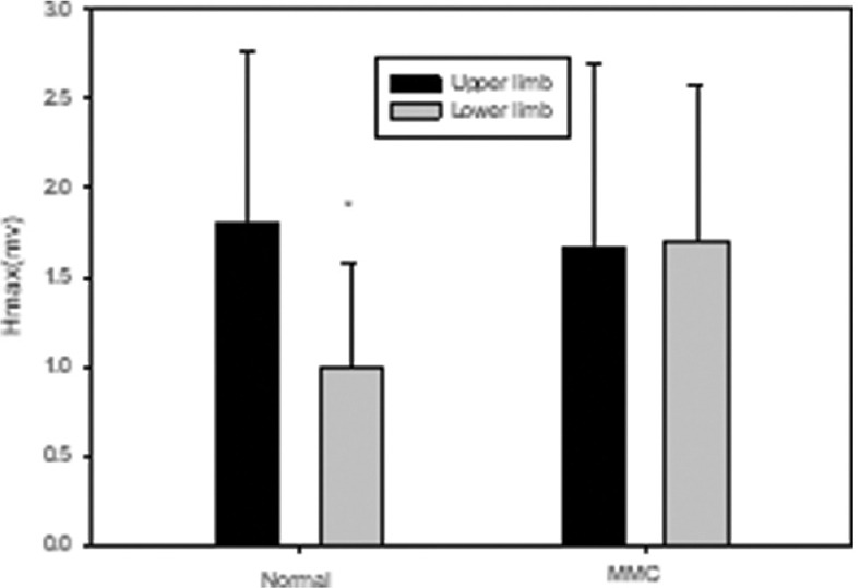
Results: H-reflex was elicited in all the control group babies. In MMC, the H-reflex was elicited in the upper limbs. However, H-reflex was not elicited in the lower limbs of a few MMC babies. The upper limb's H-reflex parameters and conduction velocity were significantly higher than those corresponding lower limbs in control babies. In MMC, where the H-reflex was elicited, such differences in the lower and upper limbs were not observed. However, the values of MNCV in the upper limb (right median nerve) were significantly less, and the values of Hmax in the lower limb (soleus muscle) were significantly more in MMC babies than in the control group.
Conclusions: The values of electrophysiological parameters were higher in the upper limbs as compared to the corresponding lower limbs in control. These values were not altered in the upper limbs than those corresponding lower limbs of MMC, suggesting that motor function development was impaired/delayed in the spinal segment cranial to MMC lesion, and motor impairment in MMC children is mostly a result of upper motor neuron dysfunction.
Keywords: Hofmann reflex; motor nerve conduction velocity; spina bifida.
Copyright: © 2023 International Journal of Applied and Basic Medical Research.
Conflict of interest statement
There are no conflicts of interest.
Figures


References
-
- Hasan KM, Eluvathingal TJ, Kramer LA, Ewing-Cobbs L, Dennis M, Fletcher JM. White matter microstructural abnormalities in children with spina bifida myelomeningocele and hydrocephalus: A diffusion tensor tractography study of the association pathways. J Magn Reson Imaging. 2008;27:700–9. - PMC - PubMed
-
- Jacobs RA, Wolfe G, Rasmuson M. Upper extremity dysfunction in children with myelomeningocele. Z Kinderchir. 1988;43(Suppl 2):19–21. - PubMed
LinkOut - more resources
Full Text Sources