. 2023 Aug 7;19(3):62.
doi: 10.3892/br.2023.1644. eCollection 2023 Sep.

Different responses of the MIO‑M1 Mueller cell line to angiotensin II under hyperglycemic or hypoxic conditions

Affiliations

Different responses of the MIO‑M1 Mueller cell line to angiotensin II under hyperglycemic or hypoxic conditions

Ansgar Beuse et al. Biomed Rep. .

Abstract

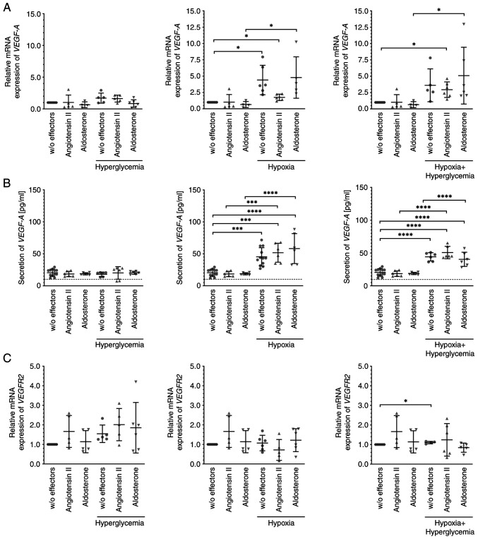
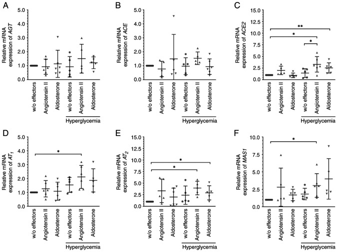
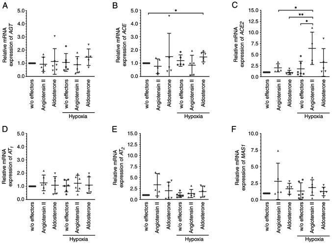
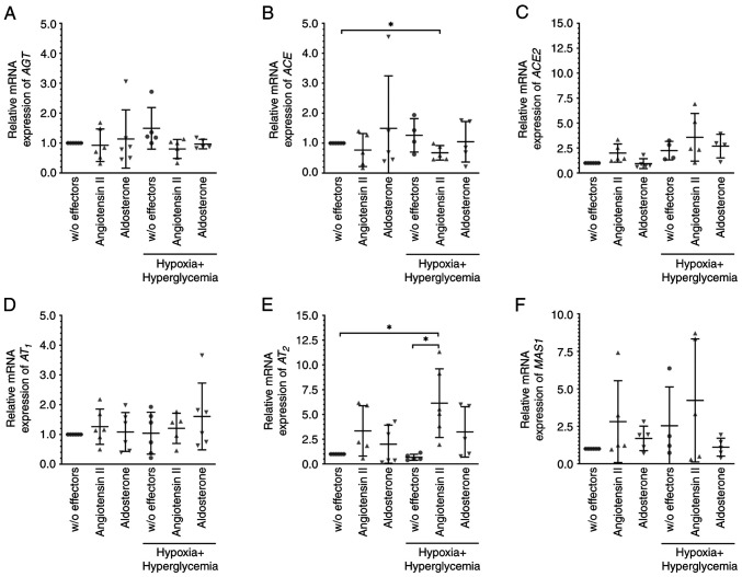
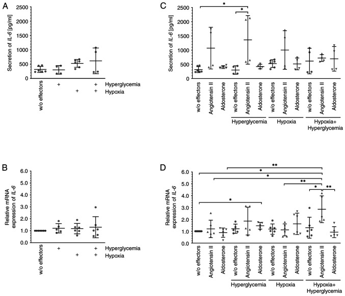
Members of the renin-angiotensin aldosterone system (RAAS) are expressed by various retinal tissues including Mueller glial cells. As the RAAS is hypothesized to play an important role in the pathogenesis of diseases that threaten vision, such as diabetic macular edema or retinal vein occlusion, the possible changes induced by exposure of the human cell line MIO-M1, an established model of Mueller cells, to angiotensin II or aldosterone for 6 h under hypoxic and/or hyperglycemic conditions were investigated. The mRNA expression levels of the members of the RAAS were assessed by reverse transcription-quantitative PCR, and the secretion of cytokines was assessed by ELISA. Under hyperglycemic conditions, the mRNA expression levels of the angiotensin-converting enzyme 2 (ACE2), angiotensin II receptors, AT1 and AT2, and the receptor of angiotensin (1-7) MAS1 were significantly higher after exposure to angiotensin II, and the expression of ACE2, AT2, and IL-6 (a marker of inflammation) was significantly increased after treatment with aldosterone; the expression of the other targets investigated remained unchanged. Significantly more IL-6 was secreted by MIO-M1 cells exposed to hyperglycemia and angiotensin. When cells were cultured in a hypoxic environment, additional treatment with aldosterone significantly increased the mRNA expression levels of ACE, but significantly more ACE2 mRNA was expressed in the presence of angiotensin II. Under hypoxic plus hyperglycemic conditions, significantly less ACE but more AT2 was expressed after treatment with angiotensin II, which also led to strongly elevated expression of IL-6. The mRNA expression levels of the angiogenic growth factor VEGF-A and secretion of the encoded protein were notably increased under hypoxic and hypoxic plus hyperglycemic conditions, irrespective of additional treatment with angiotensin II or aldosterone. These findings suggest that angiotensin II induces a pro-inflammatory response in MIO-M1 cells under hyperglycemic conditions despite activation of the counteracting ACE2/MAS1 signaling cascade. However, hypoxia results in an increased expression of angiogenic VEGF-A by these cells, which is not altered by angiotensin II or aldosterone.

Keywords: Mueller cells; aldosterone; angiotensin II; diabetic macular edema; renin-angiotensin aldosterone-system.


Conflict of interest statement

The authors declare that they have no competing interest.

Figures

Figure 1
Figure 1
Exposure to hypoxia or hypoxia plus hyperglycemia increased the expression and secretion of VEGF-A and this was not modulated by angiotensin II or aldosterone. MIO-M1 cells were cultured under normal or hypoxic conditions in the absence or presence of 10 nM angiotensin II or 10 nM aldosterone for 6 h. Hyperglycemia was induced by addition of 25 mM glucose. mRNA expression levels of (A) VEGF-A and (C) VEGFR2 or (B) secretion of the growth factor were assessed. Hypoxia and hypoxia plus hyperglycemia, but not hyperglycemia alone, enhanced the (A) mRNA expression and (B) secretion of VEGF-A, which was not modulated by angiotensin II or aldosterone. (C) mRNA expression levels of VEGFR2 remained largely unchanged. Data were normalized to the relevant control. Data are presented as scatter plots depicting the mean ± standard deviation. *P#x003C;0.05, ***P#x003C;0.001, ****P#x003C;0.0001. Only statistically significant differences are indicated. The dotted line in (B) depicts the lowest detectable concentration. VEGF, vascular endothelial growth factor; VEGFR2, VEGFR receptor 2; w/o, without.
Figure 2
Figure 2
Angiotensin II and aldosterone altered the expression of members of the RAAS under hyperglycemic conditions. Cells were treated with 10 nM angiotensin II, 10 nM aldosterone or glucose (final concentration of 30 mM) to induce hyperglycemic conditions for 6 h. The mRNA expression levels of (A) AGT, (B) ACE, or (C) ACE2, (D) AT1 and (E) AT2, and (F) the G-protein-coupled receptor MAS1 were analyzed. Angiotensin II increased the mRNA expression levels of (C) ACE2, (D) AT1, (E) AT2, and (F) MAS1, and aldosterone those of (C) ACE2 and (E) AT2. Data are presented as scatter plots depicting the mean ± standard deviation. *P#x003C;0.05, **P#x003C;0.01. RAAS, renin-angiotensin aldosterone system; AGT, angiotensinogen; ACE, angiotensin-converting enzyme; AT1, angiotensin II receptor 1; AT2, angiotensin II receptor 2; w/o, without.
Figure 3
Figure 3
Angiotensin II and aldosterone treatment altered the expression levels of members of the RAAS under hypoxic conditions. To induce a hypoxic environment, MIO-M1 cells were cultured at 0.1% O2 with 10 nM angiotensin II or 10 nM aldosterone for 6 h, and the mRNA expression levels of (A) AGT, (B) ACE, (C) ACE2, (D) AT1, (E) AT2 and (F) MAS1 were analyzed. Aldosterone slightly increased the expression of ACE, and angiotensin II increased the expression of ACE2. Data are presented as scatter plots depicting the mean ± standard deviation. *P#x003C;0.05, **P#x003C;0.01. RAAS, renin-angiotensin aldosterone system; AGT, angiotensinogen; ACE, angiotensin-converting enzyme; AT1, angiotensin II receptor 1; AT2, angiotensin II receptor 2; w/o, without.
Figure 4
Figure 4
Angiotensin II, but not aldosterone altered the expression levels of members of the RAAS under hypoxic plus hyperglycemic conditions. MIO-M1 cells were cultured at 0.1% O2 plus glucose (final concentration of 30 mM) with 10 nM angiotensin II or 10 nM aldosterone for 6 h, and the mRNA expression levels of (A) AGT, (B) ACE, (C) ACE2, (D) AT1, (E) AT2, and (F) MAS1 were analyzed. Angiotensin II lowered the levels of (B) ACE but increased those of (E) AT2. Data are presented as scatter plots depicting the means ± standard deviation. *P#x003C;0.05. RAAS, renin-angiotensin aldosterone system; AGT, angiotensinogen; ACE, angiotensin-converting enzyme; AT1, angiotensin II receptor 1; AT2, angiotensin II receptor 2; w/o, without.
Figure 5
Figure 5
Exposure to hyperglycemia increased the secretion of IL-6 in the presence of angiotensin II. MIO-M1 cells were cultured under normoxic and/or hypoxic conditions in the absence or presence of 10 nM angiotensin II or 10 nM aldosterone for 6 h; hyperglycemia was induced by the addition of 25 mM glucose. (A and C) Secretion and (B and D) mRNA expression levels of IL-6 were assessed. (A) Secretion and (B) mRNA expression levels of IL-6 were not altered by exposure of the cells to hyperglycemia, hypoxia, or both. Angiotensin II significantly increased (C) the secretion of IL-6 when cells were cultured in a hyperglycemic environment and notably increased the (D) mRNA expression of the cytokine when cells were cultured under hypoxic plus hyperglycemic conditions. Data are presented as scatter plots depicting the means ± standard deviation. *P#x003C;0.05, **P#x003C;0.01. IL-6, interleukin-6; w/o, without.

Similar articles

Cited by

References

    1. Li JQ, Welchowski T, Schmid M, Letow J, Wolpers C, Pascual-Camps I, Holz FG, Finger RP. Prevalence, incidence and future projection of diabetic eye disease in Europe: A systematic review and meta-analysis. Eur J Epidemiol. 2020;35:11–23. doi: 10.1007/s10654-019-00560-z. - DOI - PubMed
    1. King H, Aubert RE, Herman WH. Global burden of diabetes, 1995-2025: Prevalence, numerical estimates, and projections. Diabetes Care. 1998;21:1414–1431. doi: 10.2337/diacare.21.9.1414. - DOI - PubMed
    1. Ozaki H, Yu AY, Della N, Ozaki K, Luna JD, Yamada H, Hackett SF, Okamoto N, Zack DJ, Semenza GL, Campochiaro PA. Hypoxia inducible factor-1alpha is increased in ischemic retina: Temporal and spatial correlation with VEGF expression. Invest Ophthalmol Vis Sci. 1999;40:182–189. - PubMed
    1. Bringmann A, Reichenbach A, Wiedemann P. Pathomechanisms of cystoid macular edema. Ophthalmic Res. 2004;36:241–249. doi: 10.1159/000081203. - DOI - PubMed
    1. Campochiaro PA. Seeing the light: New insights into the molecular pathogenesis of retinal diseases. J Cell Physiol. 2007;213:348–354. doi: 10.1002/jcp.21213. - DOI - PubMed