Review
. 2023 Aug 8:14:1213454.
doi: 10.3389/fpls.2023.1213454. eCollection 2023.

Exploring aquaporin functions during changes in leaf water potential

Affiliations
Review

Exploring aquaporin functions during changes in leaf water potential

Caitlin S Byrt et al. Front Plant Sci. .

Abstract

Maintenance of optimal leaf tissue humidity is important for plant productivity and food security. Leaf humidity is influenced by soil and atmospheric water availability, by transpiration and by the coordination of water flux across cell membranes throughout the plant. Flux of water and solutes across plant cell membranes is influenced by the function of aquaporin proteins. Plants have numerous aquaporin proteins required for a multitude of physiological roles in various plant tissues and the membrane flux contribution of each aquaporin can be regulated by changes in protein abundance, gating, localisation, post-translational modifications, protein:protein interactions and aquaporin stoichiometry. Resolving which aquaporins are candidates for influencing leaf humidity and determining how their regulation impacts changes in leaf cell solute flux and leaf cavity humidity is challenging. This challenge involves resolving the dynamics of the cell membrane aquaporin abundance, aquaporin sub-cellular localisation and location-specific post-translational regulation of aquaporins in membranes of leaf cells during plant responses to changes in water availability and determining the influence of cell signalling on aquaporin permeability to a range of relevant solutes, as well as determining aquaporin influence on cell signalling. Here we review recent developments, current challenges and suggest open opportunities for assessing the role of aquaporins in leaf substomatal cavity humidity regulation.

Keywords: hydration; hydraulic; membrane transport; solute flux; water channel.


Conflict of interest statement

The authors declare that the research was conducted in the absence of any commercial or financial relationships that could be construed as a potential conflict of interest.

Figures

Figure 1
Figure 1
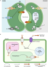
Representation of the potential for cell membrane water flux to be tuned at different cellular locations within the leaf to influence substomatal cavity humidity. (A) Each cell, including cells forming the leaf epidermis, palisade mesophyll cells, spongy mesophyll cells or cells surrounding leaf veins, or guard cells in the stomata can regulate the water flux across their plasma and tonoplast membranes, and other organelle membranes; (B) The humidity within leaf air spaces changes in response to environmental conditions, and cell signaling influences membrane water flux, here lighter shading represents lower humidity and darker shading represents higher humidity. Previous studies have reported values of relative humidity inside leaves varying from as low as 70% up to saturated; plants can modify leaf air space humidity by regulating the flux of water that moves in and out of veins and leaf mesophyll cells (following Wong et al., 2022).
Figure 2
Figure 2
Aquaporin localisation and gating influences solute flux across cell membranes. The magnitude of water flux through the aquaporins in cell membranes can be controlled by changing the abundance of different aquaporins in the cell membrane, altering the localisation of aquaporins within the cell, changing the stoichiometry of aquaporin tetramers, and gating of aquaporin channels via signaling and post-translational modifications. The stoichiometry of the aquaporin refers to which types of monomers are present in the aquaporin tetramer. Aquaporins tetramers have four monomeric channels and a central channel in the middle and all five of the channels can be gated independently. Note that differential gating of individual monomers is not represented in this figure, for additional examples see Tyerman et al. (2021). For some types of aquaporins the monomeric channels may be only permeable to water (represented by grey arrows), and the central channel may be permeable to other molecules (represented by black arrows) (Yu et al., 2006).
Figure 3
Figure 3
Aquaporins have multiple roles in guard cell (GC) membrane transport and signaling. (A) Modulation of the activity of aquaporins influences the aperture of stomata (Hachez et al., 2017). Known (left, guard cell 1) and putative (right, guard cell 2) examples of aquaporin roles in GC are represented, such as; (a) Aquaporin functions in regulating H2O flux across the plasma membrane (PM) (Bienert and Chaumont, 2014; Maurel et al., 2016); (b) Some aquaporins, such as AtPIP2;1, influence hydrogen peroxide (H2O2) flux; (c) Some aquaporins have roles in carbon dioxide (CO2) transport and can interact with carbonic anhydrase (CA; d), which converts between CO2 and HCO3 - (Wang et al., 2016; Groszmann et al., 2017; Zhang et al., 2018); (e) Water limitation can trigger increased abscisic acid (ABA) in plants, ABA influences Open Stomata 1 (OST1) kinase and the regulation of multiple proteins, including AtPIP2;1 (Grondin et al., 2015; Maurel et al., 2016); (f) Subsets of aquaporins can transport monovalent ions like potassium (K+; Byrt et al., 2017); (g)> Post-translational modifications (PTMs) such as phosphorylation/de-phosphorylation can switch some aquaporins between functioning more as ion channels rather than water channels (Qiu et al., 2020); AtPIP2;1 C-terminal (serine 280/283) PTMs influence water and transport, leaf hydraulics (involves 14-3-3 proteins, Prado et al., 2019) and light induced stomatal opening (Huang et al., 2020). The complement of PTMs, kinases and phosphatases involved are yet to be reported; (h) Further research is needed to resolve the complement of external signals, receptors and molecular components involved in signaling cascades that influence aquaporin function in GCs;(i) Knowledge gaps remain in our understanding of how tonoplast and PM aquaporin fluxes are coordinated. (B) Subsets of aquaporins have roles in influencing H2O2 flux across various cell membranes (plasma membrane, chloroplast envelope, tonoplast), they can influence H2O2 flux from chloroplasts to the cytosol and vacuole and they are likely to be part of more extensive signaling networks.
Figure 4
Figure 4
Factors to consider when investigating aquaporin contributions to cell biology. Changes in the abundance of many different types of aquaporins may occur following changes in environmental conditions. The localisation of aquaporins can change and the tetramer stoichiometry can change, for example aquaporins can move around in small vesicles. Aquaporin tetramers have four monomeric channels and a central channel, and all five of these changes can be gated. There is capacity for the different levels of aquaporin regulation to be regulated independently. Information about aquaporin gene transcript abundance (represented in purple) is not a suitable proxy for assessing protein abundance, they are not equivalent measures, and the total abundance of aquaporins in a sample extracted from plant tissues does not give any indication of the cellular or subcellular localisation of the aquaporin or the aquaporin stoichiometry, or protein-protein interaction status or state of gating (Fox et al., 2017). This means that it is insufficient to just test for aquaporin transcript levels, or only measure the total amount of aquaporin protein in the tissue, and it is important to test for aquaporin subcellular localisation and determine aquaporin stoichiometry, protein interactions and state of gating when assessing the extent to which aquaporins are contributing to cell membrane permeability (Maurel, 2007; Maurel et al., 2015).
Figure 5
Figure 5
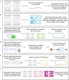
Investigation of aquaporin influence on substomatal cavity humidity requires study of many factors that can influence aquaporin function. Investigation of each of these factors needs to be cell-type specific and over a time-course following changes in VPD, examples of approaches include: (A) measuring aquaporin transcript abundance; (B) measuring aquaporin protein abundance; (C) checking for subcellular localisation of aquaporin proteins; (D) checking what post translational modifications (PTMs) occur in planta and how PTMs influence aquaporin function; (E) checking how relevant aquaporin PTMs influence localisation; and (F) studying leaf humidity in plant material with modified aquaporin function and regulation, such as aquaporin loss-of-function lines, lines over-expressing aquaporins, and potentially lines where aquaporins are edited to alter their potential for PTMs.

Similar articles

Cited by

References

    1. Andersen T. G., Liang D., Halkier B. A., White R. (2014). Grafting arabidopsis. Bio-protocol 4, e1164. doi: 10.21769/BioProtoc.1164 - DOI
    1. Ariani A., Barozzi F., Sebastiani L., di Toppi L. S., di Sansebastiano G. P., Andreucci A. (2019). AQUA1 is a mercury sensitive poplar aquaporin regulated at transcriptional and post-translational levels by zn stress. Plant Physiol. Biochem. 135, 588–600. doi: 10.1016/j.plaphy.2018.10.038 - DOI - PubMed
    1. Attia Z., Dalal A., Moshelion M. (2020). Vascular bundle sheath and mesophyll cells modulate leaf water balance in response to chitin. Plant J. 101, 1368–1377. doi: 10.1111/tpj.14598 - DOI - PubMed
    1. Azad A. K., Sawa Y., Ishikawa T., Shibata H. (2004. a). Characterization of protein phosphatase 2A acting on phosphorylated plasma membrane aquaporin of tulip petals. Bioscience Biotechnol Biochem. 68, 1170–1174. doi: 10.1271/bbb.68.1170 - DOI - PubMed
    1. Azad A. K., Sawa Y., Ishikawa T., Shibata H. (2004. b). Phosphorylation of plasma membrane aquaporin regulates temperature-dependent opening of tulip petals. Plant Cell Physiol. 45, 608–617. doi: 10.1093/pcp/pch069 - DOI - PubMed

LinkOut - more resources