

Intracranial inflammatory polyp with cerebellopontine compression and leptomeningitis secondary to chronic otitis in a red kangaroo
- PMID: 37615172
- PMCID: PMC10621546
- DOI: 10.1177/10406387231195848
Intracranial inflammatory polyp with cerebellopontine compression and leptomeningitis secondary to chronic otitis in a red kangaroo
Abstract
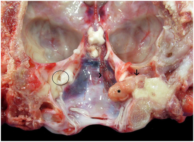
CNS lesions associated with chronic otitis have not been reported in red kangaroos (Macropus rufus), to our knowledge. Here we describe an intracranial inflammatory polyp secondary to chronic otitis in a 6-y-old female red kangaroo with right auricular discharge, loss of balance, and head tilt. Autopsy highlighted a pale-yellow, firm, intracranial polypoid growth that extended from the right tympanic cavity through the internal acoustic meatus and intracranially, with compression of the right cerebellopontine angle. Anaerobic bacterial culture yielded Bacteroides pyogenes from fresh brain and a right external ear swab. Histologically, the tympanic cavity was effaced by neutrophils and macrophages surrounded by lymphocytes and plasma cells, as well as edematous fibrovascular tissue. The epithelial lining of the mucoperiosteum was hyperplastic, with epithelial pseudoglands surrounded by fibrovascular tissue. Areas of temporal bone lysis and remodeling were associated with the inflammatory changes, which occasionally surrounded adjacent nerves. Fibrovascular tissue and inflammatory cells extended from the tympanic cavity through the internal acoustic meatus and into the intracranial cavity, forming the polypoid growth observed grossly; the polyp consisted of a dense core of fibrovascular tissue with scattered clusters of neutrophils and foamy macrophages. Lymphocytes and plasma cells surrounded the leptomeningeal perivascular spaces in the brainstem, cerebellum, and occipital lobe.
Keywords: chronic otitis; macropod; neurologic disease; pathology; red kangaroos.
Conflict of interest statement
Declaration of conflicting interestsThe authors declared no potential conflicts of interest with respect to the research, authorship, and/or publication of this article.
Figures

Similar articles
-
Magnetic resonance imaging findings in a red kangaroo (Macropus rufus) with otitis.J Zoo Wildl Med. 2008 Dec;39(4):667-70. doi: 10.1638/2008-0020.1. J Zoo Wildl Med. 2008. PMID: 19110716
-
Suppurative otitis and ascending meningoencephalitis associated with Bacteroides tectus and Porphyromonas gulae in a captive Parma wallaby (Macropus parma) with toxoplasmosis.J Vet Diagn Invest. 2014 Sep;26(5):683-8. doi: 10.1177/1040638714543676. Epub 2014 Jul 23. J Vet Diagn Invest. 2014. PMID: 25057163
-
Ventilatory accommodation of oxygen demand and respiratory water loss in kangaroos from mesic and arid environments, the eastern grey kangaroo (Macropus giganteus) and the red kangaroo (Macropus rufus).Physiol Biochem Zool. 2000 May-Jun;73(3):382-8. doi: 10.1086/316752. Physiol Biochem Zool. 2000. PMID: 10893178
-
Optimum management of the discharging ear.Drugs. 1992 Feb;43(2):219-35. doi: 10.2165/00003495-199243020-00008. Drugs. 1992. PMID: 1372220 Review.
-
Mortality trends for five species of macropods from a single institution from 1995 to 2016.Zoo Biol. 2022 Jan;41(1):44-49. doi: 10.1002/zoo.21649. Epub 2021 Aug 29. Zoo Biol. 2022. PMID: 34455635 Review.
References
-
- Allen AL, et al.. A retrospective study of brain lesions in goats submitted to three veterinary diagnostic laboratories. J Vet Diagn Invest 2013;25:482–489. - PubMed
-
- Bailey GD, et al.. Bacteroides heparinolyticus: deoxyribonucleic acid relatedness of strains from the oral cavity and oral-associated disease conditions of horses, cats, and humans. Int J Syst Bacteriol 1988;38:42–44.
-
- Giannitti F, et al.. Suppurative otitis and ascending meningoencephalitis associated with Bacteroides tectus and Porphyromonas gulae in a captive Parma wallaby (Macropus parma) with toxoplasmosis. J Vet Diagn Invest 2014;26:683–688. - PubMed
-
- Love DN, et al.. Bacteroides species from the oral cavity and oral-associated diseases of cats. Vet Microbiol 1989;19:275–281. - PubMed
MeSH terms
LinkOut - more resources
Full Text Sources
