

Preclinical and clinical trials of oncolytic vaccinia virus in cancer immunotherapy: a comprehensive review
- PMID: 37615308
- PMCID: PMC10546091
- DOI: 10.20892/j.issn.2095-3941.2023.0202
Preclinical and clinical trials of oncolytic vaccinia virus in cancer immunotherapy: a comprehensive review
Abstract
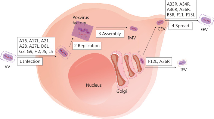
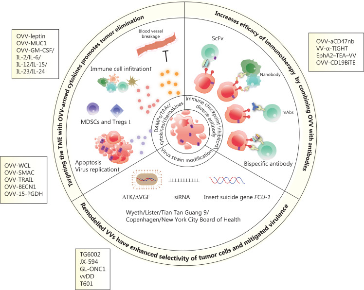
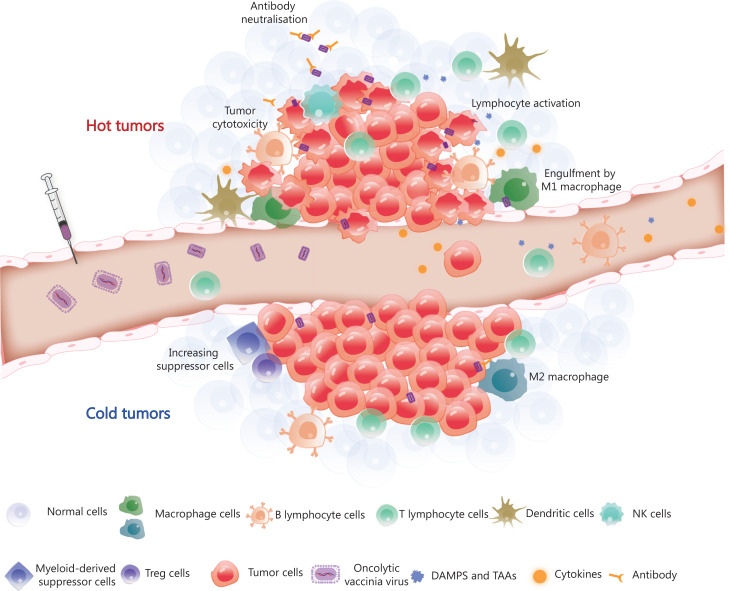
Oncolytic virotherapy has emerged as a promising treatment for human cancers owing to an ability to elicit curative effects via systemic administration. Tumor cells often create an unfavorable immunosuppressive microenvironment that degrade viral structures and impede viral replication; however, recent studies have established that viruses altered via genetic modifications can serve as effective oncolytic agents to combat hostile tumor environments. Specifically, oncolytic vaccinia virus (OVV) has gained popularity owing to its safety, potential for systemic delivery, and large gene insertion capacity. This review highlights current research on the use of engineered mutated viruses and gene-armed OVVs to reverse the tumor microenvironment and enhance antitumor activity in vitro and in vivo, and provides an overview of ongoing clinical trials and combination therapies. In addition, we discuss the potential benefits and drawbacks of OVV as a cancer therapy, and explore different perspectives in this field.
Keywords: Oncolytic virotherapy; arming strategy; engineered virus; oncolytic vaccinia virus.
Copyright © 2023 Cancer Biology & Medicine.
Conflict of interest statement
No potential conflicts of interest are disclosed.
Figures
Similar articles
-
Recent progress in combination therapy of oncolytic vaccinia virus.Front Immunol. 2024 Mar 13;15:1272351. doi: 10.3389/fimmu.2024.1272351. eCollection 2024. Front Immunol. 2024. PMID: 38558795 Free PMC article. Review.
-
Intratumoral expression of interleukin 23 variants using oncolytic vaccinia virus elicit potent antitumor effects on multiple tumor models via tumor microenvironment modulation.Theranostics. 2021 May 3;11(14):6668-6681. doi: 10.7150/thno.56494. eCollection 2021. Theranostics. 2021. PMID: 34093846 Free PMC article.
-
Oncolytic and immunologic cancer therapy with GM-CSF-armed vaccinia virus of Tian Tan strain Guang9.Cancer Lett. 2016 Mar 28;372(2):251-7. doi: 10.1016/j.canlet.2016.01.025. Epub 2016 Jan 21. Cancer Lett. 2016. PMID: 26803055
-
Oncolytic vaccinia virus armed with anti-CD47 nanobody elicit potent antitumor effects on multiple tumor models via enhancing innate and adoptive immunity.J Immunother Cancer. 2024 Dec 22;12(12):e009473. doi: 10.1136/jitc-2024-009473. J Immunother Cancer. 2024. PMID: 39794937 Free PMC article.
-
Research progress and development potential of oncolytic vaccinia virus.Chin Med J (Engl). 2025 Apr 5;138(7):777-791. doi: 10.1097/CM9.0000000000003585. Epub 2025 Mar 18. Chin Med J (Engl). 2025. PMID: 40097373 Free PMC article. Review.
Cited by
-
Combination of Oncolytic Virotherapy with Different Antitumor Approaches against Glioblastoma.Int J Mol Sci. 2024 Feb 7;25(4):2042. doi: 10.3390/ijms25042042. Int J Mol Sci. 2024. PMID: 38396720 Free PMC article. Review.
-
The Role of Natural Killer Cells in Oncolytic Virotherapy: Friends or Foes?Vaccines (Basel). 2024 Jun 28;12(7):721. doi: 10.3390/vaccines12070721. Vaccines (Basel). 2024. PMID: 39066359 Free PMC article. Review.
-
Oncolytic Viruses as a Novel Therapeutic Approach for Colorectal Cancer: Mechanisms, Current Advances, and Future Directions.Cancers (Basel). 2025 May 31;17(11):1854. doi: 10.3390/cancers17111854. Cancers (Basel). 2025. PMID: 40507337 Free PMC article. Review.
-
Immune checkpoint ligands expressed on mature high endothelial venules predict poor prognosis of NSCLC: have a relationship with CD8+ T lymphocytes infiltration.Front Immunol. 2024 Feb 8;15:1302761. doi: 10.3389/fimmu.2024.1302761. eCollection 2024. Front Immunol. 2024. PMID: 38390332 Free PMC article.
-
IL-10-Directed Cancer Immunotherapy: Preclinical Advances, Clinical Insights, and Future Perspectives.Cancers (Basel). 2025 Mar 17;17(6):1012. doi: 10.3390/cancers17061012. Cancers (Basel). 2025. PMID: 40149345 Free PMC article. Review.
References
-
- La-Beck NM, Jean GW, Huynh C, Alzghari SK, Lowe DB. Immune checkpoint inhibitors: new insights and current place in cancer therapy. Pharmacotherapy. 2015;35:963–76. - PubMed
Publication types
MeSH terms
LinkOut - more resources
Full Text Sources
Medical